

Role of 3D Coronal Ultrasound in Diagnosis of Accessory and Cavitated Uterine Mass: A rare Mullerian Anomaly
- PMID: 34898903
- PMCID: PMC8617215
- DOI: 10.1007/s13224-021-01474-1
Role of 3D Coronal Ultrasound in Diagnosis of Accessory and Cavitated Uterine Mass: A rare Mullerian Anomaly
Abstract
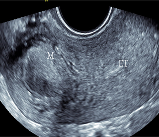
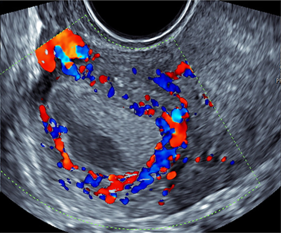
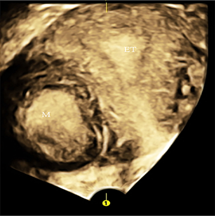
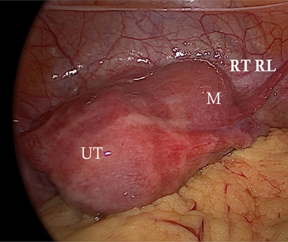
Accessory and cavitated uterine mass is rare developmental Mullerian anomaly. There is a non-communicating uterus-like mass that occurs contiguously along wall of uterus often underdiagnosed and needs expertise to identify. To raise awareness, provide information about this pathology and emphasize role of coronal 3D ultrasound in its diagnosis. A 28-year-old married female presented with dysmenorrhea and chronic pelvic pain. On ultrasound, a homogeneously isoechoic mass was noted in right lateral wall of uterus with central echogenicity. On 3D reconstruction, the main uterine cavity was normal and both cornu were visualized without any recognized Mullerian anomaly. No communication with the main endometrial cavity seen. On laparoscopy, mass was located under right round ligament insertion. Sectioning revealed chocolate colored fluid. ACUM is non-communicating uterus-like mass. It resembles uterus both macroscopically and microscopically. It represents a cavitated mass lined by endometrial glands and stroma surrounded by irregular smooth muscle cells. Criterias for diagnosing ACUM are (1) accessory cavitated mass located under round ligament; (2) normal uterus, fallopian tubes, and ovaries (3) surgical case with excised mass and pathological examination; (4) accessory cavity lined by endometrium with glands and stroma; (5) chocolate-brown fluid contents. On ultrasound, they appear solid isoechoic masses with central cystic areas separate from ovaries. 3D reconstruction can be used to rule out Mullerian anomaly. ACUM is a rare surgically treatable cause of dysmenorrhea, often underdiagnosed due to lack of knowledge about entity. 3D ultrasound can be highly accurate in making the diagnosis.
© Federation of Obstetric & Gynecological Societies of India 2021.
Conflict of interest statement
Conflict of interestThe authors declare that they have no conflict of interest.
Figures

References
-
- Betzler N, Brunes M, Anfelter P, et al. Sonografic features of accessory cavitated uterine mass (ACUM) successfully treated with robotic assisted laparoscopic surgery- a case report. Clin Obstet Gynecol Reprod Med. 2019;5:1–4. doi: 10.15761/COGRM.1000268. - DOI
Publication types
LinkOut - more resources
Full Text Sources