. 2021 Nov 24:12:750959.
doi: 10.3389/fphar.2021.750959. eCollection 2021.

A Core Omnigenic Non-coding Trait Governing Dex-Induced Osteoporotic Effects Identified Without DEXA

Affiliations

A Core Omnigenic Non-coding Trait Governing Dex-Induced Osteoporotic Effects Identified Without DEXA

Li Lu et al. Front Pharmacol. .

Abstract

Iatrogenic glucocorticoid (GC)-induced osteoporosis (GIO) is an idiosyncratic form of secondary osteoporosis. Genetic predisposition among individuals may give rise to variant degree of phenotypic changes but there has yet been a documented unified pathway to explain the idiosyncrasy. In this study, we argue that the susceptibility to epigenetic changes governing molecular cross talks along the BMP and PI3K/Akt pathway may underline how genetic background dictate GC-induced bone loss. Concordantly, osteoblasts from BALB/c or C57BL/6 neonatal mice were treated with dexamethasone for transcriptome profiling. Furthermore, we also confirmed that GC-pre-conditioned mesenchymal stem cells (MSCs) would give rise to defective osteogenesis by instigating epigenetic changes which affected the accessibility of enhancer marks. In line with these epigenetic changes, we propose that GC modulates a key regulatory network involving the scavenger receptor Cd36 in osteoblasts pre-conditioning pharmacological idiosyncrasy in GIO.

Keywords: bone loss; bone morphogenetic protein signaling pathway; cis-expression quantitative trait loci; glucocorticoid; glucocorticoid-induced osteoporosis; osteoblasts differentiation.


Conflict of interest statement

The authors declare that the research was conducted in the absence of any commercial or financial relationships that could be construed as a potential conflict of interest.

Figures
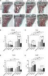
FIGURE 1
FIGURE 1
Changes of Trabecular bone of BALB/c and C57BL/6 in dexamethasone treatment. (A) Masson-Goldner Trichrome bone stains of the PTM showing the trabeculae bone loss causing by Dex. (B–E) Static parameters of PTM. *p < 0.05, **p < 0.01, significantly different from Ctrl; #p < 0.05, ##p < 0.01, significantly different between BALB/c and C57BL/6, n = 6. (%Tb.Ar, trabecular bone area percentage; Tb.N, trabecular number; Tb.Th, trabecular thickness; Tb. Sp, trabecular separation).
FIGURE 2
FIGURE 2
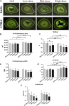
Changes of Cortical bone of BALB/c and C57BL/6 in dexamethasone treatment. (A) Images of TX transverse section fluorescence detection (injected with calcein). (B–E) Static parameters of TX. (F) Dynamic parameters of TX. *p < 0.05, **p < 0.01, significantly different from Ctrl; #p < 0.05, ##p < 0.01, significantly different between BALB/c and C57BL/6. (%Ct.Ar, percentage cortical area; %Ma.Ar, percentage marrow area; E-BFR/BS, cortical bone endosteal formation rate).
FIGURE 3
FIGURE 3
Multiple comparison of transcriptomic profiles. (A) Principal component analysis (PCA) plot for expression profiles of Day 10 ex vivo mineralizing osteoblasts from a collection of CC mice lines adapted from Khayal et al. (blue), C57/BL6 (brown/cyan) and BALB/c (red/green) mice with or without prolonged administration of Dex and differentiating Col3.1 + osteogenic stromal progenitors along osteogenic differentiation (Day 2 to Day 18) adapted from Sabik et al. (pink). (B) Expression changes in the top 50 genes contributing to the variance along PC2 listed out from top to bottom from the PCA plot. NB. Day 10 mineralizing C57/BL6 osteoblasts shared similar expression profile with mineralizing stromal progenitors upon Day 10 of their differentiation. (C) Protein-protein interaction network of co-expressed genes along PC2. (D) Expression changes in the top 50 genes contributing to the variance along PC1. NB. The top 50 genes clustered into the WGCNA module from Sabik et al. found with characteristic eigengene defined by osteogenic potential (denoted in black). (E) Expression profile of all genes in the protein-protein interaction network along PC2 in both BALB/c and C57/BL6 mice with and without prolonged administration of Dex.
FIGURE 4
FIGURE 4
Transcriptomic analysis of genes regulated by dexamethasone in mouse osteoblasts of two strains. (A) Heatmap of DEGs in Dex-treated mouse osteoblasts. (B) Scatter plot of log10 FPKM values of genes expressed in Dex-treated osteoblasts versus untreated osteoblasts. The red dots and blue dots identify that are differentially expressed at log2FoldChange ≥ with FDR ≤0.001 between untreated and Dex-treated MSCs (red dots represent the up-regulated genes, blue dots represent the down-regulated genes, gray dots represent the non-DEGs). (C) Top 20 GO Biological process terms of Gene ontology analysis of Dex-regulated genes. (D) KEGG pathway analysis enriched signaling pathways altered by Dex. (E) Gene expression shown in log2(FPKM+1) of genes relevant to PI3K-Akt and BMP signaling pathways for untreated and Dex-treated osteoblasts of BALB/c and C57BL/6.
FIGURE 5
FIGURE 5
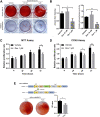
Micromolar levels of dexamethasone prevented osteogenesis and proliferation in pre-conditioned MSCs. (A,B) Alizarin Red staining and alkaline phosphatase staining of MSC-derived osteoblast culture. (C,D) MTT assay and CCK-8 assay of MSCs during the 72 h-interval of Dex preconditioning. (E) Cre-mediated silencing of NR3C1 rescued the osteogenic effect of MSCs which had subjected to Dex pre-conditioning. *p < 0.05, **p < 0.01, significant between groups.
FIGURE 6
FIGURE 6
Chromatin remodelling led to heightened enhancer accessibility to GC signaling in pre-conditioned MSCs. (A) Distribution of identified peaks MSC-derived osteoblasts with or without Dex treatment. (B) TAGLN3 and NRG1 showed differential peaks between vehicle-treated and those pre-conditioned with Dex. (C) CHIP-PCR of TAGLN3 and NR3C1 in MSC-derived osteoblasts. (D) The gene expression of TAGLN members and NRG1 along osteogenic differentiation. (E) The protein expression of TAGLN3 and NR3C1 along osteogenic differentiation. (F) Osteogenic transcription factors gene expression in pre-conditioned MSCs with or without co-treatment with SB431542 (TGF-beta inhibitor). (G,H) The proliferation and gene expression of the co-culturing of pre-conditioned MSCs with either recombinant NRG-1/EGF (NRG-1 agonists). (I) Representative differential peaks of CD36 between vehicle-treated and those pre-conditioned with Dex. (J,K) mRNA and protein expression levels of CD36 and its associated genes. *p < 0.05, *p < 0.01, significantly different from Ctrl.

Similar articles

Cited by

References

    1. Amblard D., Lafage-Proust M. H., Laib A., Thomas T., Rüegsegger P., Alexandre C., et al. (2003). Tail Suspension Induces Bone Loss in Skeletally Mature Mice in the C57BL/6J Strain but Not in the C3H/HeJ Strain. J. Bone Miner Res. 18 (3), 561–569. 10.1359/jbmr.2003.18.3.561 - DOI - PubMed
    1. Becker D. E. (2013). Basic and Clinical Pharmacology of Glucocorticosteroids. Anesth. Prog. 60 (1), 25–32. 10.2344/0003-3006-60.1.25 - DOI - PMC - PubMed
    1. Björkegren J. L. M., Kovacic J. C., Dudley J. T., Schadt E. E. (2015). Genome-Wide Significant Loci: How Important Are They? Systems Genetics to Understand Heritability of Coronary Artery Disease and Other Common Complex Disorders. J. Am. Coll. Cardiol. 65 (8), 830–845. 10.1016/j.jacc.2014.12.033 - DOI - PMC - PubMed
    1. Bouxsein M. L., Myers K. S., Shultz K. L., Donahue L. R., Rosen C. J., Beamer W. G. (2005). Ovariectomy-Induced Bone Loss Varies Among Inbred Strains of Mice. J. Bone Miner Res. 20 (7), 1085–1092. 10.1359/JBMR.050307 - DOI - PubMed
    1. Boyle E. A., Li Y. I., Pritchard J. K. (2017). An Expanded View of Complex Traits: From Polygenic to Omnigenic. Cell 169 (7), 1177–1186. 10.1016/j.cell.2017.05.038 - DOI - PMC - PubMed