Review
. 2021 Nov 26:12:785220.
doi: 10.3389/fphar.2021.785220. eCollection 2021.

Roles of Macrophages in Atherogenesis

Affiliations
Review

Roles of Macrophages in Atherogenesis

Lia Farahi et al. Front Pharmacol. .

Abstract

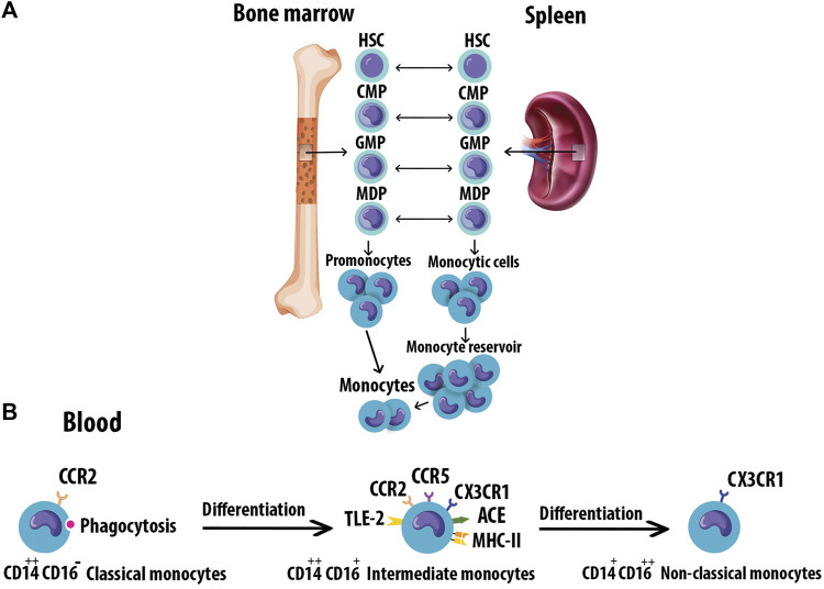
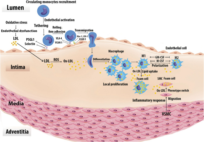
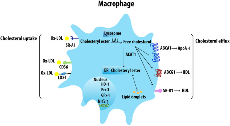
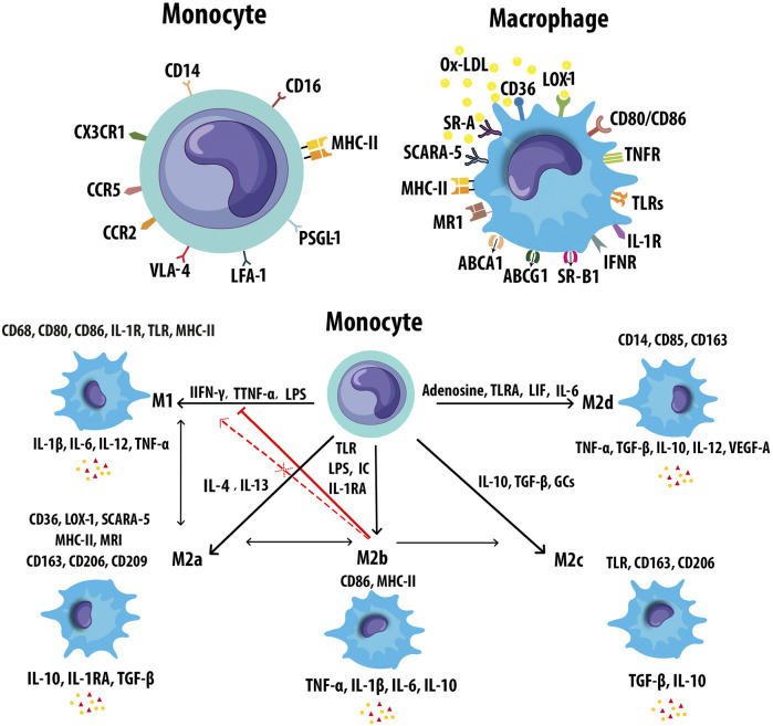
Atherosclerosis is a chronic inflammatory disease that may ultimately lead to local proteolysis, plaque rupture, and thrombotic vascular disease, resulting in myocardial infarction, stroke, and sudden cardiac death. Circulating monocytes are recruited to the arterial wall in response to inflammatory insults and differentiate into macrophages which make a critical contribution to tissue damage, wound healing, and also regression of atherosclerotic lesions. Within plaques, macrophages take up aggregated lipoproteins which have entered the vessel wall to give rise to cholesterol-engorged foam cells. Also, the macrophage phenotype is influenced by various stimuli which affect their polarization, efferocytosis, proliferation, and apoptosis. The heterogeneity of macrophages in lesions has recently been addressed by single-cell sequencing techniques. This article reviews recent advances regarding the roles of macrophages in different stages of disease pathogenesis from initiation to advanced atherosclerosis. Macrophage-based therapies for atherosclerosis management are also described.

Keywords: atherosclerosis; biomarker; inflammation; lipid; macrophages; plaque.


Conflict of interest statement

The authors declare that the research was conducted in the absence of any commercial or financial relationships that could be construed as a potential conflict of interest.

Figures

FIGURE 1
FIGURE 1
Progression of Atherosclerosis. Early fatty streak lesions are characterized by subendothelial accumulation of ApoB-LPs, the main protein constituent of lipoproteins such as VLDL and LDL which promote the recruitment of Mφs. As the atherosclerotic lesion progresses, ApoB-LP retention is amplified. Vulnerable plaques are characterized by the accumulation of ACs and defective efferocytosis, resulting in the lipid-filled necrotic core. A thinning fibrous cap reduces plaque stability and makes them susceptible to rupture and thrombus formation. Apolipoprotein B-containing lipoproteins (ApoB-LPs).
FIGURE 2
FIGURE 2
Monocyte origin and heterogeneity. (A) Blood monocytes originate from HSC-derived progenitors with myeloid restricted differentiation potential. Successive commitment steps in the bone marrow include CMPs, GMPs, and MDPs. MDPs give rise to monocytes. (B) Human monocytes are classified into three subtypes based on the differential expression of CD14 and CD16. Monocytes mature in the bone marrow and are subsequently released into the circulation. Progressively, classical monocytes CD14++CD16 give rise to non-classical monocytes CD14+CD16++ through an intermediate step of CD14++CD16+ monocytes. Hematopoietic stem cell (HSC), Common myeloid progenitors (CMPs), Granulocyte-Mφ precursors (GMPs), Mφ/Dendritic cell progenitor cells (MDPs).
FIGURE 3
FIGURE 3
Foam cell formation. Monocytes are recruited to the vascular arginase wall in response to Ox-LDL. Specific adhesion molecules such as the selectins, VCAM-1, and ICAM-1 are expressed on the surface of activated vascular ECs, mediate monocyte adhesion. Once adherent, the monocytes enter the intima and differentiate into Mφs. The differentiation process may be mediated by GM-CSF and M-CSF. Local Mφ proliferation contributes to lesion growth. Upon extensive uptake of Ox-LDL via SRs, Mφs are ultimately turned into foam cells. Chemoattractants, growth factors, and cytokines also promote SMC proliferation, uptake of Ox-LDL, and eventually conversion to foam cells. Foam cells derived from SMCs together with those derived from Mφs generate the fatty lesion.
FIGURE 4
FIGURE 4
Macrophage cholesterol uptake and efflux. Mφs uptake VLDL and Ox-LDL via SRs including SR-A1, CD36, and LOX-1. The internalized LDL is esterified by acetyl-coenzyme A acetyltransferases and stored in lipid droplets. The ester group is removed from cholesteryl by neutral cholesteryl ester hydrolase through lysosomal acid lipase (LAL) to release FC. ABCA1 transporter mediates the FC efflux from Mφs with ApoA-1. ABCG1 and SR-B1 also efflux FC to mature HDL. Acetyl-CoA acetyltransferase 1 (ACAT1).
FIGURE 5
FIGURE 5
Markers of monocyte and macrophages. (A) Monocytes and macrophages express an ample variety of receptors that modulate monocyte and Mφ activation. (B) Mφs can polarize to M1 or M2 (M2a, M2b, M2c, and M2d) in response to different inducers and secrete pro- and anti-inflammatory cytokines respectively. M2b Mφs can convert to the other M2 subtypes in response to activated factors. M2b Mφs inhibit the conversion from monocyte to M1 Mφs. M2b also cannot repolarize to M1 during exposure to M1 inducer. Leukemia inhibitory factor (LIF), mannose receptor (MR).
FIGURE 6
FIGURE 6
Macrophage polarization signaling pathways. M1 phenotypes express transcription factors such as NF-κB, STAT1, AP-1, and IRF3. M2 subtypes are stimulated by the Th2 cytokines IL-4 and IL-13 which bind to the receptor IL-4Rα and cause the activation of STAT6, IRF4, and PPAR-γ. The intracellular glucocorticoids also promote the transcription of anti-inflammatory genes. IL-10 activates STAT3 and subsequent upregulation of SOCS3. Signal transducer and activator of transcription 1 (STAT1), interferon regulatory factor -3 (IRF3), nitric oxide synthase 2 (NOS2), suppressor of cytokine signaling 3 (SOCS3), arginase 1 (Arg1).
FIGURE 7
FIGURE 7
Modes of macrophage death. Different modes of cell death including apoptosis, pyroptosis, necroptosis, parthanatos, and ferroptosis have unique activating stimuli and present different signaling pathways and distinct physiological outcomes. Inhibitors of the divalent metal transporter 1 (DMT1).
FIGURE 8
FIGURE 8
Interactions of efferocytosis signaling. Find-me signals recruit phagocytes to the sites of cell death. The dying cells will express Eat-me signals to facilitate interactions with phagocytes. Non-ACs send a Don’t-eat-me signal to avoid efferocytosis when exposed to phagocytes. Defective efferocytosis leading to inefficient clearance of ACs and subsequent necrosis and inflammation in plaques.

References

    1. Abdelaziz M. H., Abdelwahab S. F., Wan J., Cai W., Huixuan W., Jianjun C., et al. (2020). Alternatively Activated Macrophages; a Double-Edged Sword in Allergic Asthma. J. Transl Med. 18 (1), 58. 10.1186/s12967-020-02251-w - DOI - PMC - PubMed
    1. Alderson L. M., Endemann G., Lindsey S., Pronczuk A., Hoover R. L., Hayes K. C. (1986). LDL Enhances Monocyte Adhesion to Endothelial Cells In Vitro . Am. J. Pathol. 123 (2), 334–342. - PMC - PubMed
    1. Apostolopoulos J., Davenport P., Tipping P. G. (1996). Interleukin-8 Production by Macrophages from Atheromatous Plaques. Arterioscler Thromb. Vasc. Biol. 16 (8), 1007–1012. 10.1161/01.atv.16.8.1007 - DOI - PubMed
    1. Apostolov E. O., Shah S. V., Ray D., Basnakian A. G. (2009). Scavenger Receptors of Endothelial Cells Mediate the Uptake and Cellular Proatherogenic Effects of Carbamylated LDL. Arterioscler Thromb. Vasc. Biol. 29 (10), 1622–1630. 10.1161/atvbaha.109.189795 - DOI - PMC - PubMed
    1. Arango Duque G., Descoteaux A. (2014). Macrophage Cytokines: Involvement in Immunity and Infectious Diseases. Front. Immunol. 5, 491. 10.3389/fimmu.2014.00491 - DOI - PMC - PubMed