

Vitamin D Supplementation Improves Uterine Receptivity in a Rat Model of Vitamin D Deficiency: A Possible Role of HOXA-10/FKBP52 Axis
- PMID: 34899377
- PMCID: PMC8655728
- DOI: 10.3389/fphys.2021.744548
Vitamin D Supplementation Improves Uterine Receptivity in a Rat Model of Vitamin D Deficiency: A Possible Role of HOXA-10/FKBP52 Axis
Abstract
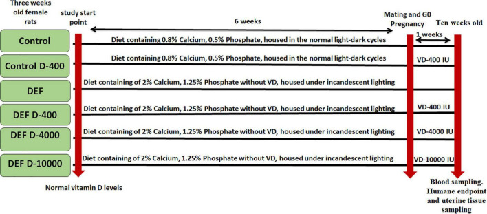
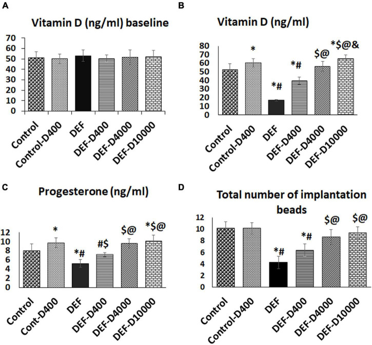
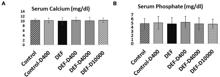
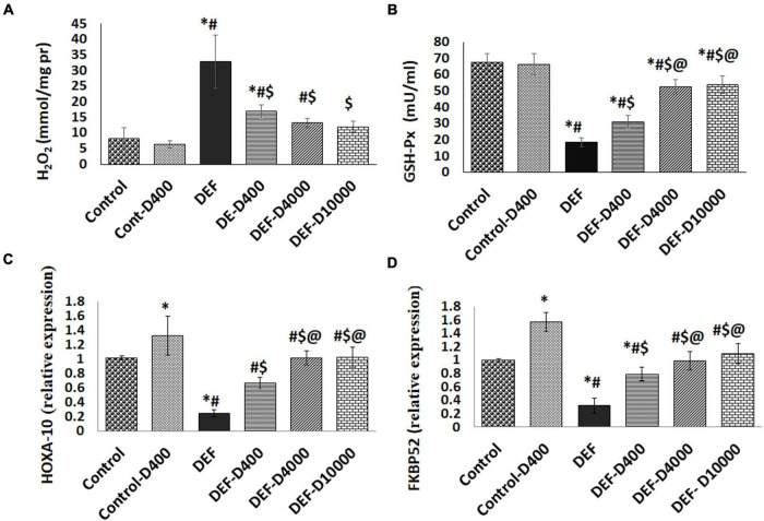
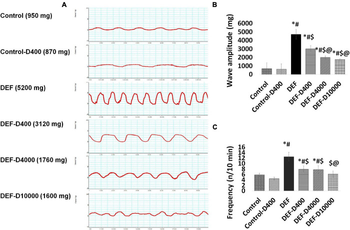
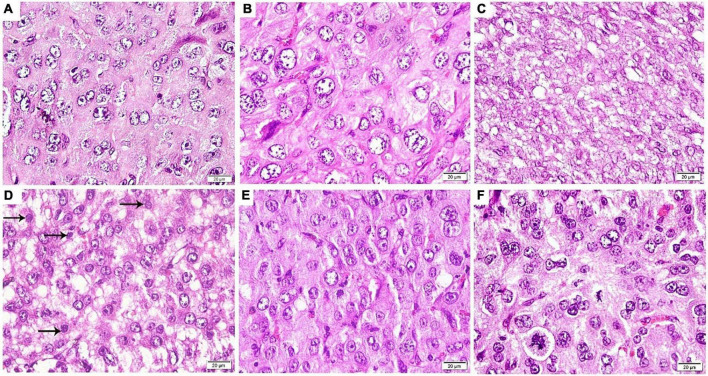
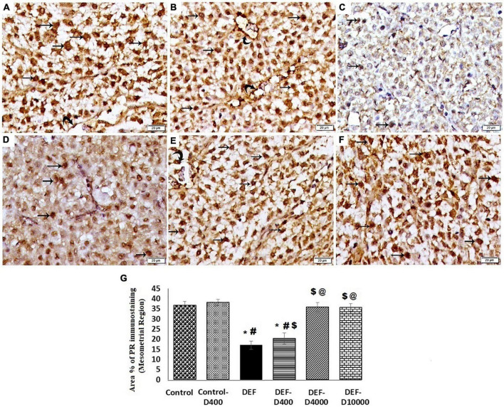
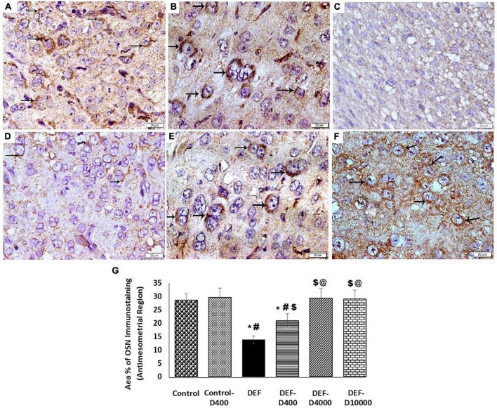
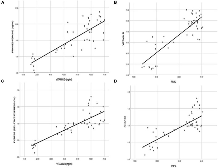
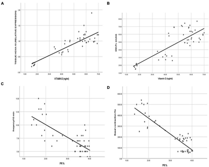
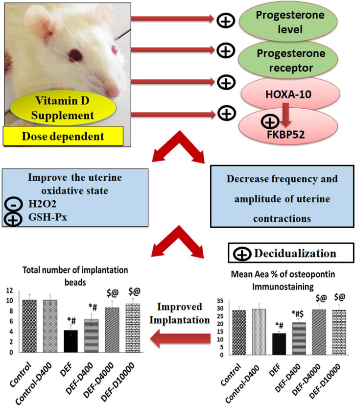
Synchronized uterine receptivity with the time of implantation is crucial for pregnancy continuity. Vitamin D (VD) deficiency has been linked to the failure of implantation. Therefore, we tested the link between the Homeobox transcription factor-10/immunophilin FK506-binding protein 52 (HOXA-10/FKBP52) axis and the uterine receptivity in VD-deficient rats. The effect of VD supplementation at different doses was also investigated. Forty-eight pregnant rats were divided into six groups (eight/group); normal control rats fed with standard chow (control), control rats supplemented with VD (equivalent dose of 400 IU/day) (control-D400). VD-deficient group (DEF) and the three VD deficiency groups with VD supplementation were equivalent to 400, 4,000, and 10,000 IU/day (DEF-D400, DEF-D4000, and DEF-D10000, respectively). The expression levels of HOXA-10/FKBP52, progesterone level, and histological evaluation of decidualization using osteopontin (OSN) and progesterone receptor (PGR) were estimated. An assessment of the uterine contractility was conducted for all rats. This study showed the downregulation of HOXA-10/FKBP52 together with increased amplitude and frequency of the uterine contractility in the DEF group compared to control. VD dose-dependent supplementation restored progesterone/receptor competency, upregulated the expressional response of HOXA-10 and its downstream FKBP52, and improved uterine receptivity and endometrial decidualization at the time of implantation that was documented by increased area% of OSN and the number of implantation beads.
Keywords: HOXA-10/FKBP52; contraction; decidualization; progesterone receptor; vitamin D.
Copyright © 2021 Ashour, Gamal, Sadek, Rashed, Hussein, Kamar, Ateyya, Mehesen and ShamsEldeen.
Conflict of interest statement
The authors declare that the research was conducted in the absence of any commercial or financial relationships that could be construed as a potential conflict of interest.
Figures
Similar articles
-
Proteomic analysis identifies immunophilin FK506 binding protein 4 (FKBP52) as a downstream target of Hoxa10 in the periimplantation mouse uterus.Mol Endocrinol. 2005 Mar;19(3):683-97. doi: 10.1210/me.2004-0332. Epub 2004 Nov 4. Mol Endocrinol. 2005. PMID: 15528267
-
Uterine FK506-binding protein 52 (FKBP52)-peroxiredoxin-6 (PRDX6) signaling protects pregnancy from overt oxidative stress.Proc Natl Acad Sci U S A. 2010 Aug 31;107(35):15577-82. doi: 10.1073/pnas.1009324107. Epub 2010 Aug 16. Proc Natl Acad Sci U S A. 2010. PMID: 20713718 Free PMC article.
-
Vitamin D supplementation blocks pulmonary structural and functional changes in a rat model of perinatal vitamin D deficiency.Am J Physiol Lung Cell Mol Physiol. 2014 Dec 1;307(11):L859-67. doi: 10.1152/ajplung.00032.2014. Epub 2014 Oct 10. Am J Physiol Lung Cell Mol Physiol. 2014. PMID: 25305247 Free PMC article.
-
Progesterone receptor requires a co-chaperone for signalling in uterine biology and implantation.Reprod Biomed Online. 2006 Nov;13(5):651-60. doi: 10.1016/s1472-6483(10)60655-4. Reprod Biomed Online. 2006. PMID: 17169175 Review.
-
The roles and expression of HOXA/Hoxa10 gene: A prospective marker of mammalian female fertility?Reprod Biol. 2022 Jun;22(2):100647. doi: 10.1016/j.repbio.2022.100647. Epub 2022 May 9. Reprod Biol. 2022. PMID: 35550944 Review.
Cited by
-
Supplementation with vitamin D improves the embryo quality in in vitro fertilization (IVF) programs, independently of the patients' basal vitamin D status.Arch Gynecol Obstet. 2024 Jun;309(6):2881-2890. doi: 10.1007/s00404-024-07473-7. Epub 2024 Apr 5. Arch Gynecol Obstet. 2024. PMID: 38580857 Free PMC article.
-
The Treatment of Complementary and Alternative Medicine on Female Infertility Caused by Endometrial Factors.Evid Based Complement Alternat Med. 2022 Sep 7;2022:4624311. doi: 10.1155/2022/4624311. eCollection 2022. Evid Based Complement Alternat Med. 2022. PMID: 36118081 Free PMC article. Review.
-
Vitamin D and reproductive disorders: a comprehensive review with a focus on endometriosis.Reprod Health. 2024 May 2;21(1):61. doi: 10.1186/s12978-024-01797-y. Reprod Health. 2024. PMID: 38698459 Free PMC article. Review.
-
Do Popular Diets Impact Fertility?Nutrients. 2024 May 31;16(11):1726. doi: 10.3390/nu16111726. Nutrients. 2024. PMID: 38892663 Free PMC article. Review.
-
Ovarian reserve modulates the impact of vitamin D deficiency on assisted reproductive outcomes in patients undergoing controlled ovarian hyperstimulation.Front Nutr. 2024 Dec 12;11:1486958. doi: 10.3389/fnut.2024.1486958. eCollection 2024. Front Nutr. 2024. PMID: 39726869 Free PMC article.
References
-
- Alaee S., Novin M. G., Yeganeh F. (2014). FKBP51 and FKBP52 as potential biomarkers for predicting endometrial receptivity and embryo implantation in assisted reproductive technologies. 2 77–81.
-
- Ashary N., Laheri S., Modi D. (2020). Homeobox genes in endometrium: from development to decidualization. Int. J. Dev. Biol. 64 227–237. - PubMed
Associated data
LinkOut - more resources
Full Text Sources
Research Materials
Miscellaneous