Review
. 2021 Nov 24:12:777997.
doi: 10.3389/fendo.2021.777997. eCollection 2021.

Appetite Regulation of TLR4-Induced Inflammatory Signaling

Affiliations
Review

Appetite Regulation of TLR4-Induced Inflammatory Signaling

Yongxiang Li et al. Front Endocrinol (Lausanne). .

Abstract

Appetite is the basis for obtaining food and maintaining normal metabolism. Toll-like receptor 4 (TLR4) is an important receptor expressed in the brain that induces inflammatory signaling after activation. Inflammation is considered to affect the homeostatic and non-homeostatic systems of appetite, which are dominated by hypothalamic and mesolimbic dopamine signaling. Although the pathological features of many types of inflammation are known, their physiological functions in appetite are largely unknown. This review mainly addresses several key issues, including the structures of the homeostatic and non-homeostatic systems. In addition, the mechanism by which TLR4-induced inflammatory signaling contributes to these two systems to regulate appetite is also discussed. This review will provide potential opportunities to develop new therapeutic interventions that control appetite under inflammatory conditions.

Keywords: SFAs; TLR4; dopamine system; hypothalamus; inflammation.


Conflict of interest statement

The authors declare that the research was conducted in the absence of any commercial or financial relationships that could be construed as a potential conflict of interest.

Figures

Graphical Abstract
Graphical Abstract
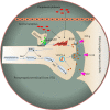
The activation of TLR4 in the regulation of appetite. Brain image was provided by smart.servier.com.
Figure 1
Figure 1
Central neuronal circuits involved in homeostatic and non-homeostatic systems. (A) Hypothalamic regulation of energy homeostasis. The Arc of the hypothalamus contains two groups of neurons: NPY/AgRP and POMC neurons. These neurons secrete orexigenic AgRP/NPY or anorexigenic POMC to the second-order neurons in the PVH and LH. In the PVN, neurons produce OT, TRH and CRH to decrease food intake. Neurons in the LH produce ORX and MCH to increase food intake. AgRP is an antagonist of anorectic MC4R expressed on neurons within the PVN. In the Arc, a local circuit exists in which POMC neurons receive GABAergic (inhibitory) input from NPY/AgRP neurons. The SF-1 nerve located in the VMH innervates POMC/CART neurons to enhance the anorexia function of POMC/CART neurons and is critically involved in the regulation of energy homeostasis. (B) The dopamine system, which contains connections between the midbrain and forebrain. The reward pathway includes dopaminergic neurons in the ventral tegmental area that project to the NAc and PFC. Dopamine, which is released from dopaminergic neurons in the VTA, binds to D1R or D2R in the NAc and D1R in the PFC, which contribute to food reward. In turn, D1R also inhibits DA and GABAergic neurons in the VTA through different GABA receptors to form negative feedback regulation. The VTA also contains GABAergic cells and Pnoc neurons that project to DA neurons to inhibit DA activity. In addition, DA neurons located in the SN also project to the DS, which potentially increases food reward behavior. Hyp, hypothalamus; Arc, arcuate nucleus; LHA, lateral hypothalamus area; PVN, paraventricular nucleus of the hypothalamus; VMH, ventromedial hypothalamus; 3V, 3rd ventricle; VTA, ventral tegmental area; NAc, nucleus accumbens; PFC, prefrontal cortex; DS, dorsal striatum; SN, substantia nigra; AgRP, agouti-related peptide; NPY, neuropeptide Y; POMC, proopiomelanocortin; Glu, glutamate; GABA, gamma-aminobutyric acid; MCH, melanin-concentrating hormone; α-MSH, α-melanocyte-stimulating hormone; CRH, corticotropin-releasing hormone; TRH, thyrotropin-releasing hormone; OT, oxytocin; DA, dopamine; D1R, dopamine 1 receptor; NOPR, nociceptin opioid peptide receptor.
Figure 2
Figure 2
Potential mechanisms by which inflammation affects DA signaling through synthesis, release, and receptor function. Inflammation and cytokines released from the periphery or produced locally by activated microglia or infiltrating macrophages contribute to oxidative stress and the production of reactive oxygen species (ROS). Increased ROS and inflammation-induced nitric oxide levels contribute to the oxidation of BH4, an essential cofactor required for the conversion of phenylalanine to tyrosine and tyrosine to L-PODA, which are necessary for the synthesis of DA. In addition, the increased glutamate release and reduced reuptake by inflammation-induced glial cells, coupled with the activation of NMDARs, may cause excitotoxicity of glutamate. In turn, these changes lead to oxidative stress and decreased DA availability. Furthermore, some evidence has shown that inflammatory cytokines reduce the expression or function of VMAT2 and/or increase the expression or function of DAT and reduce DA signaling by reducing the levels of DA D2 receptors. Dysregulation of DAT and VMAT2 increases cytosolic DA levels, leading to auto-oxidation and ROS generation. D1R, dopamine 1 receptor; D2R, dopamine 2 receptor; DDC, dopamine decarboxylase; NMDAR, N-methyl-D-aspartic acid receptor; NOS, nitric oxide synthase; ROS, reactive oxygen species; PAH, phenylalanine hydroxylase; TH, tyrosine hydroxylase; BH4, 5,6,7,8-tetrahydrobiopterin; VMAT2, vesicular monoamine transporter 2; LPODC, L-3,4-dihydroxyphenylalanine.
Figure 3
Figure 3
Summary diagram illustrating the links between inflammation in the hypothalamus and midbrain dopamine system with the deregulation of appetite. Upper panel: Lipopolysaccharide or cytokines injections, illnesses such as cancer, or infections induce high-grade inflammation. An acute increase in the local production of cytokines and chemokines is observed in the hypothalamic and midbrain dopamine systems. Cytokines, including IL-1, IL-6 and TNF-α, change the levels of neuropeptides (POMC, NPY and dopamine) involved in the hypothalamic and dopamine systems, which are associated with appetite and weight loss (the sickness behavior associated with a high-intensity infection). Lower panel: Short (3 days) or chronic (>8 weeks) HFD consumption is associated with low-grade inflammation. The increase in cytokine levels induced by a HFD also changes the levels of neuropeptides involved in the hypothalamic and dopamine systems, which appears to be associated with overeating and body weight gain and the development of obesity, leptin resistance and insulin resistance, potentially increasing appetite, and weight. However, in the first 3 days, the weight was not changed, which may protect organs from HFD-induced damage.

Similar articles

Cited by

References

    1. Kaisho T, Akira S. Toll-Like Receptors and Their Signaling Mechanism in Innate Immunity. Acta Odontol Scand (2001) 59(3):124–30. doi: 10.1080/000163501750266701 - DOI - PubMed
    1. Takeda K, Akira S. Toll Receptors and Pathogen Resistance. Cell Microbiol (2003) 5(3):143–53. doi: 10.1046/j.1462-5822.2003.00264.x - DOI - PubMed
    1. Buchanan MM, Hutchinson M, Watkins LR, Yin H. Toll-Like Receptor 4 in CNS Pathologies. J Neurochem (2010) 114(1):13–27. doi: 10.1111/j.1471-4159.2010.06736.x - DOI - PMC - PubMed
    1. Wadachi R, Hargreaves KM. Trigeminal Nociceptors Express TLR-4 and CD14: A Mechanism for Pain Due to Infection. J Dent Res (2006) 85(1):49–53. doi: 10.1177/154405910608500108 - DOI - PMC - PubMed
    1. Yirmiya R, Goshen I. Immune Modulation of Learning, Memory, Neural Plasticity and Neurogenesis. Brain Behav Immun (2011) 25(2):181–213. doi: 10.1016/j.bbi.2010.10.015 - DOI - PubMed

Publication types

LinkOut - more resources