

Regulation and Function of Interferon-Lambda (IFNλ) and Its Receptor in Asthma
- PMID: 34899691
- PMCID: PMC8660125
- DOI: 10.3389/fimmu.2021.731807
Regulation and Function of Interferon-Lambda (IFNλ) and Its Receptor in Asthma
Abstract
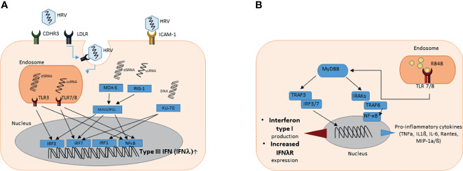
Asthma is a chronic respiratory disease affecting people of all ages, especially children, worldwide. Origins of asthma are suggested to be placed in early life with heterogeneous clinical presentation, severity and pathophysiology. Exacerbations of asthma disease can be triggered by many factors, including viral respiratory tract infections. Rhinovirus (RV) induced respiratory infections are the predominant cause of the common cold and also play a crucial role in asthma development and exacerbations. Rhinovirus mainly replicates in epithelial cells lining the upper and lower respiratory tract. Type III interferons, also known as interferon-lambda (IFNλ), are potent immune mediators of resolution of infectious diseases but they are known to be involved in autoimmune diseases as well. The protective role of type III IFNs in antiviral, antibacterial, antifungal and antiprotozoal functions is of major importance for our innate immune system. The IFNλ receptor (IFNλR) is expressed in selected types of cells like epithelial cells, thus orchestrating a specific immune response at the site of viruses and bacteria entry into the body. In asthma, IFNλ restricts the development of TH2 cells, which are induced in the airways of asthmatic patients. Several studies described type III IFNs as the predominant type of interferon increased after infection caused by respiratory viruses. It efficiently reduces viral replication, viral spread into the lungs and viral transmission from infected to naive individuals. Several reports showed that bronchial epithelial cells from asthmatic subjects have a deficient response of type III interferon after RV infection ex vivo. Toll like Receptors (TLRs) recognize pathogen-associated molecular patterns (PAMPs) expressed on infectious agents, and induce the development of antiviral and antibacterial immunity. We recently discovered that activation of TLR7/8 resulted in enhanced IFNλ receptor mRNA expression in PBMCs of healthy and asthmatic children, opening new therapeutic frontiers for rhinovirus-induced asthma. This article reviews the recent advances of the literature on the regulated expression of type III Interferons and their receptor in association with rhinovirus infection in asthmatic subjects.
Keywords: TLR7/8; asthma; epithelial cells; exacerbation; interferon; rhinovirus.
Copyright © 2021 Krammer, Sicorschi Gutu, Grund, Chiriac, Zirlik and Finotto.
Conflict of interest statement
The authors declare that the research was conducted in the absence of any commercial or financial relationships that could be construed as a potential conflict of interest.
Figures

Similar articles
-
Increased nuclear suppressor of cytokine signaling 1 in asthmatic bronchial epithelium suppresses rhinovirus induction of innate interferons.J Allergy Clin Immunol. 2015 Jul;136(1):177-188.e11. doi: 10.1016/j.jaci.2014.11.039. Epub 2015 Jan 25. J Allergy Clin Immunol. 2015. PMID: 25630941 Free PMC article.
-
Impaired type I and III interferon response to rhinovirus infection during pregnancy and asthma.Thorax. 2012 Mar;67(3):209-14. doi: 10.1136/thoraxjnl-2011-200708. Epub 2011 Sep 13. Thorax. 2012. PMID: 21917654
-
Asthmatic bronchial epithelial cells have a deficient innate immune response to infection with rhinovirus.J Exp Med. 2005 Mar 21;201(6):937-47. doi: 10.1084/jem.20041901. J Exp Med. 2005. PMID: 15781584 Free PMC article.
-
Rhinovirus-Induced Cytokine Alterations With Potential Implications in Asthma Exacerbations: A Systematic Review and Meta-Analysis.Front Immunol. 2022 Feb 15;13:782936. doi: 10.3389/fimmu.2022.782936. eCollection 2022. Front Immunol. 2022. PMID: 35242128 Free PMC article.
-
Viruses in asthma exacerbations.Curr Opin Pulm Med. 2005 Jan;11(1):21-6. doi: 10.1097/01.mcp.0000146781.11092.0d. Curr Opin Pulm Med. 2005. PMID: 15591884 Review.
Cited by
-
Altered cell function and increased replication of rhinoviruses and EV-D68 in airway epithelia of asthma patients.Front Microbiol. 2023 Mar 1;14:1106945. doi: 10.3389/fmicb.2023.1106945. eCollection 2023. Front Microbiol. 2023. PMID: 36937308 Free PMC article.
-
Innate Immune Responses by Respiratory Viruses, Including Rhinovirus, During Asthma Exacerbation.Front Immunol. 2022 Jun 20;13:865973. doi: 10.3389/fimmu.2022.865973. eCollection 2022. Front Immunol. 2022. PMID: 35795686 Free PMC article. Review.
-
Influence of polymorphic variations of IFNL, HLA, and IL-6 genes in severe cases of COVID-19.Exp Biol Med (Maywood). 2023 May;248(9):787-797. doi: 10.1177/15353702231181343. Epub 2023 Jul 15. Exp Biol Med (Maywood). 2023. PMID: 37452704 Free PMC article. Review.
-
Progress in diagnosis and treatment of difficult-to-treat asthma in children.Ther Adv Respir Dis. 2023 Jan-Dec;17:17534666231213637. doi: 10.1177/17534666231213637. Ther Adv Respir Dis. 2023. PMID: 38069568 Free PMC article. Review.
-
Type I Interferonopathies in Childhood.Balkan Med J. 2023 May 8;40(3):165-174. doi: 10.4274/balkanmedj.galenos.2023.2023-4-78. Balkan Med J. 2023. PMID: 37161741 Free PMC article.