. 2021 Nov 24:12:785222.
doi: 10.3389/fimmu.2021.785222. eCollection 2021.

Neutrophil Extracellular Traps Promote T Cell Exhaustion in the Tumor Microenvironment

Affiliations

Neutrophil Extracellular Traps Promote T Cell Exhaustion in the Tumor Microenvironment

Christof Kaltenmeier et al. Front Immunol. .

Abstract

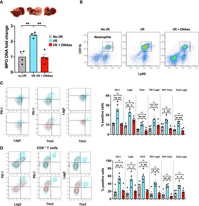
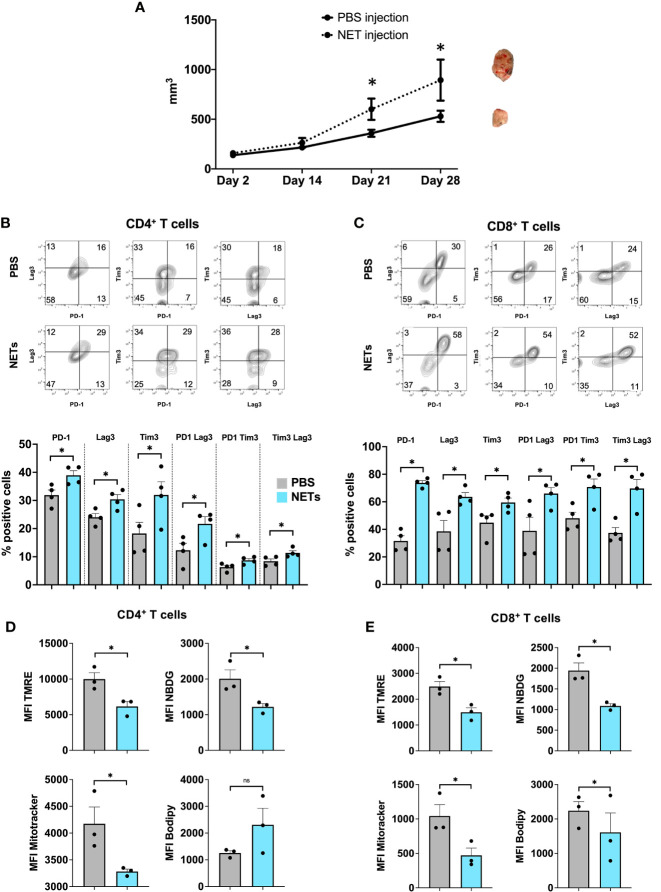
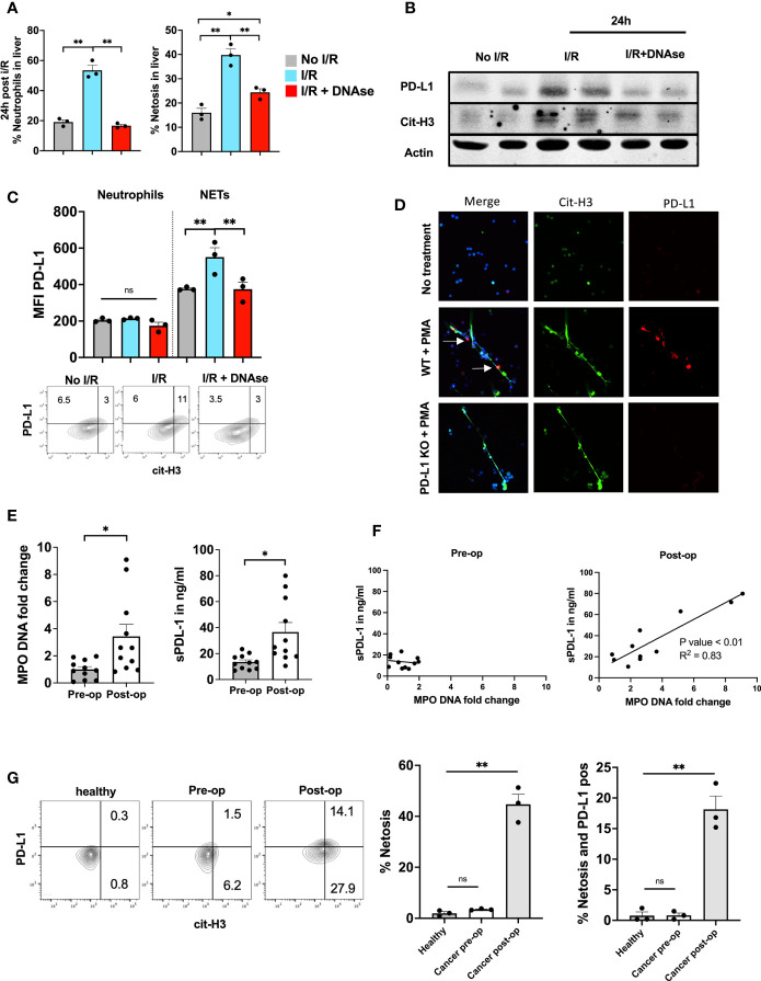
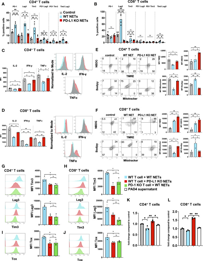
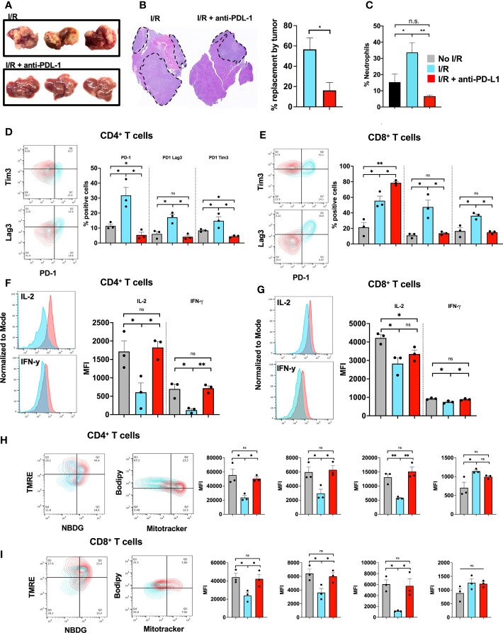
While neutrophil extracellular traps (NETs) are important for directly promoting cancer growth, little is known about their impact on immune cells within the tumor microenvironment (TME). We hypothesize that NETs can directly interact with infiltrating T cells to promote an immunosuppressive TME. Herein, to induce a NET-rich TME, we performed liver Ischemia/Reperfusion (I/R) in an established cancer metastasis model or directly injected NETs in subcutaneous tumors. In this NET-rich TME, the majority of CD4+ and CD8+ tumor infiltrating lymphocytes expressed multiple inhibitory receptors, in addition these cells showed a functional and metabolic exhausted phenotype. Targeting of NETs in vivo by treating mice with DNAse lead to decreased tumor growth, decreased NET formation and higher levels of functioning T cells. In vitro, NETs contained the immunosuppressive ligand PD-L1 responsible for T cell exhaustion and dysfunction; an effect abrogated by using PD-L1 KO NETs or culturing NETs with PD-1 KO T cells. Furthermore, we found elevated levels of sPDL-1 and MPO-DNA, a NET marker, in the serum of patients undergoing surgery for colorectal liver metastases resection. Neutrophils isolated from patients after surgery were primed to form NETs and induced exhaustion and dysfunction of human CD4+ and CD8+ T cells. We next targeted PD-L1 in vivo by injecting a blocking antibody during liver I/R. A single dose of anti-PD-L1 during surgery lead to diminished tumors at 3 weeks and functional T cells in the TME. Our data thus reveal that NETs have the capability of suppressing T cell responses through metabolic and functional exhaustion and thereby promote tumor growth. Furthermore, targeting of PD-L1 containing NETs at time of surgery with DNAse or anti-PD-L1 lead to diminished tumor growth, which represents a novel and viable strategy for sustaining immune competence within the TME.

Keywords: PD-L1; T cell dysfunction; T cell exhaustion; neutrophil extracellular traps; program death-ligand 1; tumor microenvironment.


Conflict of interest statement

The authors declare that the research was conducted in the absence of any commercial or financial relationships that could be construed as a potential conflict of interest.

Figures

Figure 1
Figure 1
T cells in a NET-rich environment exhibit a phenotype of T cell exhaustion. Mice underwent tumor inoculation followed by liver I/R on day 6 or sham treatment. Animals treated with DNAse received daily injection until tumor harvest 3 weeks later. (A) Animals that underwent I/R had elevated serum MPO-DNA levels and massive tumors. Sham mice (no I/R) and I/R mice treated with DNAse had normal levels of MPO-DNA and smaller tumors. (B) Similarly, higher levels of Neutrophils were found in the TME of I/R animals. (C, D) T cells within the TME of mice that underwent I/R express higher levels of markers for T cell exhaustion including PD1, LAG3, Tim3 and double positive markers compared to sham animals. T cell exhaustion is reversed to baseline in animals that underwent I/R and were treated with DNAse. Representative flow plots for double stained cells are shown. Bar graphs represent the mean +/- SEM of three individual experiments performed in duplicates or triplicates; *p < 0.05/**p < 0.01).
Figure 2
Figure 2
T cells in a NET-rich environment exhibit functional exhaustion. (A, B) Following I/R, T cells infiltrating the TME express reduced T cell cytokine levels; the changes return to sham levels after treatment with DNAse. Representative flow cytometry plots comparing CD4+ (A) or CD8+ (B) T cells in IR vs IR + DNAse are shown. (C, D) Following I/R, T cells in the TME have altered metabolic function measured by TMRE, Mitotracker, Bodipy and NBDG. These metabolic effects on T cell metabolism are reversed in animals treated with I/R + DNAse; Representative flow plots are shown comparing No I/R vs I/R and I/R vs I/R + DNAse. (E) T cells from mice that underwent No I/R, I/R or I/R + DNAse treatment were cultured in vitro. Proliferation was assessed following staining with a CFSE proliferation dye and 5 days of stimulation with anti-CD3/anti-CD28 beads. (F, G) Following I/R T cells infiltrating the TME express higher levels of exhaustion markers and lower levels of cytokines, these effects are reversed with DNAse. T cells in the spleen are unaffected by liver I/R or DNAse administration. (bar graphs represent the mean +/- SEM of three individual experiments performed in duplicates or triplicates: *p < 0.05/**p < 0.01). ns, not significant.
Figure 3
Figure 3
Exogenous administration of NETs leads to enhanced T cell exhaustion in the TME. (A) 8-week-old B6 mice underwent subcutaneous injection of MC38 cancer cells in addition to biweekly NETs or PBS, respectively. Mice following NET injection had larger tumors at 3-4 weeks. (B, C) Markers for T cell exhaustion were analyzed by flow cytometry, these markers were significantly upregulated on T cells in tumors that received additional injection or purified NET chromatin but not PBS. (D, E) Similarly, T cells in NET injected tumors showed decreased mitochondrial activity, decreased glucose but increased lipid uptake compared to PBS injected tumors. Bar graphs represent mean +/- SEM of two independent experiments, performed with 3 mice each. *p < 0.05, **p < 0.01.
Figure 4
Figure 4
NETs cause phenotypic, metabolic and functional T cell exhaustion in vitro. Splenic CD4+ T cells were pretreated with CD3/28 beads and then cultured for 72h in the presence of isolated WT NETs. (A, B) Markers for T cell exhaustion analyzed by flow cytometry were significantly upregulated on T cells in the presence of WT NETs. (C, D) T cell intracellular cytokines were analyzed by flow cytometry following fixation and permeabilization. T cells in the presence of WT NETs expressed significantly lower levels of intracellular cytokines. (E, F) Similarly, following culture with WT NETs, T cells were stained with metabolic markers, showing decreased metabolic activity in the presence of WT NETs. Bar graphs represent mean +/- SEM of three independent experiments, performed in duplicates or triplicates. *p < 0.05, **p < 0.01.
Figure 5
Figure 5
PD-L1 is embedded inside NET chromatin. (A) 8-week-old B6 mice underwent liver I/R without tumor injection followed by harvest of the ischemic lobe at 24h. Tissue was analyzed by flow cytometry for the presence of neutrophils and netosis. The ischemic lobe was extensively infiltrated by neutrophils and NETs unless DNAse was administered. (B) Western blot of ischemic lobes was performed showing increased levels of PD-L1 and cit-H3 in mice that underwent liver I/R This effect was reversed by treatment with DNAse. (C) Neutrophils within the ischemic lobe were stained for PD-L1 or PD-L1 and cit-H3. (D) Immunofluorescence staining shows that treatment of normal WT bone marrow derived neutrophils with PMA leads to the upregulation of PD-L1 within the extruded NET chromatin; however, no PD-L1 expression was found in PD-L1 KO NETs. Flow cytometry of isolated WT bone marrow derived Neutrophils stained with cit-H3 and PD-L1 are shown. (E) Patient serum levels of both MPO-DNA and sPD-L1 were elevated in the first post-operative days after hepatic resection. (F) MPO-DNA and sPD-L1 levels were correlated pre-/and postoperative. (G) Whole blood from healthy individuals or patients undergoing surgery was stained for the presence of neutrophil forming NETs. A greater percentage of NET forming neutrophils was found in patients on post-operative day 1 compared to pre-operative and healthy individuals. Similarly, more NETs were present and expressed PD-L1 in the post-operative patient. (n=3) Flow cytometry plots are representative of one experiment. Bar graphs represent mean +/- SEM of two independent experiments. MPO-DNA/s-PD-L1 levels (E, F) were derived from eleven healthy or postoperative patients. (G) three healthy or postoperative patients were analyzed for neutrophil NET markers and surface PD-L1 expression. *p < 0.05, **p < 0.01.
Figure 6
Figure 6
PD-L1 within NETs causes phenotypic, metabolic and functional T cell exhaustion in vitro. Splenic CD4+ and CD8+ T cells were pretreated with CD3/28 beads and then cultured for 72h in the presence of isolated WT or PD-L1 KO NETs. (A, B) Markers for T cell exhaustion were analyzed by flow cytometry were significantly upregulated on T cells in the presence of WT NET but not PD-L1 KO NETs. (C, D) T cell intracellular cytokines were analyzed by flow cytometry following fixation and permeabilization. T cells in the presence of WT NETs expressed significantly lower levels of intracellular cytokines compared to PD-L1 KO NETs. (E, F) Similarly, following culture with WT NETs, T cells were stained with metabolic markers, showing decreased mitochondrial activity, decreased glucose but increased lipid uptake in the presence of WT but not PD-L1 KO NETs. (G, H) T cells derived from PD-1 KO mice were treated in similar fashion and cocultured with WT NETs only and analyzed from Tim3 and Lag3 expression. These cells showed similar expression levels to WT T cells cultured with PD-L1 KO NETs. (I, J) Intracellular staining for TOX was performed showing decreased expression levels in T cells + PD-L1 KO NETs and PD-1 KO T cells + WT NETs. (K, L) Proliferation of CD4+ or CD8+ T cells in the presence of WT NETs or PAD4 supernatant. Bar graphs represent mean +/- SEM of at least 2 independent experiments, performed in duplicates or triplicates. *p < 0.05, **p < 0.01.
Figure 7
Figure 7
Blocking of PD-L1 at time of I/R is sufficient to abrogate tumor growth. (A) WT Mice underwent portal vein tumor infusion followed by I/R on day 6. Mice treated with I/R and anti-PD-L1 antibody had significantly decreased tumor burden at 3 weeks (B) H&E showed that animals that underwent IR and anti-PD-L1 treatment had smaller microscopic tumors. (C) Animals treated with anti-PD-L1 antibody had significantly decreased levels of neutrophils in the TME. (D, E) CD4+ and CD8+ T cells infiltrating tumors 3 weeks after liver I/R in WT mice expressed increase exhaustion markers (F, G) reduced T cell cytokine production (H, I) and altered metabolic function. Anti-PD-L1 antibody administration at time of I/R prevented these T cell changes. Bar graphs represent mean +/- SEM of two independent experiments, performed with three mice each. *p < 0.05, **p < 0.01.

References

    1. Shaul ME, Fridlender ZG. Tumour-Associated Neutrophils in Patients With Cancer. Nat Rev Clin Oncol (2019) 16(10):601–20. doi: 10.1038/s41571-019-0222-4 - DOI - PubMed
    1. Petrova V, Annicchiarico-Petruzzelli M, Melino G, Amelio I. The Hypoxic Tumour Microenvironment. Oncogenesis (2018) 7(1):10. doi: 10.1038/s41389-017-0011-9 - DOI - PMC - PubMed
    1. Brinkmann V, Reichard U, Goosmann C, Fauler B, Uhlemann Y, Weiss DS, et al. . Neutrophil Extracellular Traps Kill Bacteria. Science (2004) 303(5663):1532–5. doi: 10.1126/science.1092385 - DOI - PubMed
    1. Thålin C, Hisada Y, Lundström S, Mackman N, Wallén H. Neutrophil Extracellular Traps: Villains and Targets in Arterial, Venous, and Cancer-Associated Thrombosis. Arterioscler Thromb Vasc Biol (2019) 39(9):1724–38. doi: 10.1161/ATVBAHA.119.312463 - DOI - PMC - PubMed
    1. Singel KL, Segal BH. Neutrophils in the Tumor Microenvironment: Trying to Heal the Wound That Cannot Heal. Immunol Rev (2016) 273(1):329–43. doi: 10.1111/imr.12459 - DOI - PMC - PubMed

Publication types

MeSH terms