Review
. 2021 Nov 26:12:800242.
doi: 10.3389/fimmu.2021.800242. eCollection 2021.

How Times Have Changed! A Cornucopia of Antigens for Membranous Nephropathy

Affiliations
Review

How Times Have Changed! A Cornucopia of Antigens for Membranous Nephropathy

Tiffany N Caza et al. Front Immunol. .

Abstract

The identification of the major target antigen phospholipase A2 receptor (PLA2R) in the majority of primary (idiopathic) cases of membranous nephropathy (MN) has been followed by the rapid identification of numerous minor antigens that appear to define phenotypically distinct forms of disease. This article serves to review all the known antigens that have been shown to localize to subepithelial deposits in MN, as well as the distinctive characteristics associated with each subtype of MN. We will also shed light on the novel proteomic approaches that have allowed identification of the most recent antigens. The paradigm of an antigen normally expressed on the podocyte cell surface leading to in-situ immune complex formation, complement activation, and subsequent podocyte injury will be discussed and challenged in light of the current repertoire of multiple MN antigens. Since disease phenotypes associated with each individual target antigens can often blur the distinction between primary and secondary disease, we encourage the use of antigen-based classification of membranous nephropathy.

Keywords: antigen; autoimmune profiling; epitope spreading; mass spectrometry; membranous lupus nephritis; membranous nephropathy; serologic testing.


Conflict of interest statement

LB reports being a co-inventor on the patent “Diagnostics for Membranous Nephropathy” and receives royalty income through his employer Boston University. LB has served on advisory boards on the topic of MN and other glomerular diseases for Visterra, Ionis, Alexion and Novartis, and receives royalties from UpToDate for topics related to MN. The remaining authors declare that the research was conducted in the absence of any commercial or financial relationships that could be construed as a potential conflict of interest.

Figures

Figure 1
Figure 1
Technologies used in the identification of membranous nephropathy antigens. (A) Western blotting evaluates seroreactivity against human glomerular extract to identify podocyte antigens by mass spectrometry. (B) Laser capture microdissection (LCMD) from kidney biopsy specimens enriches for glomerular proteins, which is followed by mass spectrometry for protein identification. (C) Protein G immunoprecipitation (tissue IP) from frozen tissue enriches for immune complexes in kidney biopsy tissue for protein identification by mass spectrometry. (D) Autoimmune profiling evaluates seroreactivity against an array of peptides to identify potential autoantigens. Created with BioRender.com.
Figure 2
Figure 2
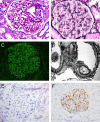
Representative histopathologic features of membranous nephropathy. (A) Glomerulus with prominent glomerular capillary loops, PAS, 400x. (B) Glomerulus with capillary loop holes/lucencies, Jones Methenamine Silver, 400x. (C) Global granular capillary loop staining, IgG immunofluorescence, 400x. (D) Electron photomicrograph of subepithelial and intramembranous electron-dense deposits. (E) Immunohistochemistry of a case of PLA2R-negative MN, demonstrating staining within podocyte cell bodies consistent with inherent low-level PLA2R expression, 200x. (F) Immunohistochemistry of a case of PLA2R-positive MN, demonstrating global granular capillary loop staining (positive result), 200x. Created with BioRender.com.
Figure 3
Figure 3
Representative histopathologic features of membranous lupus nephritis. (A) Glomerulus with mesangial expansion and prominent capillary loops, PAS, 400x. (B) Glomerulus with prominent capillary loops and endocapillary proliferation, PAS, 400x. (C) Granular mesangial and capillary loop staining, C1q immunofluorescence, 400x. (D) Electron photograph of subepithelial, intramembranous, and mesangial electron-dense deposits. (E) IgG immunofluorescence showing granular staining along tubular basement membranes, 600x. (F) IgG immunofluorescence demonstrating staining of tubular epithelial cell nuclei (‘tissue ANA’), 200x. Created with BioRender.com.
Figure 4
Figure 4
Proposed staining algorithm for phenotyping of membranous nephropathy cases. For ‘all comers’ of MN cases without a known history of systemic lupus erythematosus, we suggest staining for PLA2R, as it will identify the majority of cases (approximately 70%). For biopsies that are PLA2R negative, the clinical history, demographics, and pattern of immune reactants on biopsy could guide which antigens to evaluate. For patients with an autoimmune history (positive ANA or history of autoimmune disease), staining for EXT1/2, TGFBR3, NCAM1, and CNTN1 can together identify approximately 40% of MN cases secondary to autoimmune disease or membranous lupus nephritis. Children with MN are most commonly PLA2R-positive as adults, although SEMA3B staining will pick up approximately 10% of pediatric MN cases. In neonates, anti-neutral endopeptidase and cationic BSA are additional considerations. NELL1 and THSD7A may be enriched in patients with malignancy. The IgG pattern on biopsy is useful to choose additional antigens for staining, as THSD7A, PCDH7, and HTRA1 typically have a diffuse and global granular capillary loop pattern and NELL1 MN often shows segmental IgG staining. IgG4-related kidney disease, ANCA-associated glomerulonephritis (p-ANCA/MPO antibodies), and LRP2-associated nephropathy also often show a segmental IgG pattern along capillary loops. When MN is restricted to one light chain, cases with IgG kappa can be evaluated for SAP to identify membranous-like glomerulopathy with masked IgG kappa deposits (MGMID)*. If lambda light chain restricted or negative for SAP, IgG subclasses are helpful to identify if the MN is restricted to one subtype, for which a hematologic workup can be indicated to evaluate for an underlying lymphoproliferative disorder as a driver of disease. In patients with de novo MN following transplantation, infections, medications, graft-versus host disease, or immunodeficiency, it is common to not identify a known autoantigen at this time. *In the setting of subepithelial electron-dense deposits by electron microscopy, but no IgG staining by routine immunofluorescence microscopy, pronase digestion of FFPE tissue can ‘unmask’ immune deposits in MGMID and is required in the majority of cases. Created with BioRender.com.

References

    1. Debiec H, Guigonis V, Mougenot B, Decobert F, Haymann J, Bensman A, et al. . Antenatal Membranous Glomerulonephritis Due to Anti-Neutral Endopeptidase Antibodies. N Engl J Med (2002) 346:2053–60. doi: 10.1056/NEJMoa012895 - DOI - PubMed
    1. Madaio MP, Salant DJ, Cohen AJ, Adler S, Couser WG. Comparative Study of in Situ Immune Deposit Formation in Active and Passive Heymann Nephritis. Kidney Int (1983) 23(3):498–505. doi: 10.1038/ki.1983.47 - DOI - PubMed
    1. Hayashi N, Beck LH., Jr. Moore’s Law for Membranous Nephropathy. Kidney Int (2020) 98(5):1081–4. doi: 10.1016/j.kint.2020.06.020 - DOI - PubMed
    1. Couser WG. Membranous Nephropathy: A Long Road But Well Traveled. J Am Soc Nephrol (2005) 16(5):1184–7. doi: 10.1681/ASN.2005010087 - DOI - PubMed
    1. Sinico RA, Mezzina N, Trezzi B, Ghiggeri GM, Radice A. Immunology of Membranous Nephropathy: From Animal Models to Humans. Clin Exp Immunol (2016) 183(2):157–65. doi: 10.1111/cei.12729 - DOI - PMC - PubMed

Publication types

MeSH terms

LinkOut - more resources