. 2021 Aug 13;56(6):809-812.
doi: 10.1055/s-0041-1726064. eCollection 2021 Dec.

Use of 3D Printing in Planning the Reconstruction of Total Hip Arthroplasty: A Teaching Tool

Affiliations

Use of 3D Printing in Planning the Reconstruction of Total Hip Arthroplasty: A Teaching Tool

Marina Cornelli Girotto et al. Rev Bras Ortop (Sao Paulo). .

Abstract

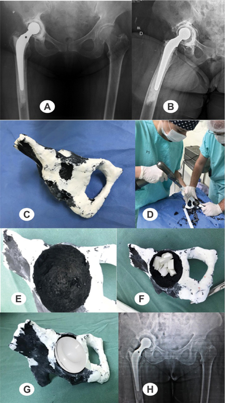
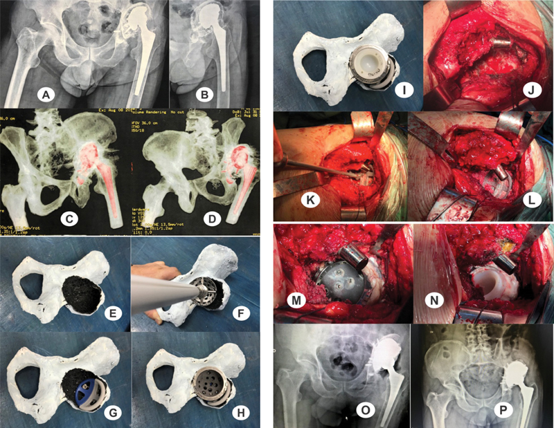
The present study aims to demonstrate how biomodels can be used as teaching tools for surgical techniques and training in a medical residency service. A case series was carried out in our orthopedics and traumatology outpatient facility using three-dimensional (3D) printing for surgical planning to contribute to the surgical teaching and training of resident physicians. Two cases were selected as examples in the present article. Biomodels enable a better understanding of the surgery by the surgical team and residents, reducing the surgical time and the risks for the patients. These models can be a good teaching method to plan reconstructions of total hip arthroplasties, evaluate and predict surgical difficulties, and optimize procedures.

Keywords: arthroplasty, replacement, hip; hip/surgery; models, anatomic; printing, three-dimensional.


Conflict of interest statement

Conflito de Interesses Os autores declaram não haver conflito de interesses.

Figures

Fig. 1
Fig. 1
Preoperative radiographs of the pelvis ( A ) and lateral view of the right hip ( B ) at the time of the review, revealing an important acetabular bone failure with loosening of the components. ( C ) View of the printed prototyping model; ( D ) residents training with prototyping models; ( E ) cavity and ( F ) acetabulum preparation with visualization of the bone defect and its filling with chopped, impacted bone; ( G ) planning of the size and position of the implant; ( H ) postoperative radiograph of the revision of the right total hip arthroplasty.
Fig. 2
Fig. 2
( A, B ) Preoperative radiographs of the pelvis and lateral view of the left hip; ( C, D ) computed tomography scans; ( E ) visualization of the extensive acetabular lesion; ( F ) study of the situation; ( G ) assembly of a new arrangement with trabeculated metal and study of its anchoring; ( H ) placement of the new acetabulum; ( I ) positioning the cemented polyethylene; ( J ) visualization of the intraoperative lesion (note the virtual absence of the acetabular roof and of the anterior and posterior walls); ( K ) assembly of the trabeculated metal structure; ( L ) placement of divergent screws and chopped, impacted bone graft; ( M ) placement of the trabeculated metal cup after deposition of a thin layer of cement between the metal components to avoid metallic contact; ( N ) acetabulum cementation at the proper position; ( O ) immediate postoperative radiograph; and ( P ) radiograph three years after the procedure (note the graft integration).
Fig. 1
Fig. 1
Radiografias de bacia ( A ) e perfil do quadril direito ( B ) pré-operatórias da paciente no momento da revisão; observa-se uma importante falha óssea acetabular com soltura deste componente. ( C ) Vista do modelo de prototipagem impresso; ( D ) treinamento de residentes com os modelos de prototipagem; ( E ) cavidade observada e ( F ) preparação do acetábulo com visualização do defeito ósseo e seu preenchimento com osso picado e impactado; ( G ) planejamento do tamanho do implante e seu posicionamento; ( H ) radiografia pós-operatória da revisão de artroplastia total do quadril direito.
Fig. 2
Fig. 2
( A, B ) radiografias pré-operatórias da bacia e de perfil do quadril esquerdo do paciente; ( C, D ) vistas da tomografia computadorizada; ( E ) visualização da extensa lesão acetabular; ( F ) estudo da situação; ( G ) montagem de uma nova disposição de uso de metal trabeculado e estudo de sua ancoragem; ( H ) colocação do novo acetábulo; ( I ) posicionamento do polietileno cimentado; ( J ) visualização da lesão intraoperatória (observar a quase ausência do teto acetabular e das paredes anterior e posterior); ( K ) montagem da estrutura de metal trabeculado; ( L ) colocação dos parafusos divergentes e enxerto ósseo picado e impactado; ( M ) colocação do acetábulo de metal trabeculado, lembrando de passar uma fina camada de cimento entre os metais para evitar contato metálico; ( N ) cimentação do acetábulo na posição adequada; ( O ) radiografia pós-operatória imediata; e ( P ) radiografia com três anos de pós-operatório (observar a integração do enxerto).

References

    1. Oliveira M F, Maia I A, Noritomi P Y, Nargi C G. Construção de Scaffolds para engenharia tecidual utilizando prototipagem rápida. Materia (Rio J) 2007;12(02):373–382.
    1. Bagaria V, Deshpande S, Rasalkar D D, Kuthe A, Paunipagar B K. Use of rapid prototyping and three-dimensional reconstruction modeling in the management of complex fractures. Eur J Radiol. 2011;80(03):814–820. - PubMed
    1. Yang S, Leong K F, Du Z, Chua C K. The design of scaffolds for use in tissue engineering. Part II. Rapid prototyping techniques. Tissue Eng. 2002;8(01):1–11. - PubMed
    1. Mothes F C, Britto A, Matsumoto F, Tonding M, Ruaro R. O uso da prototipagem tridimensional para o planejamento do tratamento das deformidades ósseas do úmero proximal. Rev Bras Ortop. 2018;53(05):595–601. - PMC - PubMed
    1. Marques T MR. Porto, Portugal: Faculdade de Engenharia da Universidade do Porto, Portugal; 2013. Definição de um modelo de planeamento pré operatório em ortopedia usando imagem digital [dissertação]