Review
. 2021 Jul 9;8(3):MMT58.
doi: 10.2217/mmt-2021-0001. eCollection 2021 Sep.

Pulmonary metastatic melanoma: current state of diagnostic imaging and treatments

Affiliations
Review

Pulmonary metastatic melanoma: current state of diagnostic imaging and treatments

Kermit S Zhang et al. Melanoma Manag. .

Abstract

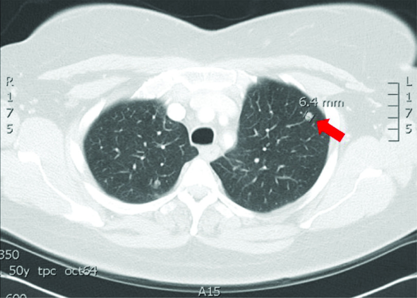
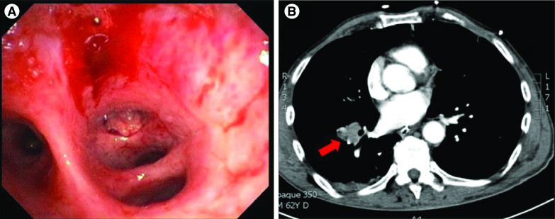
Melanoma is the deadliest form of skin cancer with an estimated incidence of over 160,000 cases annually and about 41,000 melanoma-related deaths per year worldwide. Malignant melanoma (MM) primarily occurs in the skin but has been described in other organs. Although the respiratory system is generally afflicted by tumors such as lung cancer, it is also rarely affected by primary MM. The estimated incidence of pulmonary MM of the lung accounts for 0.01% of all primary lung tumors. The current understanding of pulmonary MM of the lung pathophysiology and its management are not well established. We aim to survey current clinical modalities with a focus on diagnostic imaging and therapeutic intervention to guide providers in the management of patients with a high index of suspicion.

Keywords: diagnostics; etiology; immunotherapy; melanoma management; pulmonary malignant melanoma; targeted therapy.


Conflict of interest statement

Financial & competing interests disclosure The authors have no relevant affiliations or financial involvement with any organization or entity with a financial interest in or financial conflict with the subject matter or materials discussed in the manuscript. This includes employment, consultancies, honoraria, stock ownership or options, expert testimony, grants or patents received or pending, or royalties. No writing assistance was utilized in the production of this manuscript.

Figures

Figure 1.
Figure 1.. Pulmonary metastatic melanoma presenting as multilobulated mass.
Chest computed tomography scan of malignant melanoma in the right-upper lobe of the lungs in a 69-year-old man. (A) A round, well-demarcated, multilobulated mass approximately 5 × 3.4 cm. (B) This nodule showed strong 18F-fluorine-2-fluoro-2-deoxy-D-glucose (FDG) uptake on FDG-positron emission tomography. (C & D) Chest MRI revealed that the mass appeared to be hypointense on T1W1 and T2W1. White arrows indicate the lesion.
Figure 2.
Figure 2.. Metastatic melanoma presenting with a single pulmonary nodule.
Chest computed tomography (CT) scan with a left-upper lobe solid nodule from a 54-year-old woman who presented with 4 days of fever, nonproductive cough and scapular pain while being treated with antibiotics. The patient underwent a positron emission tomography (PET) scan 9 months after her initial CT chest scan, which demonstrated an increase in size of the known left-upper lobe noncalcified pulmonary nodule (red arrow) with focal 18F-fluorine-2-fluoro-2-deoxy-D-glucose standardized uptake value (SUV) of 3.1. She underwent video assisted mediastinal lymph node dissection and thoracoscopic resection of the enlarging left-upper lobe nodule in the anterior segment. Left-upper lobectomy was performed and pathology was consistent with metastatic malignant melanoma. Follow-up PET scan revealed several new areas of hypermetabolic activity in the right vastus intermedius muscle with SUV of 16 and a PET avid soft-tissue nodule in the left-posterior-upper-thoracic region at the level of scapular spine with maximum SUV value of 12, which was later confirmed on biopsy to be metastatic melanoma. Despite extensive workup the site of primary melanoma was not identified.
Figure 3.
Figure 3.. Metastatic melanoma presenting with a focal pulmonary parenchymal mass.
Computed tomography (CT) imaging demonstrates a 7.4 × 4.2 × 3.7 cm right-lower lobe mass (red arrow) from a 55-year-old woman, who presented to the hospital with confusion, severe right-sided headache and right facial droop. She had a history of a surgically excised melanoma on the anterior left chest 14 years prior with a positive sentinel node at that time. The patient then opted against systemic therapy. Endobronchial biopsy confirmed diagnosis of metastatic malignant melanoma.
Figure 4.
Figure 4.. Endobronchial metastasis from malignant melanoma.
This patient was a 48-year-old man who presented to the emergency department with a cough productive of yellow-green sputum. (A) Chest computed tomography (CT) scan demonstrated a soft-tissue mass measuring 5.3 × 5.9 × 5.6 cm (red arrow) in the subcarinal space extending along the azygoesophageal recess complicated with esophageal displacement toward the left side. Ultrasound-guided biopsy demonstrated atypical epithelioid cells. (B) Bronchoscopy showed gray to black colored mucosa on the lateral wall of the superior segment off the pulmonary right-lower lobe.
Figure 5.
Figure 5.. Pulmonary metastatic melanoma presenting as a focal mass with irregular margins.
(A) Bronchoscopy showing a pale exophytic lesion in the right-lower lobe anterior segmental airway in a 62-year-old man. Endobronchial cryobiopsy was significant for metastatic malignant melanoma. (B) Chest computed tomography (CT) was notable for right hilar lymphadenopathy and a 2-cm lesion with irregular margins (red arrow) was noted on the right-posterior chest wall.

References

    1. Parkin DM, Bray F, Ferlay J et al. Global cancer statistics, 2002. CA Cancer J. Clin. 55, 74–108 (2005). - PubMed
    1. Ost D, Joseph C, Menezes G. Primary pulmonary melanoma: case report and literature review. Mayo Clinic. Proc. 74, 62–66 (1999). - PubMed
    1. Dountsis A, Zisis C, Karagianni E et al. Primary malignant melanoma of the lung: a case report. World J. Surg. Oncol. 1, 26 (2003). - PMC - PubMed
    1. Jensen OA, Egedorf J. Primary malignant melanoma of the lung. Scand. J. Respir. Dis. 48, 127–135 (1967). - PubMed
    1. Jennings TA, Axiotis CA, Kress Y et al. Primary malignant melanoma of the lower respiratory tract. Am. J. Clin. Pathol. 94, 649–655 (1990). - PubMed

LinkOut - more resources