. 2021 Nov 25:9:e12559.
doi: 10.7717/peerj.12559. eCollection 2021.

The tongue of the red panda (Ailurus fulgens fulgens Cuvier, 1825)-a stereoscopy, light microscopy and ultrastructural analysis

Affiliations

The tongue of the red panda (Ailurus fulgens fulgens Cuvier, 1825)-a stereoscopy, light microscopy and ultrastructural analysis

Karolina Goździewska-Harłajczuk et al. PeerJ. .

Abstract

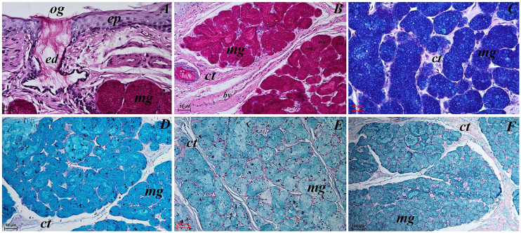
In the light of recent molecular studies, there are two phylogenetic species of the red panda (Ailurus fulgens): Ailurus fulgens fulgens and Ailurus fulgens styani. The red panda belongs to the endangered species living in the wild only in Asia and is included in the CITES list. Although the biology and diet of this species has been extensively described, the histological structure of the tongue and lingual glands has not yet been characterized in detail in relation to the lifestyle of this mammal under specific conditions and as a basis for comparative anatomical studies of the biodiversity of endemic species. Study samples were collected from two adult males of Ailurus fulgens f. held in Wrocław Zoological Garden. Both tongues were examined macroscopically; moreover, samples with lingual papillae for light microscopy and scanning electron microscopy (SEM) were collected from the apex, body and root of the tongue. Both tongues of the Ailurus fulgens f. males were approximately 9 cm long. The dorsal lingual surface was covered with mechanical and gustatory lingual papillae. Filiform papillae were observed on the apex and the body of the tongue, while small conical papillae were observed on the root of the tongue. An elongated, 1-1.5 cm long cylinder-shaped lyssa was observed in the ventral part of the apex. Moreover, most numerous and largest round in shape fungiform papillae were observed on the apex and on the border of the body and root of the tongue, located directly rostrally to 12-13 round and oval in shape vallate papillae. The SEM study showed that filiform papillae on the apex had several long secondary processes, while filiform papillae on the body of the tongue were taller and their secondary papillae were shorter than the equivalent structures on the apex of the tongue. The SEM study showed numerous taste pores on the surface of the fungiform papilla, while irregular surface of the vallate papillae, however some of them had smoother surface. Mixed glands (comprised of mucous acini and serous acini) were present within the vallum (within the connective tissue core) of the vallate papilla. Beneath the papillae more serous glands were observed, while the posterior lingual glands in the caudal part of the root of the tongue were mucoserous (mucous units were prevalent). A characteristic feature of the tongue of Ailurus fulgens f. was the presence of lyssa, which is comparable to other representatives of Carnivora, but the number of vallate papillae was individually variable. The lack of strongly developed mechanical conical papillae probably may be related to the type of plant food that is particularly dominant in red panda. Further differences between Ailurus fulgens f. and Ailurus fulgens s. cannot be excluded. The results of these studies may be useful especially for veterinarians specializing in working with exotic animals and people dealing with wildlife conservation.

Keywords: Ailurus fulgens f.; Diet; Highly selective forager; Histochemistry; Histology; Lingual papillae; Microstructure; Scanning electron microscopy.


Conflict of interest statement

The authors declare that they have no competing interests.

Figures

Figure 1
Figure 1. Photograph of the tongue of the red panda (Ailurus fulgens f.).
(A) Dorsal surface of the tongue, Bar = 1 cm. (B) Ventral surface of the apex of the tongue. (C) Magnification of the dorsal surface of the apex with numerous fungiform papillae and dominant filiform papilae. (D) Magnification of the dorsal surface of the body of the tongue. (E) Magnification of the dorsal surface of the root of the tongue. Abbreviations: a, apex of the tongue; b, body of the tongue; caudal, caudal orientation of the tongue; Fi, filiform papilla; Fu, fungiform papilla;r, root of the tongue; rostral, rostral orientation of the tongue; sCo, small conical papilla; Vp, vallate papilla.
Figure 2
Figure 2. Stereoscopic and histological analysis of the lyssa on the ventral surface of the tongue of the red panda (Ailurus fulgens f.).
(A) Transverse cross section of the lyssa. (B) Magnification of the rostral part of the lyssa. (C) Transverse cross section of the apex of the tongue with well visible location of the lyssa. (D) Magnification of the oval in shape lyssa (the rostral part of the lyssa). (E) Histomicrograph of the transverse cross section of the tongue tongue with caudal part of the lyssa. Bar = 2 mm (A–C, E); Bar = 0.2 mm (D). Abbreviations: dorsal, dorsal surface of the tongue; Fi, filiform papilla; ly, lyssa; ventral, ventral surface of the tongue.
Figure 3
Figure 3. Histological analysis of the filiform papillae and lyssa of the tongue of the of red panda (Ailurus fulgens f.).
(A) Two filiform papilla from apex of the tongue. See several processes (stratum corneum) of the keratinized stratified sqamous epithelium–blue arrows. Masson-Goldner trichrome staining. (B) Filiform papilla from body of the tongue - transverse cross section of the papilla. Masson-Goldner trichrome staining. (C) Four filiform papillae from body of the tongue with well defined interpapillary epithelium. Azan trichrome staining. (D) Magnification of the filiform papilla from body of the tongue - longitudinal cross section of the papilla. See not numerous keratohyaline granules and well visible stratum corneum of the keratinized stratified squamous epithelium (blue arrow). Masson-Goldner trichrome staining. (E) Magnification of the lyssa in the caudal part. Azan trichrome staining. Bar = 50 µm (A, B, D, E); Bar = 100 µm (C). Abbreviations: ad, adipose cells of the lyssa; bv, blood vessel; c, capsule of the lyssa; ct, connective tissue; ctc, connective tissue core; iep, interpapillary epithelium; kg, keratohyaline granules; m, muscle fibers.
Figure 4
Figure 4. Stereoscopic and SEM analysis of the apex of the tongue of the red panda (Ailurus fulgens f.).
(A) Dorsal surface of the apex of the tongue with numerous filiform papillae and round in shape fungiform papillae–SEM analysis. (B) Magnification of the filiform papilla with several (6–8) secondary processes with different length–SEM analysis. See that the cone of the main papilla is also divided. (C) Magnification of the fungiform papilla from apex of the tongue–SEM analysis. See several taste pores (blue arrow) on the surface of the fungiform papilla. (D) Magnification of the filiform papilla from lateral margin of the apex of the tongue–stereoscopic analysis. See that the cone of main papilla has several spines. Bar = 2 mm (A); Bar = 200 µm (B,C); Bar = 0.5 mm (D). Abbreviations: aj, secondary processes. Fi, filiform papilla. Fu, fungiform papilla. mp, main part of the filiform papilla.
Figure 5
Figure 5. Stereoscopic and SEM analysis of the body of the tongue of the red panda (Ailurus fulgens f.).
(A) Four fungiform papillae within the numerous filiform papillae–SEM analysis. (B) Magnification of the round in shape fungiform papilla with several taste pores on the dorsal surface of this papilla–black arrow-SEM analysis. (C) Filiform papillae and one fungiform papilla from body of the tongue. See the several secondary processes of the filiform papillae–stereoscopic analysis. (D) Numerous filiform papillae from caudal dorsal surface of the body of the tongue (directly rostral to the vallate papillae)-stereoscopic analysis. See two big fungiform papillae. Bar = 500 µm (A); Bar = 200 µm (B); Bar = 0.2 mm (C and D). Abbreviations: caudal, caudal orientation of the tongue; ec, exfoliated cell; Fi, filiform papilla; Fu, fungiform papilla; rostral, rostral orientation of the tongue.
Figure 6
Figure 6. Sstereoscopic and histological analysis of the fungiform papillae of the tongue of the red panda (Ailurus fulgens f.).
(A) Single fungiform papilla between four filiform papillae–see the elongate shape of the connective tissue core of the fungiform papilla. Masson-Goldner trichrome staining. (B) Magnification of the single fungiform papilla. Azan trichrome staining. (C) Magnification of the dorsal surface of the fungiform papillae with two taste buds within the papilary epithelium. See a thin superficial layer (stratum corneum) of the keratinized stratified squamous epithelium–blue arrow. Masson-Goldner trichrome staining. (D) Two fungiform papillae between filiform papillae–longitudinal cross section of the body of the tongue–stereoscopic analysis. Bar = 200 µm (A); Bar = 50 µm (B); Bar = 20 µm (C); Bar = 0.2 mm (D). Abbreviations: ctc, connective tissue core; ep, epithelium; Fi, filiform papilla; Fu, fungiform papilla; iep, interpapillary epithelium; tb, taste bud.
Figure 7
Figure 7. Stereoscopic and histological analysis of the vallate papillae of the tongue of the red panda (Ailurus fulgens f.).
(A) Single vallate papilla with irregular dorsal surface and with prominent papillary groove and annular pad around of the papilla–stereoscopic analysis. (B) Vallum of the papilla with well defined mixed glands within the connective tissue core of the papilla. See the prominent papillary groove (yellow asterisk). Azan trichrome staining. (C) Magnification of the lateral wall of the vallate papilla with several taste buds. Masson-Goldner trichrome staining. (D) Magnification of the elongate taste buds with their taste pores. Masson-Goldner trichrome staining. Bar = 0.2 mm (A); Bar = 200 µm (B); Bar = 50 µm (C); Bar = 20 µm (D). Abbreviations: ap, annular pad; ad, adipose cells; ctc, connective tissue core; ep, epithelium; mg, mucoserous glands; sa, serous acini; sg, serous glands; tb, taste bud; tp, taste pore; Vp, vallate papilla. * yellow asterisk, groove of the vallate papilla.
Figure 8
Figure 8. Stereoscopic and SEM analysis of the vallate papillae of red panda (Ailurus fulgens f.).
(A) Dorsal surface of the root of the tongue with all 13 vallate papillae of male 2 of red panda. (B) Microstructure of the two vallate papillae–SEM analysis. (C) Magnification of the biggest oval in shape valate papilla with clear papillary groove around of the papilla. See the irregular dorsal surface of the vallum of the papilla. (D) Longitudinal cross section of the vallum of papilla with clear and deep papillary groove. Bar = 2 mm (A and B); Bar = 0.2 mm (C and D). Abbreviations: ap, annular pad with irregular surface; Fi, filiform papilla; Fu, fungiform papila; sCo, small conical papilla; Vp, vallate papilla; *yellow asterisk, groove of the vallate papilla.
Figure 9
Figure 9. SEM analysis of the small filiform papillae from the area of the vallate papillae of the tongue of the red panda (Ailurus fulgens f.).
(A) Small filiform papillae with several short secondary conical processes: SEM analysis. (B) Magnification of the two small filiform papillae with well defined exfoliated cells: SEM analysis. Bar = 500 µm (A); Bar = 200 µm (B). Abbreviations: aj, secondary processes; ec, exfoliated cell; sFi, small filiform papilla.
Figure 10
Figure 10. Stereoscopic and SEM analysis of the root of the tongue of the red panda (Ailurus fulgens f.).
(A) Microstructure of the small conical papillae: SEM analysis. (B) Irregular dorsal surface of the root of tongue - SEM analysis. (C) Magnification of the irregular dorsal surface of the root of the tongue: SEM analysis. (D) Numerous small conical papillae: stereoscopic analysis. Bar = 500 µm (A–C); Bar = 0.5 mm (D). Abbreviations: ec, exfoliated cell; caudal, caudal orientation of the tongue; sCo, small conical papilla; rostral, rostral orientation of the tongue.
Figure 11
Figure 11. Histochemical visualization of the lingual glands concomitant to vallate papillae (gustatory gland) of the tongue of the red panda (Ailurus fulgens f.).
(A) Strong positive reaction (+++) in mucous acini (rounded secretory units) (magenta) and weakly positive reaction (+) in serous acini. PAS staining. (B) Strong strong positive reaction (+++) in mucous acini (dark blue) and weakly positive reaction (+) in serous acini (light blue). AB pH2.5 staining. (C) Magnification of mucous acini with strong positive reaction (+++) (magenta). PAS staining. (D) Magnification of mucous acini with strong positive reaction (+++) (dark blue). PAS-AB pH2.5 staining. (E) Positive reaction (++) (blue) in mucous acini. HDI staining. (F) positive reaction (++) (blue) in mucous acini. AB pH1.0 staining. (G) Positive reaction (++) (magenta) in serous acini (rounded secretory units) and positive reaction (++) - dark blue color in some of cells. PAS-AB pH2.5 staining. (H) weakly positive reaction (+) – light blue color in serous acini. HDI staining. Bar = 200 µm (A,B); Bar = 50 µm (C–H). Abbreviations: ad, adipose cells. bv, blood vessel. ep, epithelium. ct, connective tissue. ma, mucous acini; mg, mucoserous glands with dominant mucous acini (rounded secretory units); my, myoepithelial cells; sa, serous acini; sg, serous glands.
Figure 12
Figure 12. Histochemical visualization of the posterior lingual glands of the tongue of the red panda (Ailurus fulgens f.).
(A) Magnification of the excretory duct. PAS staining. (B) Mucoserous glands (with dominant of mucous acini). Strong positive reaction (+++) in mucous acini. PAS staining. (C) Strong positive reaction (+++) – dark blue color in mucous acini. PAS-AB pH2.5 staining. (D) Strong positive reaction (+++) – blue color in mucous acini. AB pH2.5 staining. (E) positive reaction (++) (blue color) in mucous acini. AB pH1.0 staining. (F) Strong positive reaction (+++) (blue color) in mucous acini. HDI staining. Bar = 20 µm (A and C); Bar = 50 µm (B, D and E); Bar = 100 µm (F). Abbreviations: bv, blood vessel; ed, excretory duct; ep, epithelium; ct, connective tissue; ma, mucous acini (round secretory units); mg, mucoserous glands with dominant mucous acini; og, opening of the excretory duct.

References

    1. Besoluk K, Eken E, Sur E. Morphological studies on lyssa in cats and dogs. Veterinární Medicína. 2006;51(10):385–489. doi: 10.17221/5582-VETMED. - DOI
    1. Boshell JL, Wilborn WH, Singh BB. Filiform papillae of cat tongue. Acta Anatomica (Basel) 1982;114(2):97–105. doi: 10.1159/000145583. - DOI - PubMed
    1. Correa AF, Sestari CE, Guimaraes GC, de Oliveira FS. Anatomical description of the crab-eating raccoon tongue-(Procyon cancrivorus) Ciência Rural. 2012;42(10):1840–1843.
    1. Čížek P, Hamouzová P, Goździewska-Harłajczuk K, Klećkowska-Nawrot J, Kvapil P. Microscopic structure of the tongue in the lesser hedgehog tenrec (Echinops telfairi, Afrosoricida) and its relation to phylogenesis. Anatomical Science International. 2020;95(3):313–322. doi: 10.1007/s12565-019-00522-1. - DOI - PubMed
    1. Dangol B. Habitat and distribution of red panda: a case study from Ranchuli VDC, Kalikot District, Nepal. 2014. MS thesis, Tribhuvan University, Central Department of Environmental Science, Tribhuvan University. Kathmandu, Nepal.

LinkOut - more resources