. 2021 Nov;11(11):3465-3480.
doi: 10.1016/j.apsb.2021.05.027. Epub 2021 May 27.

Sinomenine ester derivative inhibits glioblastoma by inducing mitochondria-dependent apoptosis and autophagy by PI3K/AKT/mTOR and AMPK/mTOR pathway

Affiliations

Sinomenine ester derivative inhibits glioblastoma by inducing mitochondria-dependent apoptosis and autophagy by PI3K/AKT/mTOR and AMPK/mTOR pathway

Xiangjin Zheng et al. Acta Pharm Sin B. 2021 Nov.

Abstract

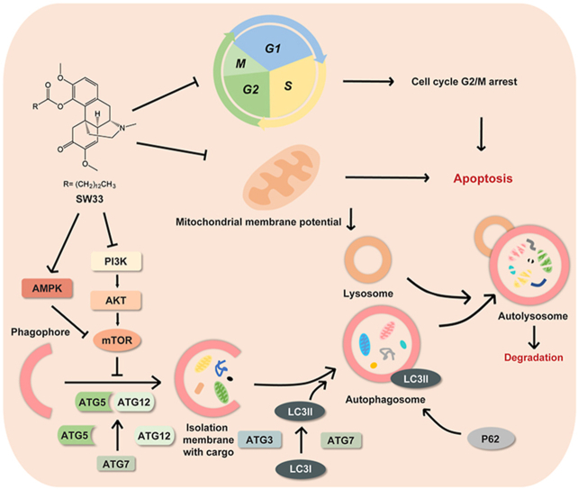
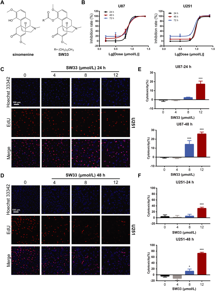
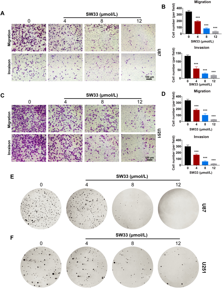
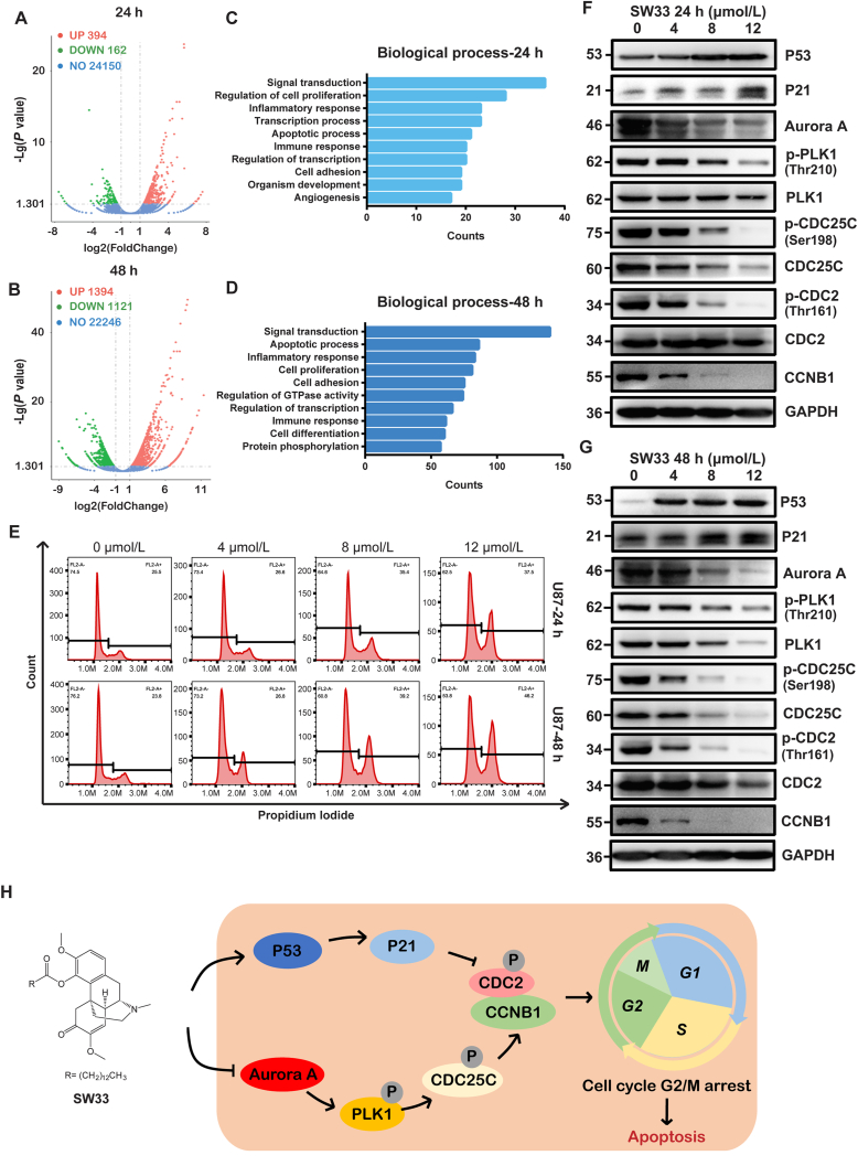
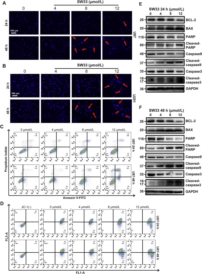
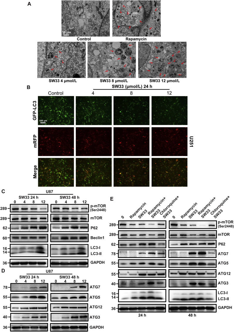
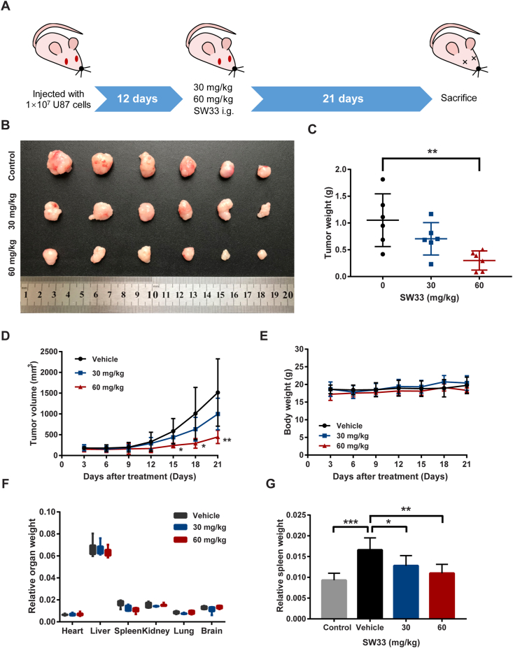
Glioblastoma multiforme (GBM) in the central nervous system is the most lethal advanced glioma and currently there is no effective treatment for it. Studies of sinomenine, an alkaloid from the Chinese medicinal plant, Sinomenium acutum, showed that it had inhibitory effects on several kinds of cancer. Here, we synthesized a sinomenine derivative, sino-wcj-33 (SW33), tested it for antitumor activity on GBM and explored the underlying mechanism. SW33 significantly inhibited proliferation and colony formation of GBM and reduced migration and invasion of U87 and U251 cells. It also arrested the cell cycle at G2/M phase and induced mitochondria-dependent apoptosis. Differential gene enrichment analysis and pathway validation showed that SW33 exerted anti-GBM effects by regulating PI3K/AKT and AMPK signaling pathways and significantly suppressed tumorigenicity with no obvious adverse effects on the body. SW33 also induced autophagy through the PI3K/AKT/mTOR and AMPK/mTOR pathways. Thus, SW33 appears to be a promising drug for treating GBM effectively and safely.

Keywords: Anti-inflammation; Apoptosis; Autophagy; G2/M phase; GBM; SW33; Safety; mTOR.


Figures

Image 1
Graphical abstract
Figure 1
Figure 1
SW33 inhibited viability and proliferation of U87 and U251 cells. (A) Structures of SIN and SW33. (B) IC50 of SW33 on U87 and U251 cells at 24, 48, and 72 h. SW33 inhibited DNA synthesis in U251 cells at 24 (C) and 48 h (D) as measured by ethynyl deoxyuridine (EdU) incorporation. Scale bar = 100 μm. (E) Toxicity of SW33 on U87 cells at 24 and 48 h by LDH assay. (F) Toxicity of SW33 on U251 cells at 24 and 48 h by LDH assay. Data are expressed as mean ± SD (n = 3). ∗P < 0.05, ∗∗P < 0.01, ∗∗∗P < 0.001 vs. Control.
Figure 2
Figure 2
SW33 inhibited migration, invasion and colony formation of U87 and U251 cells. (A) and (B) Transwell assays showed SW33 inhibited migration and invasion ability of U87 cells. Scale bar = 100 μm. (C) and (D) Transwell assays showed SW33 inhibited migration and invasion ability of U251 cells. Scale bar = 100 μm. (E) SW33 inhibited colony formation of U87 cells by soft agar assay. (F) SW33 inhibited colony formation of U251 cells by soft agar assay. Data are expressed as mean ± SD (n = 3). ∗P < 0.05, ∗∗P < 0.01, ∗∗∗P < 0.001 vs. Control.
Figure 3
Figure 3
SW33 induced cell cycle arrest at G2/M phase in U87 and U251 cells. Visualization of differential genes by volcano plot of U251 cells treated with SW33 for 24 (A) and 48 h (B). Red points for up-regulated genes, green for down-regulated genes, and blue for unchanged genes (|log2FoldChange|>1, P < 0.05). Top ten enriched biological processes at 24 (C) and 48 h (D). (E) Flow cytometry analysis showed that SW33 arrested cell cycle at G2/M phase in U87 cells. Protein level of G2/M checkpoint by Western blotting at 24 (F) and 48 h (G). Expression of P53 and P21 was up-regulated while expression of Aurora A, p-PLK1, PLK1, p-CDC25C, CDC25C, p-CDC2, CDC2 and CCNB1 was down-regulated. (H) Schematic model for mechanism of cell cycle arrest at G2/M by SW33. Experiments were performed in triplicate.
Figure 4
Figure 4
SW33-induced apoptosis of U87 and U251 cells via mitochondrial disruption. Morphological changes in nuclei of U87 (A) and U251 (B) treated with SW33. Scale bar = 100 μm. (C) Flow cytometry using PI/Annexin V-FITC double stain showed SW33 induced apoptosis of U87 at 24 and 48 h. (D) Flow cytometry analysis using 5,5′,6,6′-tetrachloro-1,1′,3,3′-tetraethylbenzimidazolocarbocyanine iodide (JC-1) stain showed that SW33 reduced mitochondrial membrane potential of U87 at 24 and 48 h. Western blotting detection of proteins associated with apoptosis in U87 cells treated with 0, 4, 8, 12 μmol/L SW33 for 24 (E) and 48 h (F). Expression of BAX was up-regulated while ratios of cleaved PARP/PARP, cleaved caspase 9/caspase 9, cleaved caspase 3/caspase 3 and expression of BCL-2 were reduced. Experiments were performed in triplicate.
Figure 5
Figure 5
SW33 induced autophagy by regulating mTOR signaling pathway. (A) Transmission electron microscopy showing increase in phagosomes and phagolysosomes in U251 cells with increasing SW33 concentration. Scale bar = 10 μm. (B) mRFP-GFP-LC3 dual fluorescent autophagy indicators showed the ratio of GFP to mRFP was reduced in U87 cells as concentration of SW33 was increased. Scale bar = 100 μm. (C) and (D) Proteins associated with autophagy in SW33-treated U87 cells quantitated by Western blotting. Ratio of p-mTOR/mTOR was decreased and the levels of P62, Beclin1, LC3-II, ATG7, ATG5, ATG12 and ATG3 increased. (E) Autophagy protein levels in U87 cells treated with SW33, rapamycin and chloroquine for 24 and 48 h. p-mTOR/mTOR was consistently reduced after treatment with SW33 and rapamycin. A greater decrease of p-mTOR/mTOR was seen with combined SW33 and rapamycin treatment. Chloroquine reversed the SW33-mediated reduction in p-mTOR/mTOR. The level of P62 and LC3-II, as well as ATG7, ATG5, ATG12 and ATG3, increased after treatment with SW33 and rapamycin, and this was also reversed by chloroquine. Experiments were performed in triplicate.
Figure 6
Figure 6
SW33 induced autophagy of GBM cells by regulating PI3K/AKT and AMPK signaling pathways. KEGG pathway analysis of differentially expressed genes at 24 (A) and 48 h (B). (C) and (D) Protein levels of p-PI3K, PI3K, p-AKT, AKT, p-AMPK, AMPK in SW33-treated U87 and U251 cells detected by Western blotting. The ratio of p-PI3K/PI3K and p-AKT/AKT decreased while p-AMPK/AMPK increased. (E) Diagram of autophagy induced by SW33 via PI3K/AKT/mTOR and AMPK/mTOR signaling pathways. Experiments were performed in triplicate.
Figure 7
Figure 7
SW33 suppressed glioma growth in vivo. (A) Schematic diagram of GBM xenograft mouse model treated with SW33 in vivo. (B) Image of tumors in different groups treated with SW33 for three weeks. (C) Tumor weight after three weeks of treatment with SW33. (D) Tumor volume during administration of SW33. (E) Body weight during administration of SW33. (F) Relative organ weights after three weeks of treatment with SW33. (G) Changes in spleen weight of different groups after SW33 treatment for three weeks. Data are expressed as mean ± SD (n = 6). ∗P < 0.05, ∗∗P < 0.01, ∗∗∗P < 0.001 (D: vs. Vehicle).

References

    1. Ostrom Q.T., Gittleman H., Stetson L., Virk S.M., Barnholtz-Sloan J.S. Epidemiology of gliomas. Cancer Treat Res. 2015;163:1–14. - PubMed
    1. Chen R., Smith-Cohn M., Cohen A.L., Colman H. Glioma subclassifications and their clinical significance. Neurotherapeutics. 2017;14:284–297. - PMC - PubMed
    1. Bush N.A., Chang S.M., Berger M.S. Current and future strategies for treatment of glioma. Neurosurg Rev. 2017;40:1–14. - PubMed
    1. Alexander B.M., Cloughesy T.F. Adult glioblastoma. J Clin Oncol. 2017;35:2402–2409. - PubMed
    1. Ostrom Q.T., Bauchet L., Davis F.G., Deltour I., Fisher J.L., Langer C.E., et al. The epidemiology of glioma in adults: a "state of the science" review. Neuro Oncol. 2014;16:896–913. - PMC - PubMed

LinkOut - more resources