Review
. 2021 Nov 25:11:777709.
doi: 10.3389/fcimb.2021.777709. eCollection 2021.

A Review on Host- Leptospira Interactions: What We Know and Future Expectations

Affiliations
Review

A Review on Host- Leptospira Interactions: What We Know and Future Expectations

Brenda B Daroz et al. Front Cell Infect Microbiol. .

Abstract

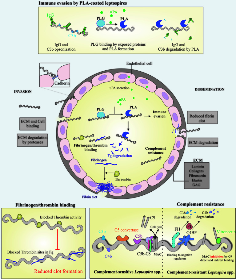
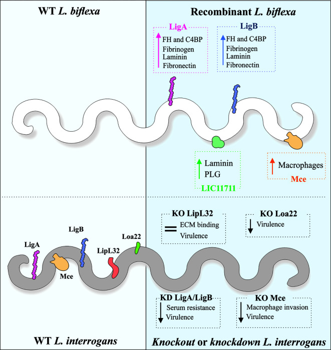
Leptospirosis is a widespread zoonosis caused by pathogenic Leptospira spp. It is considered a neglected infectious disease of human and veterinary concern. Our group has been investigating proteins annotated as hypothetical, predicted to be located on the leptospiral surface. Because of their location, these proteins may have the ability to interact with various host components, which could allow establishment of the infection. These proteins act as adherence factors by binding to host receptor molecules, such as the extracellular matrix (ECM) components laminin and glycosaminoglycans to help bacterial colonization. Leptospira also interacts with the host fibrinolytic system, which has been demonstrated to be a powerful tool for invasion mechanisms. The interaction with fibrinogen and thrombin has been shown to reduce fibrin clot formation. Additionally, the degradation of coagulation cascade components by secreted proteases or by acquired surface plasmin could also play a role in reducing clot formation, hence facilitating dissemination during infection. Interaction with host complement system regulators also plays a role in helping bacteria to evade the immune system, facilitating invasion. Interaction of Leptospira to cell receptors, such as cadherins, can contribute to investigate molecules that participate in virulence. To achieve a better understanding of the host-pathogen interaction, leptospiral mutagenesis tools have been developed and explored. This work presents several proteins that mediate binding to components of the ECM, plasma, components of the complement system and cells, to gather research achievements that can be helpful in better understanding the mechanisms of leptospiral-host interactions and discuss genetic manipulation for Leptospira spp. aimed at protein function validation.

Keywords: cadherins; components of complement system; extracellular matrix components; fibrinogen,; host-pathogen interactions; leptospiral mutagenesis tools; leptospiral proteins; plasminogen-plasmin.


Conflict of interest statement

The authors declare that the research was conducted in the absence of any commercial or financial relationships that could be construed as a potential conflict of interest.

Figures

Figure 1
Figure 1
Schematic view of colonization, invasion and evasion mechanisms displayed by pathogenic Leptospira. Leptospires can penetrate the host via breached skin or intact mucosa, taking advantage of many surface exposed proteins that are able to interact with a broad range of host components, including the extracellular matrix (ECM) components and glycosaminoglycans (GAGs). During the invasion process, leptospires can directly bind to ECM components and cell receptors, as cadherins, the latter favoring cell-cell integrity disruption (center). Leptospires interact with host plasminogen (PLG) (top and center) and induce the endothelial secretion of urokinase-type PLG activator (uPA), which in turn converts leptospires-bound PLG to its active form, plasmin (PLA). The latter, a broad-spectrum serine protease, is capable of degrading ECM components and immune mediators, as IgG and C3b, reducing opsonophagocytosis (top). One of the host mechanisms to block pathogen dissemination to other sites after endothelial lesion is the formation of fibrin clot, as a result of fibrinogen (Fg) cleavage by thrombin. In addition to Fg degradation by PLA, pathogenic Leptospira can also bind both Fg and thrombin, causing a bilateral obstruction of the fibrin clot reaction, favoring the dissemination step (left bottom), in association with ECM degradation by endogenous proteases and surface-associated PLA (center). Once in the bloodstream, leptospires must overcome one of the first lines of host defense, the complement, and this is achieved by a multitude of mechanisms, including binding to the negative complement regulators Factor H (FH) and C4 binding protein (C4BP), which participate in the degradation of C3b and C4b, respectively. Binding to terminal components C7, C8, C9 and vitronectin, would decrease membrane attack complex (MAC) formation (right bottom). Taken together, it is anticipated that these mechanisms will facilitate invasion and dissemination of Leptospira through the hosts.
Figure 2
Figure 2
Genetic tools and mutant evaluation of Leptospira. As the saprophytic L. biflexa lacks most of the virulence-associated proteins, it is used as a surrogate for the expression of pathogen-specific proteins and gain-of-function phenotype evaluation. Increased binding to ECM and plasma components has been observed in recombinant L. biflexa expressing L. interrogans proteins. Contrarily to this strategy, knockout (KO) or knockdown (KD) in the pathogenic L. interrogans has been used to evaluate loss-of-function phenotypes, in comparison to the wild-type strain. Reduced virulence in animal model was observed for KD double LigA/LigB, KO Loa22 and KO Mce mutants. KO of LipL32, the major lipoprotein of pathogenic leptospires, did not alter virulence or ECM binding.

References

    1. Adler B., de la Peña Moctezuma A. (2010). Leptospira and Leptospirosis. Vet. Microbiol. 140(3–4), 287–296. doi: 10.1016/j.vetmic.2009.03.012 - DOI - PubMed
    1. Andrade G. I., Brown P. D. (2012). A Comparative Analysis of the Attachment of Leptospira Interrogans and L. Borgpetersenii to Mammalian Cells. FEMS Immunol. Med. Microbiol. 65, 105–115. doi: 10.1111/j.1574-695X.2012.00953.x - DOI - PubMed
    1. Atzingen M. V., Barbosa A. S., De Brito T., Vasconcellos S. A., de Morais Z. M., Lima D. M., et al. . (2008). Lsa21, a Novel Leptospiral Protein Binding Adhesive Matrix Molecules and Present During Human Infection. BMC Microbiol. 8 (8), 70. doi: 10.1186/1471-2180-8-70 - DOI - PMC - PubMed
    1. Atzingen M. V., Gómez R. M., Schattner M., Pretre G., Gonçales A. P., de Morais Z. M., et al. . (2009). Lp95, a Novel Leptospiral Protein That Binds Extracellular Matrix Components and Activates E-Selectin on Endothelial Cells. J. Infect. 59, 264–276. doi: 10.1016/j.jinf.2009.07.010 - DOI - PubMed
    1. Ballard S. A., Williamson M., Adler B., Vinh T., Faine S. (1986). Interactions of Virulent and Avirulent Leptospires With Primary Cultures of Renal Epithelial Cells. J. Med. Microbiol. 21, 59–67. doi: 10.1099/00222615-21-1-59 - DOI - PubMed

Publication types