

The Femoral Tunnel Drilling Angle at 45° Coronal and 45° Sagittal Provided the Lowest Peak Stress and Strain on the Bone Tunnels and Anterior Cruciate Ligament Graft
- PMID: 34900975
- PMCID: PMC8661475
- DOI: 10.3389/fbioe.2021.797389
The Femoral Tunnel Drilling Angle at 45° Coronal and 45° Sagittal Provided the Lowest Peak Stress and Strain on the Bone Tunnels and Anterior Cruciate Ligament Graft
Abstract
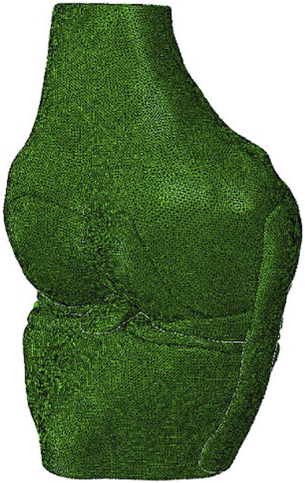
Purpose: The aims of this study were to 1) investigate the effects of femoral drilling angle in coronal and sagittal planes on the stress and strain distribution around the femoral and tibial tunnel entrance and the stress distribution on the graft, following anterior cruciate ligament reconstruction (ACLR), 2) identify the optimal femoral drilling angle to reduce the risk of the tunnel enlargement and graft failure. Methods: A validated three-dimensional (3D) finite element model of a healthy right cadaveric knee was used to simulate an anatomic ACLR with the anteromedial (AM) portal technique. Combined loading of 103.0 N anterior tibial load, 7.5 Nm internal rotation moment, and 6.9 Nm valgus moment during normal human walking at joint flexion of 20° was applied to the ACLR knee models using different tunnel angles (30°/45°/60° and 45°/60° in the coronal and sagittal planes, respectively). The distribution of von Mises stress and strain around the tunnel entrances and the graft was calculated and compared among the different finite element ACLR models with varying femoral drilling angles. Results: With an increasing coronal obliquity drilling angle (30° to 60°), the peak stress and maximum strain on the femoral and tibial tunnel decreased from 30° to 45° and increased from 45° to 60°, respectively. With an increasing sagittal obliquity drilling angle (45° to 60°), the peak stress and the maximum strain on the bone tunnels increased. The lowest peak stress and maximum strain at the ACL tunnels were observed at 45° coronal/45° sagittal drilling angle (7.5 MPa and 7,568.3 μ-strain at the femoral tunnel entrance, and 4.0 MPa and 4,128.7 μ-strain at the tibial tunnel entrance). The lowest peak stress on the ACL graft occurred at 45° coronal/45° sagittal (27.8 MPa) drilling angle. Conclusions: The femoral tunnel drilling angle could affect both the stress and strain distribution on the femoral tunnel, tibial tunnel, and graft. A femoral tunnel drilling angle of 45° coronal/ 45° sagittal demonstrated the lowest peak stress, maximum strain on the femoral and tibial tunnel entrance, and the lowest peak stress on the ACL graft.
Keywords: anterior cruciate ligament reconstruction; bone tunnel enlargement; femoral and tibial tunnel; femoral tunnel drilling angle; finite element analysis; graft failure.
Copyright © 2021 Cheng, Wang, Jiang, Dimitriou, Cheng and Tsai.
Conflict of interest statement
The authors declare that the research was conducted in the absence of any commercial or financial relationships that could be construed as a potential conflict of interest.
Figures


Similar articles
-
Central femoral tunnel placement can reduce stress and strain around bone tunnels and graft more than anteromedial femoral tunnel in anterior cruciate ligament reconstruction.Int J Numer Method Biomed Eng. 2022 May;38(5):e3590. doi: 10.1002/cnm.3590. Epub 2022 Mar 22. Int J Numer Method Biomed Eng. 2022. PMID: 35289106
-
The 45° and 60° of sagittal femoral tunnel placement in anterior cruciate ligament reconstruction provide similar knee stability.Knee Surg Sports Traumatol Arthrosc. 2024 Nov;32(11):3031-3038. doi: 10.1002/ksa.12341. Epub 2024 Jul 8. Knee Surg Sports Traumatol Arthrosc. 2024. PMID: 38973630
-
Transtibial versus anteromedial portal drilling for anterior cruciate ligament reconstruction: a cadaveric study of femoral tunnel length and obliquity.Arthroscopy. 2010 Mar;26(3):342-50. doi: 10.1016/j.arthro.2009.12.006. Arthroscopy. 2010. PMID: 20206044
-
Medial portal drilling: effects on the femoral tunnel aperture morphology during anterior cruciate ligament reconstruction.J Bone Joint Surg Am. 2011 Nov 16;93(22):2063-71. doi: 10.2106/JBJS.J.01705. J Bone Joint Surg Am. 2011. PMID: 22262377 Review.
-
Comparing the Use of Flexible and Rigid Reaming Systems Through an Anteromedial Portal for Femoral Tunnel Creation During Anterior Cruciate Ligament Reconstruction: A Systematic Review.Orthop J Sports Med. 2021 Oct 4;9(10):23259671211035741. doi: 10.1177/23259671211035741. eCollection 2021 Oct. Orthop J Sports Med. 2021. PMID: 34631903 Free PMC article. Review.
Cited by
-
Hourglass-shaped grafts are superior to conventional grafts for restoring knee stability and graft force at knee flexion angle of 30° following anterior cruciate ligament reconstruction: A finite element analysis.Front Bioeng Biotechnol. 2022 Dec 19;10:967411. doi: 10.3389/fbioe.2022.967411. eCollection 2022. Front Bioeng Biotechnol. 2022. PMID: 36601393 Free PMC article.
-
Quantification and classification of lumbar disc herniation on axial magnetic resonance images using deep learning models.Radiol Med. 2025 Jun;130(6):795-804. doi: 10.1007/s11547-025-01996-y. Epub 2025 Mar 24. Radiol Med. 2025. PMID: 40126796 Free PMC article.
-
Clinical Outcomes of a Novel Hybrid Transtibial Technique for Femoral Tunnel Drilling in Anterior Cruciate Ligament Reconstruction: A Large Single-Center Case Series With a Minimum 2-Year Follow-up.Orthop J Sports Med. 2024 Jun 4;12(6):23259671241242778. doi: 10.1177/23259671241242778. eCollection 2024 Jun. Orthop J Sports Med. 2024. PMID: 39131489 Free PMC article.
-
Model Properties and Clinical Application in the Finite Element Analysis of Knee Joint: A Review.Orthop Surg. 2024 Feb;16(2):289-302. doi: 10.1111/os.13980. Epub 2024 Jan 4. Orthop Surg. 2024. PMID: 38174410 Free PMC article. Review.
-
Increased Bone Plug Depth From the Joint Increases Tunnel Enlargement in Anterior Cruciate Ligament Reconstruction Using Bone-Patellar Tendon-Bone Autograft With Suspensory Femoral Fixation.Arthrosc Sports Med Rehabil. 2023 Jul 22;5(4):100755. doi: 10.1016/j.asmr.2023.100755. eCollection 2023 Aug. Arthrosc Sports Med Rehabil. 2023. PMID: 37520501 Free PMC article.
References
-
- Alentorn-Geli E., Samitier G., Álvarez P., Steinbacher G., Cugat R. (2010). Anteromedial portal versus Transtibial Drilling Techniques in ACL Reconstruction: a Blinded Cross-Sectional Study at Two- to Five-Year Follow-Up. Int. Orthopaedics (Sicot) 34 (5), 747–754. 10.1007/s00264-010-1000-1 - DOI - PMC - PubMed
LinkOut - more resources
Full Text Sources
Research Materials