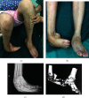
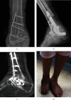
The Novel Application of Three-Dimensional Printing Assisted Patient-Specific Instrument Osteotomy Guide in the Precise Osteotomy of Adult Talipes Equinovarus
- PMID: 34901265
- PMCID: PMC8660203
- DOI: 10.1155/2021/1004849
The Novel Application of Three-Dimensional Printing Assisted Patient-Specific Instrument Osteotomy Guide in the Precise Osteotomy of Adult Talipes Equinovarus
Abstract
Objective: This current research is aimed at assessing clinical efficacy and prognosis of three-dimensional (3D) printing assisted patient-specific instrument (PSI) osteotomy guide in precise osteotomy of adult talipes equinovarus (ATE).
Methods: We included a total of 27 patients of ATE malformation (including 12 males and 15 females) from June 2014 to June 2018 in the current research. The patients were divided into the routine group (n = 12) and 3D printing group (n = 15) based on different operative methods. The parameters, including the operative time, intraoperative blood loss, complications, time to obtain bony fusion, functional outcomes based on American Orthopedic Foot and Ankle Society (AOFAS), and International Congenital Clubfoot Study group (ICFSG) scoring systems between the two groups were observed and recorded regularly.
Results: The 3D printing group exhibits superiorities in shorter operative time, less intraoperative blood loss, higher rate of excellent, and good outcomes presented by ICFSG score at last follow-up (P < 0.001, P < 0.001, P = 0.019) than the routine group. However, there was no significant difference exhibited in the AOFAS score at the last follow-up and total rate of complications between the two groups (P = 0.136, P = 0.291).
Conclusion: Operation assisted by 3D printing PSI osteotomy guide for correcting the ATE malformation is novel and feasible, which might be an effective method to polish up the precise osteotomy of ATE malformation and enhance the clinical efficacy.
Copyright © 2021 Yuan-Wei Zhang et al.
Conflict of interest statement
The authors declare there are no conflicts of interest regarding the publication of this paper.
Figures


Similar articles
-
Clinical application of 3D printing-assisted patient-specific instrument osteotomy guide in stiff clubfoot: preliminary findings.J Orthop Surg Res. 2023 Nov 7;18(1):843. doi: 10.1186/s13018-023-04341-z. J Orthop Surg Res. 2023. PMID: 37936150 Free PMC article.
-
Efficacy evaluation of three-dimensional printing assisted osteotomy guide plate in accurate osteotomy of adolescent cubitus varus deformity.J Orthop Surg Res. 2019 Nov 9;14(1):353. doi: 10.1186/s13018-019-1403-7. J Orthop Surg Res. 2019. PMID: 31706346 Free PMC article.
-
[Short-term effectiveness of total knee arthroplasty assisted by three-dimensional printing osteotomy navigation template].Zhongguo Xiu Fu Chong Jian Wai Ke Za Zhi. 2018 Jul 15;32(7):899-905. doi: 10.7507/1002-1892.201802013. Zhongguo Xiu Fu Chong Jian Wai Ke Za Zhi. 2018. PMID: 30129315 Free PMC article. Chinese.
-
Three-dimensional printing combined with open reduction and internal fixation versus open reduction and internal fixation in the treatment of acetabular fractures: A systematic review and meta-analysis.Chin J Traumatol. 2021 May;24(3):159-168. doi: 10.1016/j.cjtee.2021.02.007. Epub 2021 Feb 27. Chin J Traumatol. 2021. PMID: 33678536 Free PMC article.
-
The efficacy of 3D printing-assisted surgery in treating distal radius fractures: systematic review and meta-analysis.J Comp Eff Res. 2020 Sep;9(13):919-931. doi: 10.2217/cer-2020-0099. Epub 2020 Sep 24. J Comp Eff Res. 2020. PMID: 32969712
Cited by
-
Clinical application of 3D printing-assisted patient-specific instrument osteotomy guide in stiff clubfoot: preliminary findings.J Orthop Surg Res. 2023 Nov 7;18(1):843. doi: 10.1186/s13018-023-04341-z. J Orthop Surg Res. 2023. PMID: 37936150 Free PMC article.
-
Clinical added value of 3D printed patient-specific guides in orthopedic surgery (excluding knee arthroplasty): a systematic review.Arch Orthop Trauma Surg. 2025 Mar 3;145(1):173. doi: 10.1007/s00402-025-05775-2. Arch Orthop Trauma Surg. 2025. PMID: 40025308 Free PMC article.
-
The efficacy and safety of platelet-rich plasma in the tendon-exposed wounds: a preliminary study.J Orthop Surg Res. 2022 Nov 19;17(1):497. doi: 10.1186/s13018-022-03401-0. J Orthop Surg Res. 2022. PMID: 36403073 Free PMC article.
-
Preoperative 3D printing planning technology combined with orthopedic surgical robot-assisted minimally invasive screw fixation for the treatment of pelvic fractures: a retrospective study.PeerJ. 2024 Dec 12;12:e18632. doi: 10.7717/peerj.18632. eCollection 2024. PeerJ. 2024. PMID: 39677955 Free PMC article.
References
MeSH terms
LinkOut - more resources
Full Text Sources
Research Materials

