

Transcatheter occlusion of the vertical vein in a partial anomalous pulmonary venous connection with dual Drainage, case series with literature review
- PMID: 34901378
- PMCID: PMC8640871
- DOI: 10.1016/j.ijcha.2021.100889
Transcatheter occlusion of the vertical vein in a partial anomalous pulmonary venous connection with dual Drainage, case series with literature review
Abstract
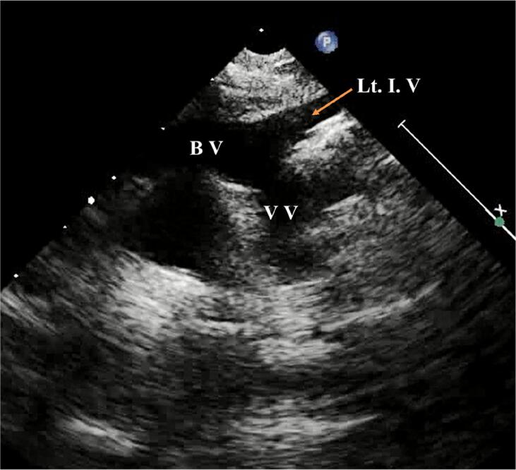
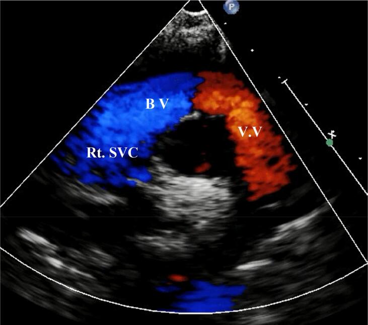
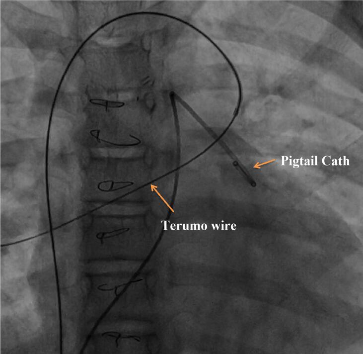
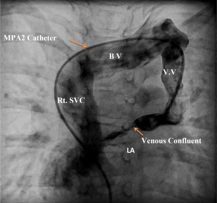
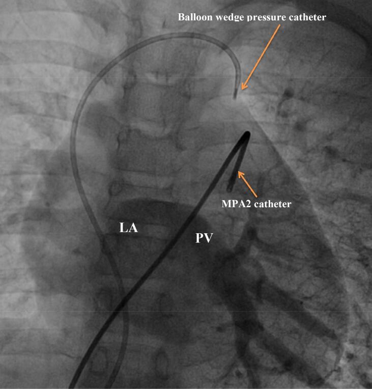
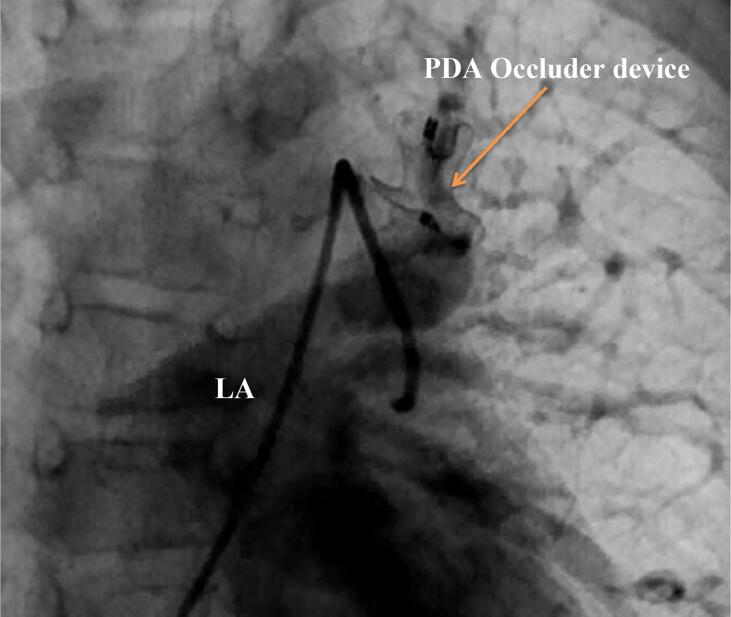
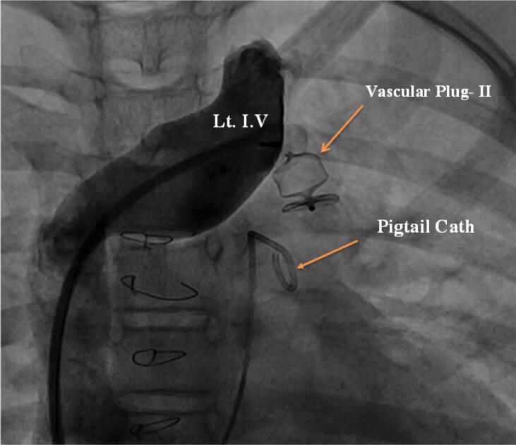
Partial anomalous pulmonary venous return (PAPVR) is an uncommon type of congenital heart disease occurring due to abnormal drainage of one or more, but not all the pulmonary veins to the systemic veins or directly to the right atrium. The PAPVR might have single (to the systemic veins) or dual drainage (to the systemic as well as left atrium). Management depends on the shunt impact on the heart and lungs, and it is usually surgical correction. In case of PAPVR with dual drainage, there is a new trend of percutaneous occlusion of the vertical vein with dual drainage anatomy, so that the blood is obliged to flow to the left atrium as in normal hearts. The scope of this manuscript is to highlight the availability of this alternative option and to present our experience and outcome in 6 PAPVR patients with dual drainage treated using this percutaneous approach.
Keywords: Device closure; Dual drainage; Partial anomalous pulmonary venous connection.
© 2021 Published by Elsevier B.V.
Conflict of interest statement
The authors report no relationships that could be construed as a conflict of interest.
Figures
References
-
- Christos Tourmousoglou, Christina Kalogeropoulou, Efstratios Koletsis, Nikolaos Charoulis, Christos Prokakis, Panagiotis Alexopoulos, Emmanoil Margaritis, and Dimitrios Dougenis, Hindawi Publishing Corporation. Right Upper Lobe Partial Anomalous Pulmonary. Venous Connection, case report, Vasc. Med. Vol. 2014, Article ID 249896, 3 pages. - PMC - PubMed
-
- Alsoufi Bahaaldin, Cai Sally, Van Arsdell Glen S., Williams William G., Caldarone Christopher A., Coles John G. Outcomes after surgical treatment of children with partial anomalous pulmonary venous connection. Ann. Thorac. Surg. 2007;84:2020–2026. - PubMed
-
- Alejandro R. Peirone, Alejandro E. Contreras, Carolina Carrizo, Mailén Konicoff and Raúl O. Cayre, Percutaneous occlusion of right partial anomalous pulmonary venous connection with dual drainage to the innominate vein and the left atrium: a unique anatomical finding, DOI: 10.32604/CHD.2020.013199. - DOI
-
- Atiq Mehnaz, Younis Muhammad Kamran, Amanullah Muneer, Transcatheter treatment of coarctation of aorta and dually connected anomalous vertical pulmonary vein as a combined procedure, DOI: 10.26717/BJSTR.2019.19.003372. - DOI
LinkOut - more resources
Full Text Sources
