

Updated Diagnostic Criteria and Classification of Mast Cell Disorders: A Consensus Proposal
- PMID: 34901755
- PMCID: PMC8659997
- DOI: 10.1097/HS9.0000000000000646
Updated Diagnostic Criteria and Classification of Mast Cell Disorders: A Consensus Proposal
Abstract
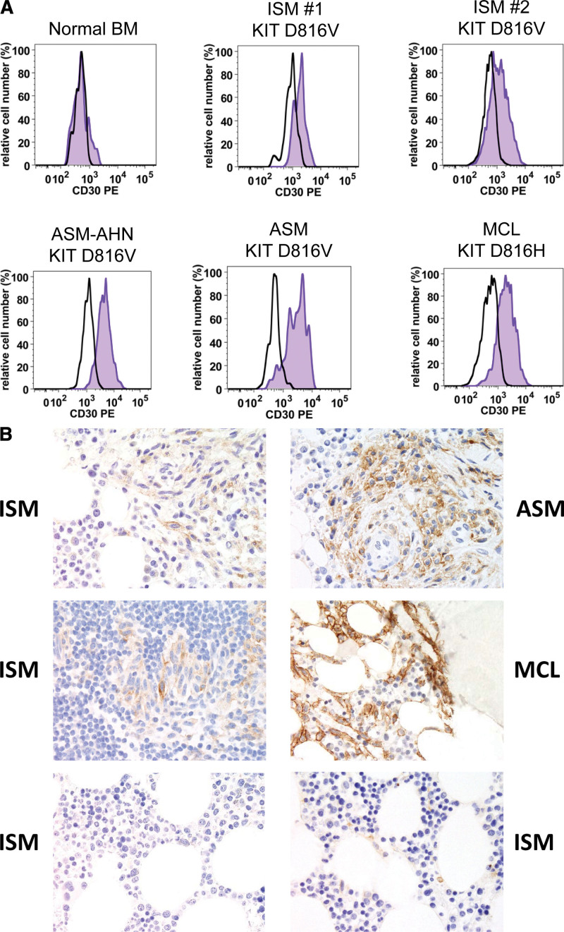
Mastocytosis is a hematologic neoplasm characterized by expansion and focal accumulation of neoplastic mast cells (MC) in diverse organs, including the skin, bone marrow (BM), spleen, liver, and gastrointestinal tract. The World Health Organization classification divides the disease into prognostically distinct variants of cutaneous mastocytosis (CM) and systemic mastocytosis (SM). Although this classification remains valid, recent developments in the field and the advent of new diagnostic and prognostic parameters created a need to update and refine definitions and diagnostic criteria in MC neoplasms. In addition, MC activation syndromes (MCAS) and genetic features predisposing to SM and MCAS have been identified. To discuss these developments and refinements in the classification, we organized a Working Conference comprised of experts from Europe and the United States in August 2020. This article reports on outcomes from this conference. Of particular note, we propose adjustments in the classification of CM and SM, refinements in diagnostic criteria of SM variants, including smoldering SM and BM mastocytosis (BMM), and updated criteria for MCAS and other conditions involving MC. CD30 expression in MC now qualifies as a minor SM criterion, and BMM is now defined by SM criteria, absence of skin lesions and absence of B- and C-findings. A basal serum tryptase level exceeding 20 ng/mL remains a minor SM criterion, with recognition that hereditary alpha-tryptasemia and various myeloid neoplasms may also cause elevations in tryptase. Our updated proposal will support diagnostic evaluations and prognostication in daily practice and the conduct of clinical trials in MC disorders.
Copyright © 2021 the Author(s). Published by Wolters Kluwer Health, Inc. on behalf of the European Hematology Association.
Figures
References
-
- Horny HP, Sotlar K, Valent P. Mastocytosis: state of the art. Pathobiology. 2007;74:121–132. - PubMed
-
- George TI, Horny HP. Systemic mastocytosis. Hematol Oncol Clin North Am. 2011;25:1067–1083, vii. - PubMed
-
- Theoharides TC, Valent P, Akin C. Mast cells, mastocytosis, and related disorders. N Engl J Med. 2015;373:163–172. - PubMed
Publication types
Grants and funding
LinkOut - more resources
Full Text Sources