. 2022 Jan 1;163(1):bqab237.
doi: 10.1210/endocr/bqab237.

Concordant Androgen-Regulated Expression of Divergent Rhox5 Promoters in Sertoli Cells

Affiliations

Concordant Androgen-Regulated Expression of Divergent Rhox5 Promoters in Sertoli Cells

Anjana Bhardwaj et al. Endocrinology. .

Abstract

Concordant transcriptional regulation can generate multiple gene products that collaborate to achieve a common goal. Here we report a case of concordant transcriptional regulation that instead drives a single protein to be produced in the same cell type from divergent promoters. This gene product-the RHOX5 homeobox transcription factor-is translated from 2 different mRNAs with different 5' untranslated regions (UTRs) transcribed from alternative promoters. Despite the fact that these 2 promoters-the proximal promoter (Pp) and the distal promoter (Pd)-exhibit different patterns of tissue-specific activity, share no obvious sequence identity, and depend on distinct transcription factors for expression, they exhibit a remarkably similar expression pattern in the testes. In particular, both depend on androgen signaling for expression in the testes, where they are specifically expressed in Sertoli cells and have a similar stage-specific expression pattern during the seminiferous epithelial cycle. We report evidence for 3 mechanisms that collaborate to drive concordant Pp/Pd expression. First, both promoters have an intrinsic ability to respond to androgen receptor and androgen. Second, the Pp acts as an enhancer to promote androgen-dependent transcription from the Pd. Third, Pd transcription is positively autoregulated by the RHOX5 protein, which is first produced developmentally from the Pp. Together, our data support a model in which the Rhox5 homeobox gene evolved multiple mechanisms to activate both of its promoters in Sertoli cells to produce Rhox5 in an androgen-dependent manner during different phases of spermatogenesis.

Keywords: ARE; Rhox5; Sertoli; androgen receptor; gene regulation; transcription factors.


Figures

Figure 1.
Figure 1.
The expression pattern of the Rhox5 distal (Pd) and proximal (Pp) promoters. (A) Rhox5 exon-intron structure and position of its 2 tissue-specific promoters. Also shown is the position of the probe used for RNase protection analysis, as well as the protected mRNA fragments predicted to be generated from Pp- and Pd-derived mRNAs (of note, 40 nt of exon 2 is also predicted to be protected but is obscured by the loading control β-actin bands). (B) RNase protection analysis of total cellular RNA from adult mice testes, placenta, and the SL12.4 T-lymphoma cell line using the probe shown in panel A. (C) qPCR analysis of the indicated adult mouse tissues. The RNase protection and qPCR experiments were repeated at least 3 times. Shown are average values + SEM (standard error).
Figure 2.
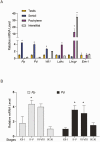
Figure 2.
The Pd and Pp are coexpressed during the seminiferous epithelial cycle in Sertoli cells in vivo. (A) qPCR analysis of mouse adult testes and cells enriched for the indicated testicular cell types. Wt1, Ldhc, and Lhcgr are testicular subset marker genes. Emr1 is a macrophage marker that serves as a negative control. The level of each transcript in the cell fractions are relative to that of unfractionated testes, which was set to “1.” (B) qPCR analysis of dissected adult testes sections enriched for different stages of the seminiferous epithelial cycle. The primers used for Fig. 1C were used to analyze Pd and Pp expression, with the stage harboring the lowest Pp expression Ct value set to “1” (left); the same approach was used for portraying Pd expression values (right). All results in this figure were obtained from 3 replicates. Shown are average values + SEM (*P < 0.05).
Figure 3.
Figure 3.
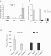
Both the Pd and Pp are androgen regulated in vivo. (A-C) qPCR analysis of testes from adult mice. (A) Hypophysectomized (Hpx) C57/BL6 mice injected with or without LH or sham-treated control mice. Pp and Pd mRNA levels in the Hpx and Hpx + LH samples are relative to the Sham-treatment group, which was set to “1.” (B) AR Knockout (ARKO), Sertoli Cell-specific AR Knockout (SCARKO), and the corresponding Cre control mice. All mRNA levels are relative to Pp transcript levels in PGK-Cre mice, which was set to 100. (C) SPecificity-Affecting AR Knock-In (SPARKI) and control (wt) mice. All mRNA levels are relative to Pd expression in control mice, which was set to “1.” All results in this figure are from 3 mice per group, with 2 repeats of each experiment. Shown are average values + SEM (*P < 0.05).
Figure 4.
Figure 4.
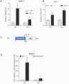
Both the Pd and Pp are androgen regulated in vitro. (A, B) qPCR analysis of the Sertoli cell lines, MSC-1 and 15P-1, transfected with an AR expression vector and incubated with R1881 (T), as indicated. (C) Schematic of a construct harboring Pd region sequences upstream of the luciferase (LUC) coding region, followed by a polyadenylation (pA) site. (D) Luciferase analysis of the Sertoli cell line, MSC-1, transiently transfected with either the Pd-LUC constructs shown in panel C or an equivalent Pp-LUC construct (32, 34). The indicated cells were cotransfected with an AR expression and incubated with R1881 (T). Shown are average Luciferase activity values + SEM obtained from at least 3 replicates (*P < 0.05). The values in panels A, B, and D are relative to cells incubated without AR and T, which was set to “1.”
Figure 5.
Figure 5.
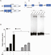
The Pd is directly regulated by AR. (A) Schematic of wild-type (WT) Pd- and ARE-mutant (mARE)-luciferase construct, the former which is also shown in Fig. 4C. (B) Luciferase analysis of the Sertoli cell line, MSC-1, transiently transfected with the constructs shown in panel A. The cell cultures indicated were cotransfected with an AR expression and/or incubated with androgen (T). Shown are average Luciferase activity values + SEM obtained from at least 3 replicates (* P < 0.05; # P < 0.01). (C) EMSA showing AR binding to the Pd. Pd probe harbor ARE sequence (underlined). The control sequence is the Tubb3 AR-binding sequence (69). The arrow indicates the shifted complex.
Figure 6.
Figure 6.
The Pd and Pp are differentially methylated in vitro and in vivo. (A) Schematic of the Rhox5 gene, indicating the location of 3 CpG-rich regions: loci 1-3. (B) Bisulfite sequencing analysis of cultured Sertoli cell lines (top panel) and purified Sertoli cells isolated from adult testes (bottom). Closed circles indicate the presence of a methylated cytosine at the indicated positions in the bar above the circles (the chromosomal location of the CpGs is drawn to scale). The average percentage of methylation is indicated below each respective panel. (C) Bisulfite sequencing analysis of 15P-1-AR cells incubated with DMSO or 10 nM R1881 (T), depicted as in panel B.
Figure 7.
Figure 7.
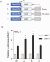
Evidence that Pp is a Pd enhancer. (A) Schematic of constructs containing the Pd upstream of luciferase (LUC), followed by a polyadenylation (pA) site. The top construct (wild-type [Wt]) has only Pd sequences, while constructs A-C have also have Pp sequences (0.3 kb) or a random DNA fragment (Stuffer; 0.3 kb) inserted (in the sense or antisense direction with respect to the Pd) downstream of the pA site. (B) Luciferase analysis of the Sertoli cell line, MSC-1, transiently transfected with the constructs shown in panel A. As indicated, some cells were cotransfected with an AR expression and/or incubated with R1881 (T). Shown are average luciferase activity values + SEM obtained from at least 3 replicates (* P < 0.05).
Figure 8.
Figure 8.
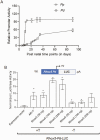
Differential induction of Pp and Pd during testicular postnatal development. (A) qPCR analysis of Pd- and Pp-derived transcripts at the indicated postnatal time points. All mRNA values are relative to the time point with the lowest expression, which was set to “1.” (B) Luciferase analysis of the Sertoli cell line, MSC-1, transiently cotransfected with the construct shown (described in Fig. 4) and an empty expression vector (lane 1), an empty expression vector + an AR expression vector (lane 2), or a Rhox5 expression vector (lanes 3-8). The cells in lane 2 were also incubated with R1881 (T). Shown are average luciferase activity values + SEM obtained from at least 3 replicates (* P < 0.05). All values are relative to cells transfected with empty vector, which was set to “1”.
Figure 9.
Figure 9.
Model: The Pp and Pd are alternative promoters driving expression of the RHOX5 transcription factor. Shown are the cells and tissues that transcribe these 2 promoters, as well as cis-regulatory elements known to regulate these promoters in a regulated manner, as shown herein and in earlier studies (31-35, 51). Not shown is the Pp and Pd are transcriptionally induced in Sertoli cells on P10 and P23, respectively. The size and location of AR- and other transcription factor-binding sites are not drawn to scale.

Similar articles

Cited by

References

    1. Kimura K, Wakamatsu A, Suzuki Y, et al. . Diversification of transcriptional modulation: large-scale identification and characterization of putative alternative promoters of human genes. Genome Res. 2006;16(1):55-65. - PMC - PubMed
    1. Davuluri RV, Suzuki Y, Sugano S, Plass C, Huang TH. The functional consequences of alternative promoter use in mammalian genomes. Trends Genet. 2008;24(4):167-177. - PubMed
    1. Demircioğlu D, Cukuroglu E, Kindermans M, et al. . A pan-cancer transcriptome analysis reveals pervasive regulation through alternative promoters. Cell. 2019;178(6):1465-1477.e17. - PubMed
    1. Carninci P, Sandelin A, Lenhard B, et al. . Genome-wide analysis of mammalian promoter architecture and evolution. Nat Genet. 2006;38(6):626-635. - PubMed
    1. Sandelin A, Carninci P, Lenhard B, Ponjavic J, Hayashizaki Y, Hume DA. Mammalian RNA polymerase II core promoters: insights from genome-wide studies. Nat Rev Genet. 2007;8(6):424-436. - PubMed

Publication types

MeSH terms