Review
. 2022 Jul;72(4):333-352.
doi: 10.3322/caac.21713. Epub 2021 Dec 13.

Molecular imaging in oncology: Current impact and future directions

Affiliations
Review

Molecular imaging in oncology: Current impact and future directions

Steven P Rowe et al. CA Cancer J Clin. 2022 Jul.

Abstract

The authors define molecular imaging, according to the Society of Nuclear Medicine and Molecular Imaging, as the visualization, characterization, and measurement of biological processes at the molecular and cellular levels in humans and other living systems. Although practiced for many years clinically in nuclear medicine, expansion to other imaging modalities began roughly 25 years ago and has accelerated since. That acceleration derives from the continual appearance of new and highly relevant animal models of human disease, increasingly sensitive imaging devices, high-throughput methods to discover and optimize affinity agents to key cellular targets, new ways to manipulate genetic material, and expanded use of cloud computing. Greater interest by scientists in allied fields, such as chemistry, biomedical engineering, and immunology, as well as increased attention by the pharmaceutical industry, have likewise contributed to the boom in activity in recent years. Whereas researchers and clinicians have applied molecular imaging to a variety of physiologic processes and disease states, here, the authors focus on oncology, arguably where it has made its greatest impact. The main purpose of imaging in oncology is early detection to enable interception if not prevention of full-blown disease, such as the appearance of metastases. Because biochemical changes occur before changes in anatomy, molecular imaging-particularly when combined with liquid biopsy for screening purposes-promises especially early localization of disease for optimum management. Here, the authors introduce the ways and indications in which molecular imaging can be undertaken, the tools used and under development, and near-term challenges and opportunities in oncology.

Keywords: magnetic resonance imaging (MRI); nuclear medicine; optical imaging; positron emission tomography (PET); single-photon emission computed tomography (SPECT); theranostics.


Figures

Figure 1.
Figure 1.
The use of indocyanine green for surgical guidance during a lung segmentectomy. The intersegmental plane was difficult to identify with traditional techniques (top image), but was visualized much more clearly with the use of indocyanine green (red arrows, bottom image). Image reproduced from Liu, et al., Journal of Cardiothoracic Surgery, 2020; 15: 303 [21].
Figure 2.
Figure 2.
25-year-old man with anaplastic astrocytoma. The images are axial DGE difference images at 5.3-second temporal resolution. Note the differential and heterogeneous enhancement centered in the region of the right insula and extending into the right frontal and temporal lobes. Although the contrast between the abnormal right side and the normal left side is less than might be encountered with some other molecular imaging modalities, it is nonetheless impressive that tumor visualization can take place through the exogenous application of glucose. Image re-printed from Xu, et al., Tomography, 2015; 1: 105–114 [71].
Figure 3.
Figure 3.
Proposed work-up algorithm for indeterminate renal masses incorporating molecular imaging with 99mTc-sestamibi SPECT and a genomic classifier (ONEX, [84]). This algorithm is derived from a previously-published figure in [85].
Figure 4.
Figure 4.
24-year-old man with Epstein-Barr Virus-associated lymphoma before and after systemic therapy. (A) Maximum intensity projection (MIP) 18F-FDG PET image prior to the initiation of therapy demonstrates numerous sites of abnormal radiotracer uptake throughout lymph nodes and skeletal structures, consistent with widespread lymphomatous involvement. (B) Axial 18F-FDG, (C) CT, and (D) fused 18F-FDG PET/CT images through the pelvis demonstrate a particularly prominent right external iliac lymph node with intense uptake, consistent with a site of disease (red arrows). (E) MIP 18F-FDG PET image after completion of therapy. All of the abnormal uptake has resolved (note, the apparent focus of uptake in the left arm is a result of motion artifact). (F) Axial 18F-FDG, (G) CT, and (H) fused 18F-FDG PET/CT images through the pelvis show that the left external iliac node has decreased in size, although it remains enlarged (red arrows). Despite the residual anatomic abnormality, the uptake has been reduced to blood pool levels, consistent with a complete metabolic response. This example demonstrates the ability of 18F-FDG PET to characterize residual anatomic lesions after therapy.
Figure 5.
Figure 5.
74-year-old woman with metastatic small bowel neuroendocrine tumor. (A) MIP 68Ga-DOTATATE PET image shows numerous sites of abnormal uptake in lymph nodes and bones. (B) Axial 68Ga-DOTATATE PET, (B) CT, and (D) fused 68Ga-DOTATATE PET/CT images demonstrate that many of the bone lesions are easily visible on the PET but are occult on the corresponding CT anatomic images (red arrows). This case demonstrates the high sensitivity that is achievable with optimized PET radiotracers and that normal-appearing anatomic structures can harbor disease that is well visualized with molecular imaging.
Figure 6.
Figure 6.
64-year-old man who was 11 years post-prostatectomy for Gleason 4 + 5 = 9, grade group 5, PCa and presented for PSMA PET with a PSA of 2.2 ng/mL. (A) MIP 18F-DCFPyL PSMA-targeted PET image demonstrates subtle uptake at multiple sites of small, morphologically normal lymph nodes (red arrows). (B) Axial 18F-DCFPyL PET, (B) CT, and (D) fused 18F-DCFPyL PET/CT images demonstrate focal uptake in a 2 mm left supraclavicular (Virchow) node (red arrows), consistent with low-volume systemic nodal disease. Conventional imaging with bone scan and CT had not indicated a site of disease.
Figure 7.
Figure 7.
As theranostics becomes an ever-increasing aspect of cancer therapy, our ability to image relevant targets becomes more important. The images in this figure are from a 46-year-old woman with newly diagnosed metastatic lung cancer. Not only does [68Ga]FAPI PET (right panel) demonstrate higher uptake, but it also suggests that FAP-directed therapy may be effective for some patients with metastatic cancers. Reprinted from [133].
Figure 8.
Figure 8.
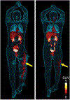
Whole-body 18F-FDS images of a 33-year-old man who had left leg osteomyelitis. The left panel was from an imaging study obtained at baselines, whereas the right panel was after attempted therapy. The yellow arrows show that uptake at the site of infection decreased, but did not resolve; clinically, the patient had persistent infection after therapy. These images suggest 18F-FDS PET may be a means of following, and determining adequacy of, anti-microbial therapy. Reprinted with permission from [143].

References

    1. About nuclear medicine and molecular imaging. Available at: https://www.snmmi.org/AboutSNMMI/Content.aspx?ItemNumber=6433. Last accessed: March 13, 2021.
    1. Chan KW, Bulte JW, McMahon MT. Diamagnetic chemical exchange saturation transfer (diaCEST) liposomes: physicochemical properties and imaging applications. Wiley Interdiscip Rev Nanomed Nanobiotechnol. 2014; 6: 111–124. - PMC - PubMed
    1. Choi CH, Iordanishvili E, Shah NJ, Binkofski F. Magnetic resonance spectroscopy with transcranial direct current stimulation to explore the underlying biochemical and physiological mechanism of the human brain: a systematic review. Hum Brain Mapp. 2021; 42: 2642–2671. - PMC - PubMed
    1. Wu M, Shu J. Multimodal molecular imaging: current status and future directions. Contrast Media Mol Imaging. 2018; 2018: 1382183. - PMC - PubMed
    1. Anderson CJ, Lewis JS. Current status and future challenges for molecular imaging. Philos Trans A Math Phys Eng Sci. 2017; 375: 20170023. - PubMed

Publication types