. 2021 Dec 13;19(1):417.
doi: 10.1186/s12951-021-01165-z.

A novel biomimetic nanomedicine system with anti-inflammatory and anti-osteoporosis effects improves the therapy efficacy of steroid-resistant nephrotic syndrome

Affiliations

A novel biomimetic nanomedicine system with anti-inflammatory and anti-osteoporosis effects improves the therapy efficacy of steroid-resistant nephrotic syndrome

Jian Li et al. J Nanobiotechnology. .

Abstract

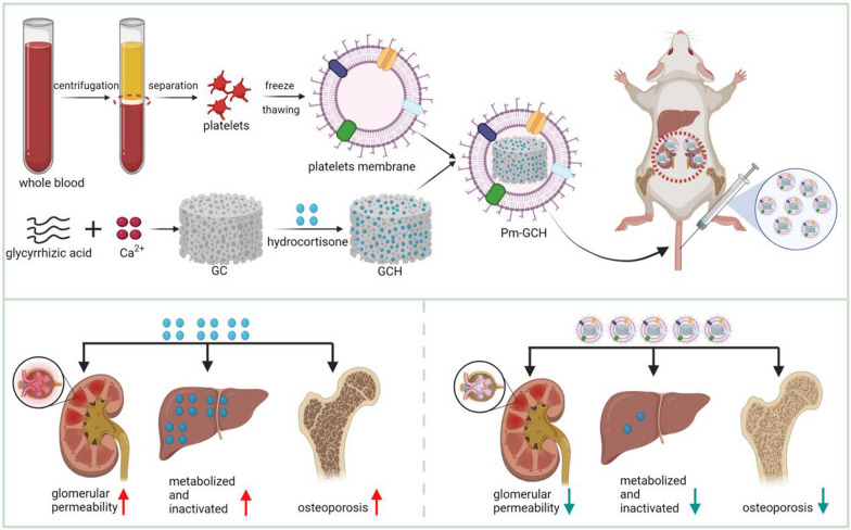
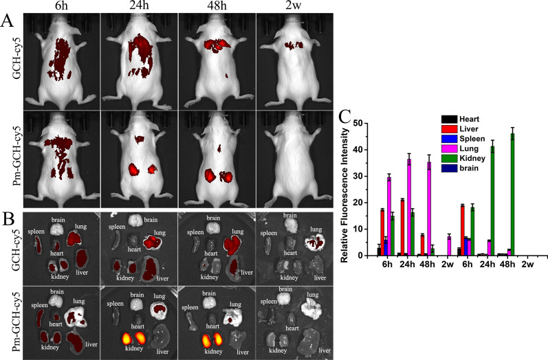
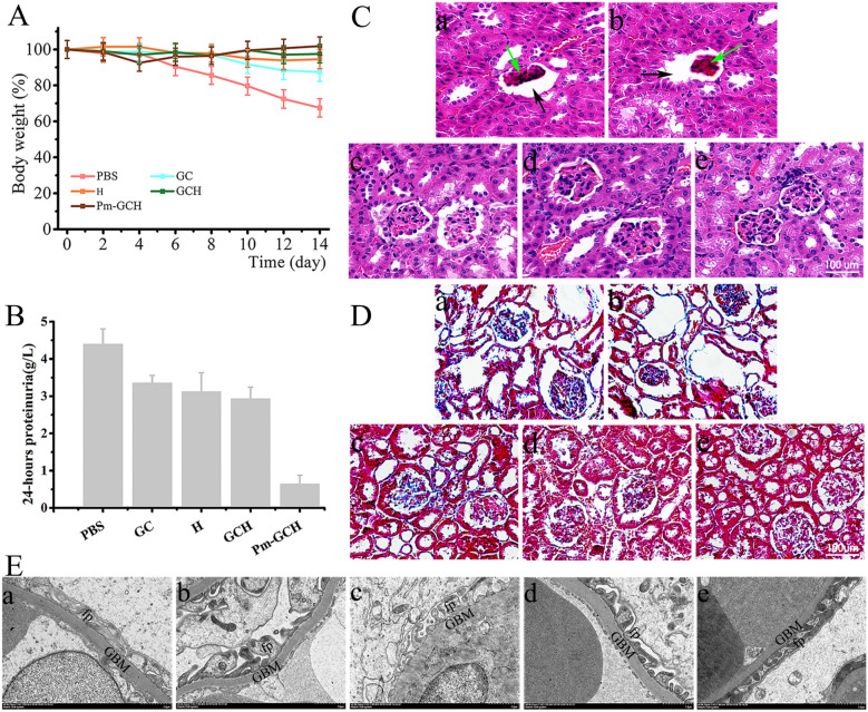
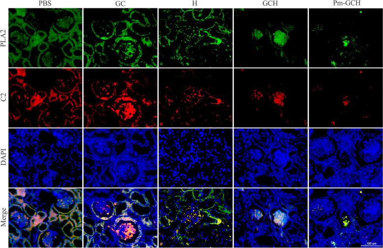
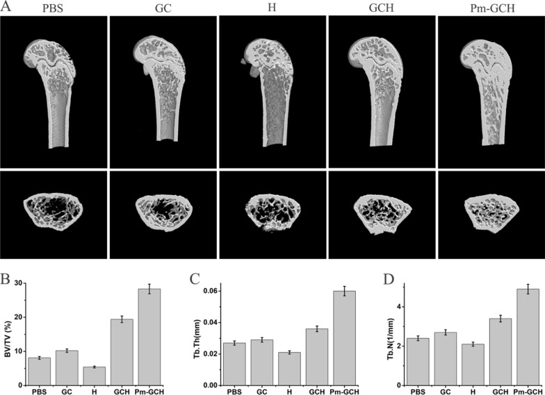
Clinically, steroid-resistant nephrotic syndrome (SRNS) is always prolonged and difficult to treat and easily develops into end-stage renal disease, resulting in a low survival rate. Strategies to reverse steroid resistance and reduce the long-term use of high doses of steroid medicines are urgently needed. In this study, a novel nanoparticle drug system (Pm-GCH) with a core-shell structure was designed. Metal-organic frameworks, synthesized by glycyrrhizic acid (G) and calcium ions (Ca2+) loaded with hydrocortisone (H) were the core of the nanoparticles. Platelet membrane vesicles were the shells. The natural platelet membrane endows Pm-GCH with good biocompatibility and the ability to promote immune escape. In addition, under the chemotaxis of inflammatory factors, platelet membranes assist Pm-GCH in nonspecific targeting of the inflammatory sites of the kidney. Under an inflammatory acid environment, GCH slowly degrades and releases glycyrrhizic acid and hydrocortisone. Glycyrrhizic acid inhibits the inactivation of hydrocortisone, jointly inhibits the activity of phospholipase A2 (PLA2) and the classic activation pathway of complement C2, blocks the production of inflammatory factors, plays an anti-inflammatory role, and enhances the efficacy of hydrocortisone in the treatment of SRNS. Moreover, glycyrrhizic acid alleviates osteoporosis induced by long-term use of glucocorticoids. These results indicate that Pm-GCH is a promising treatment strategy for SRNS.

Keywords: Biomimetic; Glycyrrhizic acid; Inflammatory; Steroid-resistant nephrotic syndrome.


Conflict of interest statement

The authors declare that they have no competing interests.

Figures

Fig. 1
Fig. 1
Construction of Pm-GCH and its targeted therapeutic mechanism in SRNS by inhibitting inflammation and osteoporosis
Fig. 2
Fig. 2
Characterization of Pm-GCH. A TEM-image analysis of GCH (a), Pm vesicles (b), and Pm-GCH (c). Scale bar: 200 nm. B Hydrodynamic size and C zeta potential of GCH, Pm and Pm-GCH. D The size change of Pm-GCH over 14 days in PBS, DMEM and 10% FBS. E UV–vis spectra of Pm, H, G and Pm-GCH. F The profile of platelet membrane proteins was detected by SDS-PAGE analysis
Fig. 3
Fig. 3
The hemocompatibility and immune escape effect of Pm-GCH. A The hemolysis rate of RBCs with different concentrations of GCH and Pm-GCH. B The fluorescence images of macrophages aftter treated with GCH and Pm-GCH for 6 h. C The fluorescence intensity of macrophage lysates after treated with GCH-Cy5 and Pm-GCH-Cy5 for 6 h
Fig. 4
Fig. 4
A The EE and LE of hydrocortisone. B Cumulative release ratio of H from GCH and Pm-GCH at different pH values (6.5, 7.4). C Cumulative release ratio of GA from GCH and Pm-GCH at different pH values (6.5, 7.4)
Fig. 5
Fig. 5
A The viability of 293T and HKC-8 cells was detected by CCK8 assays after administration of PBS, GC, H, GCH and Pm-GCH for 24, 48 and 72 h. B The viability of 293T/D cells was detected by CCK8 assays after administration of PBS, GC, H, GCH and Pm-GCH for 24 h. C Live/dead staining of 293T/D cells after various treatments (PBS, GC, H, GCH and Pm-GCH) for 24 h. D Semiquantitative ratio of cell death
Fig. 6
Fig. 6
In vivo targeting potential of Pm-GCH. A The biodistribution images of the whole body of mice. B Fluorescence images of main organs (brain, heart, liver, spleen, lung, and kidney) were measured at 6 h, 24 h, 48 h and 2 week after intravenous injection of GCH and Pm-GCH in SRNS mice. B Semiquantitative assessment of fluorescent signals in brain, heart, liver, spleen, lung, and kidney
Fig. 7
Fig. 7
A Body weight alterations in the mice with SRNS during GC, H, GCH and Pm-GCH treatment. B Quantification of 24-h proteinuria after GC, H, GCH and Pm-GCH treatment. Representative images of kidneys after H&E staining (C) and Masson staining (D) after intravenous injection of (a) PBS, (b) GC, (c) H, (d) GCH and (e) Pm-GCH, respectively. Black arrows, enlarged Bowman’s space; green arrows, glomerular sclerosis. E The ultrastructure of the glomerular filtration barrier was assessed by transmission electron microscopy. fp foot process, GBM glomerular basement membrane
Fig. 8
Fig. 8
Expression pattern of inflammatory cytokines (PLA2 and C2) analyzed by immunofluorescence staining in the kidney
Fig. 9
Fig. 9
Effect of Pm-GCH on the femur of the mice with SRNS. A Three-dimensional reconstruction images of femurs obtained by micro-CT. Bone parameters including BV/TV (B), Tb. Th (C) and Tb. N (D) were calculated
Fig. 10
Fig. 10
A Hematological and hematobiochemical analysis of peripheral blood from the mice with SRNS administrated with PBS, GC, H, GCH and Pm-GCH. (a) RBC, (b) Hb, (c) WBC, (d) PLT, (e) ALT, (f) AST, (g) BUN and (h) CREA. B HE staining images of mice heart, liver, spleen and lung slices from different treatment groups at the 14th day after different treatments

References

    1. Agrawal S, Zaritsky JJ, Fornoni A, Smoyer WE. Dyslipidaemia in nephrotic syndrome: mechanisms and treatment. Nat Rev Nephrol. 2018;14(1):57–70. - PMC - PubMed
    1. Zhao F, Zhu J, Richman A, Fu Y, Huang W, Chen N, et al. Mutations in are implicated in steroid-resistant nephrotic syndrome. J Am Soc Nephrol. 2019;30(5):840–853. - PMC - PubMed
    1. Chotiyarnwong P, McCloskey E. Pathogenesis of glucocorticoid-induced osteoporosis and options for treatment. Nat Rev Endocrinol. 2020;16(8):437–447. - PubMed
    1. Sosa M, Gómez de Tejada M. Glucocorticoid-induced osteoporosis. N Engl J Med. 2019;380(14):1378–1379. - PubMed
    1. Siji A, Karthik KN, Pardeshi VC, Hari PS, Vasudevan A. Targeted gene panel for genetic testing of south Indian children with steroid resistant nephrotic syndrome. BMC Med Genet. 2018;19(1):200. - PMC - PubMed

MeSH terms

LinkOut - more resources