Review
. 2021 Dec;9(12):e003430.
doi: 10.1136/jitc-2021-003430.

Immunogenic ferroptosis and where to find it?

Affiliations
Review

Immunogenic ferroptosis and where to find it?

Robin Demuynck et al. J Immunother Cancer. 2021 Dec.

Abstract

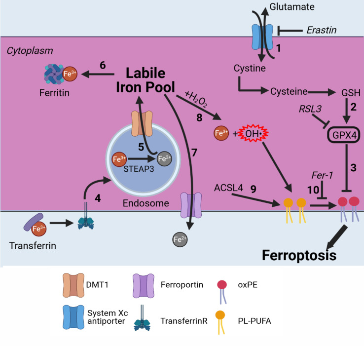
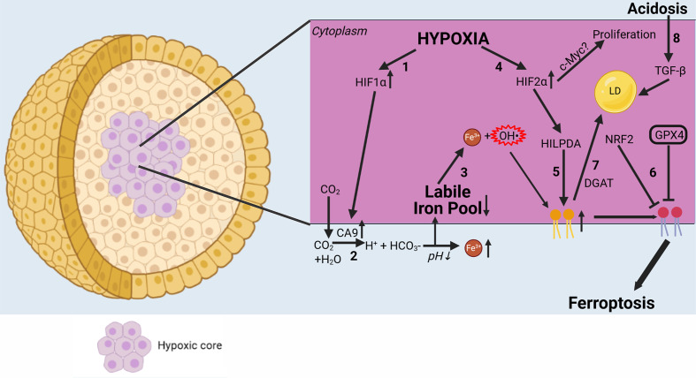
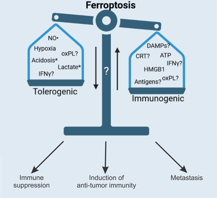
Ferroptosis is a recently discovered form of regulated cell death that is morphologically, genetically, and biochemically distinct from apoptosis and necroptosis, and its potential use in anticancer therapy is emerging. The strong immunogenicity of (early) ferroptotic cancer cells broadens the current concept of immunogenic cell death and opens up new possibilities for cancer treatment. In particular, induction of immunogenic ferroptosis could be beneficial for patients with cancers resistant to apoptosis and necroptosis. However, ferroptotic cancer cells may be a rich source of oxidized lipids, which contribute to decreased phagocytosis and antigen cross-presentation by dendritic cells and thus may favor tumor evasion. This could explain the non-immunogenicity of late ferroptotic cells. Besides the presence of lactate in the tumor microenvironment, acidification and hypoxia are essential factors promoting ferroptosis resistance and affecting its immunogenicity. Here, we critically discuss the crucial mediators controlling the immunogenicity of ferroptosis that modulate the induction of antitumor immunity. We emphasize that it will be necessary to also identify the tolerogenic (ie, immunosuppressive) nature of ferroptosis, which can lead to tumor evasion.

Keywords: immunogenicity; immunomodulation; immunotherapy; phagocytosis; tumor microenvironment; vaccine.


Conflict of interest statement

Competing interests: None declared.

Figures

Figure 1
Figure 1
Ferroptosis pathway in brief. Ferroptosis is an iron-dependent cell death modality. (1) Cystine is imported into the cell in exchange for glutamate by the system XC antiporter. Cystine is converted into cysteine and used for GSH production. (2) GSH is an essential cofactor for GPX4. By blocking the system XC‾ antiporter (eg, with erastin), GSH is depleted and GPX4 can no longer function. (3) GPX4 inhibits lipid peroxidation; directly inhibiting it (eg, by RSL3) induces ferroptosis. (4) Fe3+ bound to transferrin is transported into endosomes by binding to the transferrin receptor. (5) Fe3+ is converted to Fe2+ by STEAP3. Fe2+ is then transported to the labile iron pool by DMT1. (6) Fe2+ can be stored by binding to ferritin. (7) Fe2+ can be transported outside the cell by ferroportin. (8) Fe2+ is oxidized back to Fe3+ via the Fenton reaction, leading to production of hydroxyl radicals. (9) ACSl4 selectively enriches PUFAs in the plasma membrane. (10) Radicals formed from the Fenton reaction react with PUFAs, leading to oxPE formation and cell death. This can be inhibited by Fer-1. ACSl4, acyl–CoA synthetase long-chain family member 4; CoA, coenzyme A; DMT1, divalent metal transporter 1; Fer-1, ferrostatin-1; GSH, glutathione; GPX4, glutathione peroxidase 4; oxPE, oxidized phosphatidylethanolamine; PUFA, polyunsaturated fatty acid; RSL3, Ras-selective lethal 3.
Figure 2
Figure 2
Hallmarks of ICD. The immunogenicity of cancer cell death is dependent on two main elements: adjuvanticity (green) and antigenicity (blue). HMGB1 binds to TLR4 on DCs, causing their activation and maturation. CRT is expressed on the surface of dying (apoptotic) cells and will bind to LRP1 on DCs. ATP is either actively secreted or passively released by dying cancer cells in a specific spatiotemporal pattern and will bind to P2X7 receptors on DCs, causing their activation and maturation and leading to inflammasome activation. Proinflammatory cytokines act as adjuvants and can be seen as iDAMPs activating the immune system cells. Anti-inflammatory cytokines cause inhibition of immune cells and are considered protumorigenic. Antigens are presented by DCs on MHC-I molecules to T cells in a process called cross-presentation. After antigen recognition, cytotoxic CD8+ T cells are formed and migrate to the tumor, where they eliminate antigen-expressing cancer cells. Memory T cells may be formed, establishing long-lasting antitumor immune responses and reducing the chance of tumor reoccurrence. CRT, calreticulin; DAMP, danger-associated molecular pattern; DC, dendritic cell; HMGB1, high-mobility group 1; ICD, immunogenic cell death; iDAMP, inducible danger-associated molecular pattern; LRP1, lipoprotein receptor-related protein 1; TCR, T-cell receptor.
Figure 3
Figure 3
Immunogenic potential of ferroptosis. Several factors might influence the immunogenicity of ferroptosis. Early, but not late, ferroptotic cancer cells are immunogenic in vitro and in vivo, and HMGB1 and ATP are released by early ferroptotic cancer cells. it is unknown if CRT is exposed on early ferroptotic cells, and this requires investigation. Notably, HMGB1 and ATP are not released by late ferroptotic cells. HMGB1 might act through the AGER/RAGE or TLR2/TLR4/TLR9 pathway and activate innate and adaptive immunity. Moreover, knockdown of the HMGB1 gene decreases erastin-induced ferroptosis, therefore pointing to the regulatory role of HMGB1 in ferroptosis (*). Another component potentially modifying the immunogenicity of ferroptotic cell death might hide behind oxPLs and other oxidized products generated during ferroptosis. OxPLs (eg, oxPE) can play a dual role in ferroptosis, inducing either tumor promotion or tumor suppression. However, the role of oxidized oxPLS in the immunogenicity of ferroptosis is not known. CRT, calreticulin; HMGB1, high-mobility group 1; IL, interleukin; oxPE, oxidized phosphatidylethanolamine; oxPL, oxidized phospholipid; oxPS, oxidized phosphatidylserine.
Figure 4
Figure 4
Complex interplay of ferroptosis with the tumor microenvironment. Hypoxia has an important influence on ferroptosis. (1) HIF-1α is upregulated in hypoxic conditions. (2) In turn, HIF-1α causes CA9 to be upregulated. CA9 then converts CO2 to H+ and HCO3−, further acidifying the extracellular environment and leading to increased iron solubility. (3) Increased extracellular iron leads to a decrease in the labile iron pool via increased intracellular iron storage. (4) HIF-2α is also upregulated in hypoxia, leading to stronger expression of HILPDA. (5) HILPDA then selectively enriches PUFAs in the plasma membrane, creating more substrate for lipid peroxidation. (6) In contrast, NRF2 and GPX4 protect cancer cells from ferroptosis by blocking lipid peroxidation. Due to hypoxia, many cancer types become dependent on GPX4 for their survival. (7) However, PUFAs can be stored in LDS by DGAT, making peroxidation impossible and thereby reducing sensitivity to ferroptosis. (8) Acidosis causes autocrine TGF-β secretion, leading to increased LD formation. CA9, carbonic anhydrase 9; DGAT, diacylglycerol O-acyltransferase; GPX4, glutathione peroxidase 4; HIF1α, hypoxia-inducible factor 1α; HIF2α, hypoxia-inducible factor 2α; HILPDA, hypoxia-inducible, lipid droplet-associated protein; NRF2, nuclear factor erythroid 2-related factor 2; LD, lipid droplet; oxPE, oxidized phosphatidylethanolamine; PUFA, polyunsaturated fatty acid.
Figure 5
Figure 5
Future perspectives. Ferroptosis has proven to be an attractive alternative to apoptosis and necroptosis for use in cancer therapy. However, more research is needed to understand the full therapeutic potential of ferroptosis. The full repertoire of spatiotemporal release of both DAMPs and cytokines (ie, iDAMPs) needs to be investigated and linked to the immunogenic potential of ferroptosis. As ferroptotic cancer cells contain large numbers of various oxidized and peroxidized lipids and PLs, it is possible to speculate about their effects on the anticancer immune response. Nevertheless, studies are needed to clarify whether ferroptosis-derived oxPLs are responsible for modulating ferroptosis immunogenicity, which may explain the non-immunogenicity of the later stages of ferroptotic cell death.65 Importantly, the modulation of antigenicity by ferroptosis is still poorly understood, and there is a need to also investigate the formation of memory T cells, which is an important contributor to the therapeutic potential of ferroptosis. In addition, IFN-γ released from CD8+ T cells has been reported to promote cancer cell lipid peroxidation and ferroptosis.. in addition, expression of the protumor PDL1 is increased on tumor cells due to increased secretion of IFN-γ by CD8+T cells. these findings might also explain the tolerogenic (ie, immune suppressive) phenotype of late ferroptotic cells reported in Efimova et al. Finally, it is important to understand how ferroptotic cancer cells affect metastatic capacity. *Lactate and acidification do not have a direct effect on immunogenicity (or it is not known yet), but they increase cell death resistance, thereby reducing therapeutic efficacy. CRT, calreticulin; DAMP, damage-associated molecular pattern; HMGB1, high-mobility group 1; iDAMP, inducible danger-associated molecular pattern; IFN-γ, interferon gamma; NO·, nitric oxide; oxPL, oxidized phospholipid; PDL1, prodeath ligand 1; PL, phospholipid.

References

    1. Dixon SJ, Lemberg KM, Lamprecht MR, et al. . Ferroptosis: an iron-dependent form of nonapoptotic cell death. Cell 2012;149:1060–72. 10.1016/j.cell.2012.03.042 - DOI - PMC - PubMed
    1. Ingold I, Berndt C, Schmitt S, et al. . Selenium utilization by GPX4 is required to prevent hydroperoxide-induced ferroptosis. Cell 2018;172:409–22. 10.1016/j.cell.2017.11.048 - DOI - PubMed
    1. D'Herde K, Krysko DV. Ferroptosis: oxidized PEs trigger death. Nat Chem Biol 2017;13:4–5. 10.1038/nchembio.2261 - DOI - PubMed
    1. Friedmann Angeli JP, Krysko DV, Conrad M. Ferroptosis at the crossroads of cancer-acquired drug resistance and immune evasion. Nat Rev Cancer 2019;19:405–14. 10.1038/s41568-019-0149-1 - DOI - PubMed
    1. Kagan VE, Mao G, Qu F, et al. . Oxidized arachidonic and adrenic PEs navigate cells to ferroptosis. Nat Chem Biol 2017;13:81–90. 10.1038/nchembio.2238 - DOI - PMC - PubMed

Publication types