. 2022 Feb 15;82(4):695-707.
doi: 10.1158/0008-5472.CAN-20-3209.

HDAC2 Facilitates Pancreatic Cancer Metastasis

Affiliations

HDAC2 Facilitates Pancreatic Cancer Metastasis

Lukas Krauß et al. Cancer Res. .

Abstract

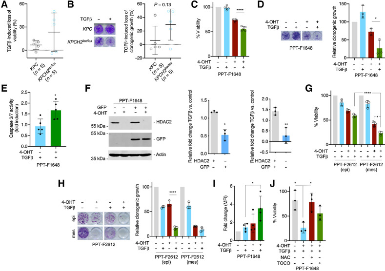
The mortality of patients with pancreatic ductal adenocarcinoma (PDAC) is strongly associated with metastasis, a multistep process that is incompletely understood in this disease. Although genetic drivers of PDAC metastasis have not been defined, transcriptional and epigenetic rewiring can contribute to the metastatic process. The epigenetic eraser histone deacetylase 2 (HDAC2) has been connected to less differentiated PDAC, but the function of HDAC2 in PDAC has not been comprehensively evaluated. Using genetically defined models, we show that HDAC2 is a cellular fitness factor that controls cell cycle in vitro and metastasis in vivo, particularly in undifferentiated, mesenchymal PDAC cells. Unbiased expression profiling detected a core set of HDAC2-regulated genes. HDAC2 controlled expression of several prosurvival receptor tyrosine kinases connected to mesenchymal PDAC, including PDGFRα, PDGFRβ, and EGFR. The HDAC2-maintained program disabled the tumor-suppressive arm of the TGFβ pathway, explaining impaired metastasis formation of HDAC2-deficient PDAC. These data identify HDAC2 as a tractable player in the PDAC metastatic cascade. The complexity of the function of epigenetic regulators like HDAC2 implicates that an increased understanding of these proteins is needed for implementation of effective epigenetic therapies.

Significance: HDAC2 has a context-specific role in undifferentiated PDAC and the capacity to disseminate systemically, implicating HDAC2 as targetable protein to prevent metastasis.


Figures

Figure 1. Hdac2 knockout induced reduced growth and arrest in the G2–M phase of the cell cycle. A, Top, depiction of the floxed Hdac2 allele. 4-OHT treatment leads to the deletion of exon2 to exon4. Bottom, brightfield microscopy shows the morphology of PPT-F1648 cells. Scale bar, 100 μm. B, PPT-F1648 cells were treated for the indicated time points with 600 nmol/L 4-OHT or were left as vehicle-treated controls. Western blot controls the expression of HDAC2. Actin, loading control. C, Quantification of B. n = 3. *, P < 0.05 (paired t test). D, PPT-F1648 cells were treated for eight days with 600 nmol/L 4-OHT or were left as vehicle-treated controls. Quantitative PCR determines Hdac2 mRNA expression. Three independent experiments were analyzed. E, PPT-F1648 cells were treated for 8 days with 4-OHT or vehicle control. Afterwards, cells were plated in 96 wells and growth was determined by MTT assay over the indicated time points. Assay was performed in triplicate with three technical replicates. F, PPT-F1648 cells were treated for 8 days with 4-OHT or vehicle control. Afterwards, 3,000 cells were plated in 6-well plates and growth was determined by cell counting over the indicated time points. The assay was performed in triplicate with three technical replicates. G, 2,000 cells were seeded in 24 wells and clonogenic growth was analyzed after seven days. Left, representative clonogenic growth assay. Quantification (counting of Giemsa-stained colonies) of three independent experiments conducted as technical triplicate. H, PI cell-cycle FACS analysis after eight days of treatment as indicated. Three independent experiments were performed. I, RNA-seq of Hdac2-deficient and -proficient PPT-F1648 cells was analyzed by GSEA. Enrichment plots for the depicted HALLMARK signatures are shown. NES, normalized enrichment score; P, nominal P value; q, FDR. *, P < 0.05; **, P < 0.01; t test.
Figure 1.
Hdac2 knockout induced reduced growth and arrest in the G2–M phase of the cell cycle. A, Top, depiction of the floxed Hdac2 allele. 4-OHT treatment leads to the deletion of exon2 to exon4. Bottom, brightfield microscopy shows the morphology of PPT-F1648 cells. Scale bar, 100 μm. B, PPT-F1648 cells were treated for the indicated time points with 600 nmol/L 4-OHT or were left as vehicle-treated controls. Western blot controls the expression of HDAC2. Actin, loading control. C, Quantification of B. n = 3. *, P < 0.05 (paired t test). D, PPT-F1648 cells were treated for eight days with 600 nmol/L 4-OHT or were left as vehicle-treated controls. Quantitative PCR determines Hdac2 mRNA expression. Three independent experiments were analyzed. E, PPT-F1648 cells were treated for 8 days with 4-OHT or vehicle control. Afterwards, cells were plated in 96 wells and growth was determined by MTT assay over the indicated time points. Assay was performed in triplicate with three technical replicates. F, PPT-F1648 cells were treated for 8 days with 4-OHT or vehicle control. Afterwards, 3,000 cells were plated in 6-well plates and growth was determined by cell counting over the indicated time points. The assay was performed in triplicate with three technical replicates. G, 2,000 cells were seeded in 24 wells and clonogenic growth was analyzed after seven days. Left, representative clonogenic growth assay. Quantification (counting of Giemsa-stained colonies) of three independent experiments conducted as technical triplicate. H, PI cell-cycle FACS analysis after eight days of treatment as indicated. Three independent experiments were performed. I, RNA-seq of Hdac2-deficient and -proficient PPT-F1648 cells was analyzed by GSEA. Enrichment plots for the depicted HALLMARK signatures are shown. NES, normalized enrichment score; P, nominal P value; q, FDR. *, P < 0.05; **, P < 0.01; t test.
Figure 2. HDAC2 is relevant in undifferentiated PDAC cells. A, Brightfield microscopy of parental PPT-F2612 and the corresponding epithelial and mesenchymal sublines established by differential trypsinization. Scale bar, 100 μm. B and C, Adequate fractionation of epithelial (epi) and mesenchymal (mes) PPT-F2612 cells was controlled by Western blotting (B) of E-cadherin and vimentin. Actin, loading control. One representative Western blot out of three is depicted. C, Quantitative analysis of the mRNA expression of Cdh1 and Vim in epithelial and mesenchymal PPT-F2612 cells. Four independent biological experiments were conducted. **, P < 0.01 (t test). D, 3,000 epithelial or mesenchymal PPT-F2612 cells were plated in 6-well plates and growth was determined by cell counting over the indicated time points. The assay was performed in triplicate with three technical replicates. E, HDAC2 Western blot in the mesenchymal and epithelial fraction of PPT-F2612 cell treated with 4-OHT or vehicle control over eight days. Actin, loading control. F, The mesenchymal and epithelial fraction of PPT-F2612 cell treated with 4-OHT or vehicle control over eight days. Afterwards, 2,000 cells were seeded in a 96-well plate and viability was measured after 3 days by MTT assay. Three independent experiments conducted as technical triplicate were analyzed. Shown is the percent reduction of viability induced by the Hdac2 knockout. **, P < 0.01 (unpaired t test). G and H, Mesenchymal and epithelial fractions of PPT-F2612 cell were treated as described in E. 2,000 cells were seeded in a 24-well plate and clonogenic growth was analyzed after seven days. G, Representative clonogenic growth assay. H, Quantification of three independent experiments conducted as three technical replicates. Growth of epithelial control–treated cells were arbitrarily set to 1. **, P < 0.01 (unpaired t test). I, Mesenchymal and epithelial fraction of PPT-F2612 cells was treated as in E. Afterwards, 2,000 cells were transferred to a low attachment plate and viability was measured after 3 days using MTT assay. Four independent biological experiments are depicted. ****, P < 0.0001 (unpaired t test).
Figure 2.
HDAC2 is relevant in undifferentiated PDAC cells. A, Brightfield microscopy of parental PPT-F2612 and the corresponding epithelial and mesenchymal sublines established by differential trypsinization. Scale bar, 100 μm. B and C, Adequate fractionation of epithelial (epi) and mesenchymal (mes) PPT-F2612 cells was controlled by Western blotting (B) of E-cadherin and vimentin. Actin, loading control. One representative Western blot out of three is depicted. C, Quantitative analysis of the mRNA expression of Cdh1 and Vim in epithelial and mesenchymal PPT-F2612 cells. Four independent biological experiments were conducted. **, P < 0.01 (t test). D, 3,000 epithelial or mesenchymal PPT-F2612 cells were plated in 6-well plates and growth was determined by cell counting over the indicated time points. The assay was performed in triplicate with three technical replicates. E, HDAC2 Western blot in the mesenchymal and epithelial fraction of PPT-F2612 cell treated with 4-OHT or vehicle control over eight days. Actin, loading control. F, The mesenchymal and epithelial fraction of PPT-F2612 cell treated with 4-OHT or vehicle control over eight days. Afterwards, 2,000 cells were seeded in a 96-well plate and viability was measured after 3 days by MTT assay. Three independent experiments conducted as technical triplicate were analyzed. Shown is the percent reduction of viability induced by the Hdac2 knockout. **, P < 0.01 (unpaired t test). G and H, Mesenchymal and epithelial fractions of PPT-F2612 cell were treated as described in E. 2,000 cells were seeded in a 24-well plate and clonogenic growth was analyzed after seven days. G, Representative clonogenic growth assay. H, Quantification of three independent experiments conducted as three technical replicates. Growth of epithelial control–treated cells were arbitrarily set to 1. **, P < 0.01 (unpaired t test). I, Mesenchymal and epithelial fraction of PPT-F2612 cells was treated as in E. Afterwards, 2,000 cells were transferred to a low attachment plate and viability was measured after 3 days using MTT assay. Four independent biological experiments are depicted. ****, P < 0.0001 (unpaired t test).
Figure 3. HDAC2 controls a route to metastasis in vivo. A, Kaplan–Meier plots of KPC (black), KPCH2lox/+(red), and KPCH2lox/lox (blue) mice. B, Hematoxylin and eosin staining of murine PDAC from KPC and KPCH2lox/lox mice. G1, well-differentiated; G2, moderately differentiated; G3, poorly differentiated; G4, undifferentiated PDAC. Scale bar, 500 μm. C and D, Quantification of Ki67 stainings (C) and cleaved caspase-3 (D) stainings in murine PDAC from KPC (n = 4) and KPCH2lox/lox (n = 5) mice. E, Quantification of gradings from KPC and KPCH2lox/lox mice. F, Morphology of murine PDAC cell lines from KPC (n = 8) and KPCH2lox/lox mice (n = 7) was evaluated and stratified as epithelial (epi; blue), mesenchymal (mes; red), or as intermediate/mixed (mixed; green). G, Metastasis frequency of KPC and KPCH2lox/lox mice. **, P < 0.01 (Fisher exact test). H, Hematoxylin and eosin staining of liver and lung metastasis of KPC and KPCH2lox/lox mice. Scale bar, 500 μm. I, HDAC2 IHC of liver and lung metastasis of KPCH2lox/lox mice. Scale bar, 200 μm.
Figure 3.
HDAC2 controls a route to metastasis in vivo. A, Kaplan–Meier plots of KPC (black), KPCH2lox/+(red), and KPCH2lox/lox (blue) mice. B, Hematoxylin and eosin staining of murine PDAC from KPC and KPCH2lox/lox mice. G1, well-differentiated; G2, moderately differentiated; G3, poorly differentiated; G4, undifferentiated PDAC. Scale bar, 500 μm. C and D, Quantification of Ki67 stainings (C) and cleaved caspase-3 (D) stainings in murine PDAC from KPC (n = 4) and KPCH2lox/lox (n = 5) mice. E, Quantification of gradings from KPC and KPCH2lox/lox mice. F, Morphology of murine PDAC cell lines from KPC (n = 8) and KPCH2lox/lox mice (n = 7) was evaluated and stratified as epithelial (epi; blue), mesenchymal (mes; red), or as intermediate/mixed (mixed; green). G, Metastasis frequency of KPC and KPCH2lox/lox mice. **, P < 0.01 (Fisher exact test). H, Hematoxylin and eosin staining of liver and lung metastasis of KPC and KPCH2lox/lox mice. Scale bar, 500 μm. I, HDAC2 IHC of liver and lung metastasis of KPCH2lox/lox mice. Scale bar, 200 μm.
Figure 4. HDAC2 maintains RTK-driven survival signaling. A, HDAC2 regulated genes in microarray of KPC (n = 3) and KPCH2lox/lox (n = 3) cancer cell lines (i) and 4-OHT–treated compared with vehicle-treated PPT-F1648 cells (ii) were analyzed in a Venn diagram including genes regulated with a log FC ± 0.58 and a P < 0.05. B, Genes consistently downregulated in Hdac2 knockout models were analyzed by the MolecularSignatureDatabase (MSigDB). HALLMARK signatures with an FDR < 0.05 are depicted. The FDR is color coded, number of genes contributing are coded by size. The ratio gene number to number of genes in the signature is depicted. C and D, Genes consistently downregulated in Hdac2 knockout models were analyzed by the Enrichr web tool using the libraries Kyoto Encyclopedia of Genes and Genomes (KEGG) 2021 and ARCH4 kinase coexpression. Top five signatures ranked according to the combined score. Padj value is color coded and the combined score is depicted. E, PPT-F1648 cells were treated for eight days with 4-OHT (600 nmol/L) or were left as vehicle treated controls. Western blot of PDGFRα, PDGFRβ, EGFR, phospho-AKT (T308 and S473), pan-AKT, phospho-GSK3 (S9), phospho-S6 (S235/236), phospho-ERK (T202/Y204), and HDAC2. Actin, loading control. One representative Western blot out of three is depicted F, Quantification of three independent experiments according to E. *, P < 0.05; **, P < 0.01 (t test). G, Differential analysis of reads in HDAC2 peaks between Hdac2-proficient and Hdac2-deficient PPT-F1648. Blue, significant peaks (FDR < 0.05, n = 2754). H, Density plot (top) and heatmap (bottom) of HDAC2 ChIP-seq reads around the proximal promoter in Hdac2-proficient and Hdac2-deficient PPT-F1648 (read counts per million). I, Overlap of significant HDAC2 peaks around the TSS (± 3,000 bp; Padj < 0.05) with significant differentially expressed genes in Hdac2-deficient PPT-F1648 cells (Padj < 0.05). J, Volcano plot of differentially regulated genes in Hdac2-deficient PPT-F1648 cells. Blue, genes with HDAC2 ChIP-seq peaks; gray, significant differential genes (Padj < 0.05). K, Homer motif analysis of all significant HDAC2 ChIP-seq peaks in promoter regions (TSS ± 3,000 bp; Padj < 0.05; n = 2,360); known motifs are depicted. L, Homer motif analysis of significant HDAC2 ChIP-seq peaks with downregulation in RNA-seq. De novo motif results are depicted (TSS ± 3,000 bp; Padj < 0.05; n = 102).
Figure 4.
HDAC2 maintains RTK-driven survival signaling. A, HDAC2 regulated genes in microarray of KPC (n = 3) and KPCH2lox/lox (n = 3) cancer cell lines (i) and 4-OHT–treated compared with vehicle-treated PPT-F1648 cells (ii) were analyzed in a Venn diagram including genes regulated with a log FC ± 0.58 and a P < 0.05. B, Genes consistently downregulated in Hdac2 knockout models were analyzed by the MolecularSignatureDatabase (MSigDB). HALLMARK signatures with an FDR < 0.05 are depicted. The FDR is color coded, number of genes contributing are coded by size. The ratio gene number to number of genes in the signature is depicted. C and D, Genes consistently downregulated in Hdac2 knockout models were analyzed by the Enrichr web tool using the libraries Kyoto Encyclopedia of Genes and Genomes (KEGG) 2021 and ARCH4 kinase coexpression. Top five signatures ranked according to the combined score. Padj value is color coded and the combined score is depicted. E, PPT-F1648 cells were treated for eight days with 4-OHT (600 nmol/L) or were left as vehicle treated controls. Western blot of PDGFRα, PDGFRβ, EGFR, phospho-AKT (T308 and S473), pan-AKT, phospho-GSK3 (S9), phospho-S6 (S235/236), phospho-ERK (T202/Y204), and HDAC2. Actin, loading control. One representative Western blot out of three is depicted F, Quantification of three independent experiments according to E. *, P < 0.05; **, P < 0.01 (t test). G, Differential analysis of reads in HDAC2 peaks between Hdac2-proficient and Hdac2-deficient PPT-F1648. Blue, significant peaks (FDR < 0.05, n = 2754). H, Density plot (top) and heatmap (bottom) of HDAC2 ChIP-seq reads around the proximal promoter in Hdac2-proficient and Hdac2-deficient PPT-F1648 (read counts per million). I, Overlap of significant HDAC2 peaks around the TSS (± 3,000 bp; Padj < 0.05) with significant differentially expressed genes in Hdac2-deficient PPT-F1648 cells (Padj < 0.05). J, Volcano plot of differentially regulated genes in Hdac2-deficient PPT-F1648 cells. Blue, genes with HDAC2 ChIP-seq peaks; gray, significant differential genes (Padj < 0.05). K, Homer motif analysis of all significant HDAC2 ChIP-seq peaks in promoter regions (TSS ± 3,000 bp; Padj < 0.05; n = 2,360); known motifs are depicted. L, Homer motif analysis of significant HDAC2 ChIP-seq peaks with downregulation in RNA-seq. De novo motif results are depicted (TSS ± 3,000 bp; Padj < 0.05; n = 102).
Figure 5. HDAC2 protects from TGFβ-dependent tumor suppression. A and B, Five murine PDAC cell lines from KPC mice and five lines from KPCH2lox/lox mice were treated with TGFβ (5 ng/mL) or left as untreated control. A, Viability was determined by MTT assay 72 hours after the treatment. Mean viability of a line was determined by three independent biological experiments. B, Clonogenic assays were performed and analyzed after 7 days of treatment. Left, macroscopic picture of a clonogenic assay of a KPC and a KPCH2lox/lox line. Right, quantification. Mean clonogenic growth of a line was determined by three independent biological experiments conducted as technical triplicates. P value of an unpaired t test is depicted. C, PPT-F1648 cells were treated for eight days with 4-OHT (600 nmol/L) or were left as vehicle-treated controls. Afterwards, cells were replated and 24 hours later treated with TGFβ (5 ng/mL) for 72 hours. Viability was determined by MTT assay. ****, P < 0.0001 (ANOVA). D, PPT-F1648 cells were treated as in C. After eight days of 4-OHT treatment, cells were replated in 24 wells and treated after 24 hours with TGFβ (5 ng/mL) for 7 days. Left, macroscopic picture of a clonogenic assay. Right, quantification of three independent experiments conducted as technical triplicates. *, P < 0.05 (ANOVA). E, PPT-F1648 cells were treated as in C. Afterwards, cells were plated in 96 wells and 24 hours later treated with TGFβ (5 ng/mL) for 72 hours. Caspase activity (measured by Caspase-Glo 3/7 Assay) was normalized to cell viability (determined by CellTiter-Glo Assay) and fold induction upon TGFβ treatment compared with control was calculated. *, P < 0.05 (t test). F, PPT-F1648 cells were transduced with a GFP vector and then treated for eight days with 4-OHT (600 nmol/L) or were left as vehicle-treated controls. Afterwards, cells were replated and 24 hours later treated with TGFβ (5 ng/mL) for 96 hours. Left, Western blotting controls expression of GFP and HDAC2. Actin, loading control; right, relative fold change in the depicted conditions as determined by FACS. *, P < 0.05; **, P < 0.01 (t test). G, Epithelial (epi) and mesenchymal (mes) fraction of PPT-F2612 cells were treated for eight days with 4-OHT (600 nmol/L) or were left as vehicle-treated controls. Afterwards, cells were replated and 24 hours later treated with TGFβ (5 ng/mL) for 72 hours. Viability was determined by MTT assay. *, P < 0.05 (ANOVA); green bars were analyzed with an unpaired t test (****, P < 0.0001). H, Epithelial and mesenchymal fraction of PPT-F2612 cells as in D. After eight days of 4-OHT treatment, cells were replated in 24 wells and after 24 hours treated with TGFβ (5 ng/mL) for 7 days. Left, macroscopic picture of a clonogenic assay. Right, quantification of three independent experiments. ****, P < 0.0001 (ANOVA). I, ROS levels of control and 4-OHT (600 nmol/L)-treated PPT-F1648 cells after addition of TGFβ (5 ng/mL) for 72 hours. FACS measurements are plotted as fold change of the mean fluorescence intensity (MFI). J, Viability of control and 4-OHT (600 nmol/L)-treated PPT-F1648 cells treated with TGFβ (5 ng/mL) or control treatment in addition to ROS scavengers, 2 mmol/L NAC or 2 mmol/L α-tocopherol. Cell viability was measured after 72 hours by CellTiter-Glo Assay and viability of untreated Hdac2-proficient PPT-F1648 cells was set to 100%. *, P < 0.05 (ANOVA).
Figure 5.
HDAC2 protects from TGFβ-dependent tumor suppression. A and B, Five murine PDAC cell lines from KPC mice and five lines from KPCH2lox/lox mice were treated with TGFβ (5 ng/mL) or left as untreated control. A, Viability was determined by MTT assay 72 hours after the treatment. Mean viability of a line was determined by three independent biological experiments. B, Clonogenic assays were performed and analyzed after 7 days of treatment. Left, macroscopic picture of a clonogenic assay of a KPC and a KPCH2lox/lox line. Right, quantification. Mean clonogenic growth of a line was determined by three independent biological experiments conducted as technical triplicates. P value of an unpaired t test is depicted. C, PPT-F1648 cells were treated for eight days with 4-OHT (600 nmol/L) or were left as vehicle-treated controls. Afterwards, cells were replated and 24 hours later treated with TGFβ (5 ng/mL) for 72 hours. Viability was determined by MTT assay. ****, P < 0.0001 (ANOVA). D, PPT-F1648 cells were treated as in C. After eight days of 4-OHT treatment, cells were replated in 24 wells and treated after 24 hours with TGFβ (5 ng/mL) for 7 days. Left, macroscopic picture of a clonogenic assay. Right, quantification of three independent experiments conducted as technical triplicates. *, P < 0.05 (ANOVA). E, PPT-F1648 cells were treated as in C. Afterwards, cells were plated in 96 wells and 24 hours later treated with TGFβ (5 ng/mL) for 72 hours. Caspase activity (measured by Caspase-Glo 3/7 Assay) was normalized to cell viability (determined by CellTiter-Glo Assay) and fold induction upon TGFβ treatment compared with control was calculated. *, P < 0.05 (t test). F, PPT-F1648 cells were transduced with a GFP vector and then treated for eight days with 4-OHT (600 nmol/L) or were left as vehicle-treated controls. Afterwards, cells were replated and 24 hours later treated with TGFβ (5 ng/mL) for 96 hours. Left, Western blotting controls expression of GFP and HDAC2. Actin, loading control; right, relative fold change in the depicted conditions as determined by FACS. *, P < 0.05; **, P < 0.01 (t test). G, Epithelial (epi) and mesenchymal (mes) fraction of PPT-F2612 cells were treated for eight days with 4-OHT (600 nmol/L) or were left as vehicle-treated controls. Afterwards, cells were replated and 24 hours later treated with TGFβ (5 ng/mL) for 72 hours. Viability was determined by MTT assay. *, P < 0.05 (ANOVA); green bars were analyzed with an unpaired t test (****, P < 0.0001). H, Epithelial and mesenchymal fraction of PPT-F2612 cells as in D. After eight days of 4-OHT treatment, cells were replated in 24 wells and after 24 hours treated with TGFβ (5 ng/mL) for 7 days. Left, macroscopic picture of a clonogenic assay. Right, quantification of three independent experiments. ****, P < 0.0001 (ANOVA). I, ROS levels of control and 4-OHT (600 nmol/L)-treated PPT-F1648 cells after addition of TGFβ (5 ng/mL) for 72 hours. FACS measurements are plotted as fold change of the mean fluorescence intensity (MFI). J, Viability of control and 4-OHT (600 nmol/L)-treated PPT-F1648 cells treated with TGFβ (5 ng/mL) or control treatment in addition to ROS scavengers, 2 mmol/L NAC or 2 mmol/L α-tocopherol. Cell viability was measured after 72 hours by CellTiter-Glo Assay and viability of untreated Hdac2-proficient PPT-F1648 cells was set to 100%. *, P < 0.05 (ANOVA).

References

    1. Mueller S, Engleitner T, Maresch R, Zukowska M, Lange S, Kaltenbacher T, et al. . Evolutionary routes and KRAS dosage define pancreatic cancer phenotypes. Nature 2018;554:62–8. - PMC - PubMed
    1. Chan-Seng-Yue M, Kim JC, Wilson GW, Ng K, Figueroa EF, O'Kane GM, et al. . Transcription phenotypes of pancreatic cancer are driven by genomic events during tumor evolution. Nat Genet 2020;52:231–40. - PubMed
    1. Makohon-Moore AP, Zhang M, Reiter JG, Bozic I, Allen B, Kundu D, et al. . Limited heterogeneity of known driver gene mutations among the metastases of individual patients with pancreatic cancer. Nat Genet 2017;49:358–66. - PMC - PubMed
    1. McDonald OG, Li X, Saunders T, Tryggvadottir R, Mentch SJ, Warmoes MO, et al. . Epigenomic reprogramming during pancreatic cancer progression links anabolic glucose metabolism to distant metastasis. Nat Genet 2017;49:367–76. - PMC - PubMed
    1. Roe J-S, Hwang C-I, Somerville TDD, Milazzo JP, Lee EJ, Silva BD, et al. . Enhancer Reprogramming Promotes Pancreatic Cancer Metastasis. Cell 2017;170:875–88. - PMC - PubMed

Publication types

MeSH terms

Substances