. 2022 Feb;15(2):327-337.
doi: 10.1038/s41385-021-00472-w. Epub 2021 Dec 13.

LIGHT controls distinct homeostatic and inflammatory gene expression profiles in esophageal fibroblasts via differential HVEM and LTβR-mediated mechanisms

Affiliations

LIGHT controls distinct homeostatic and inflammatory gene expression profiles in esophageal fibroblasts via differential HVEM and LTβR-mediated mechanisms

Mario C Manresa et al. Mucosal Immunol. 2022 Feb.

Abstract

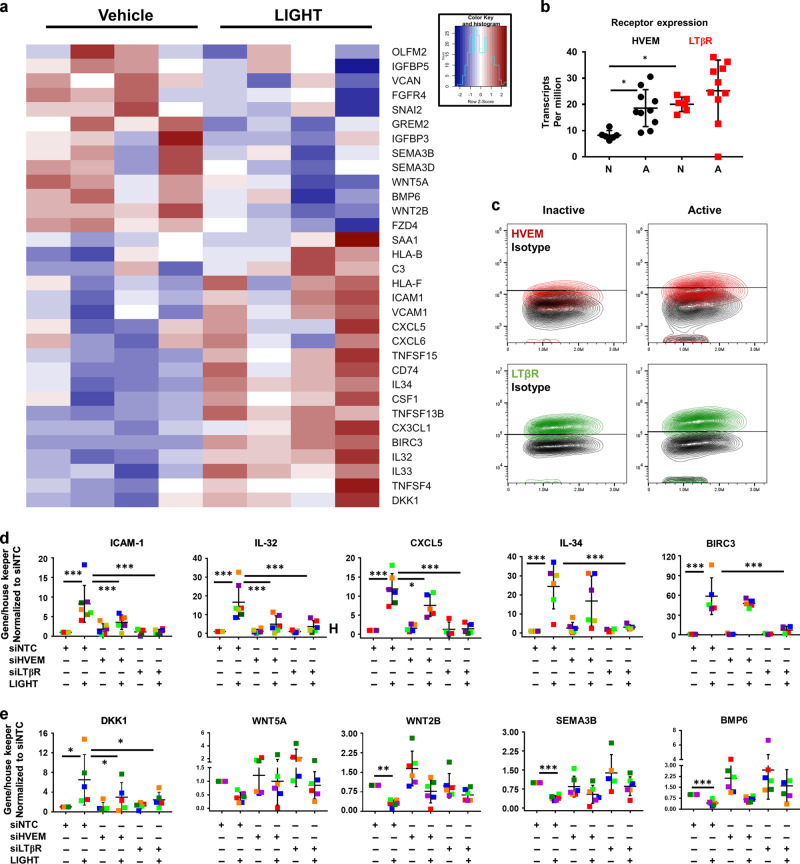
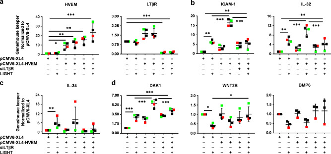
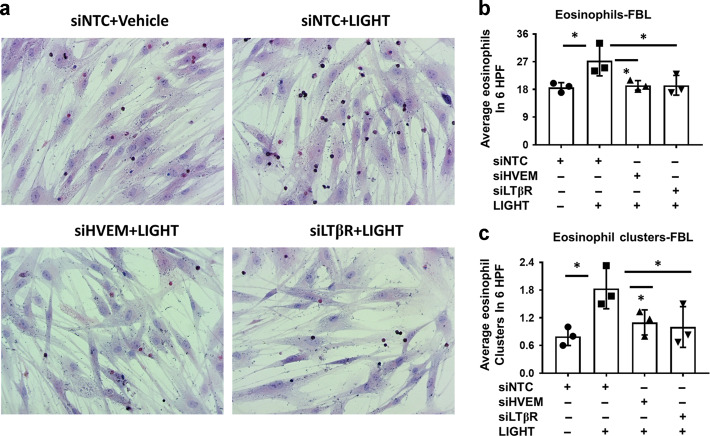
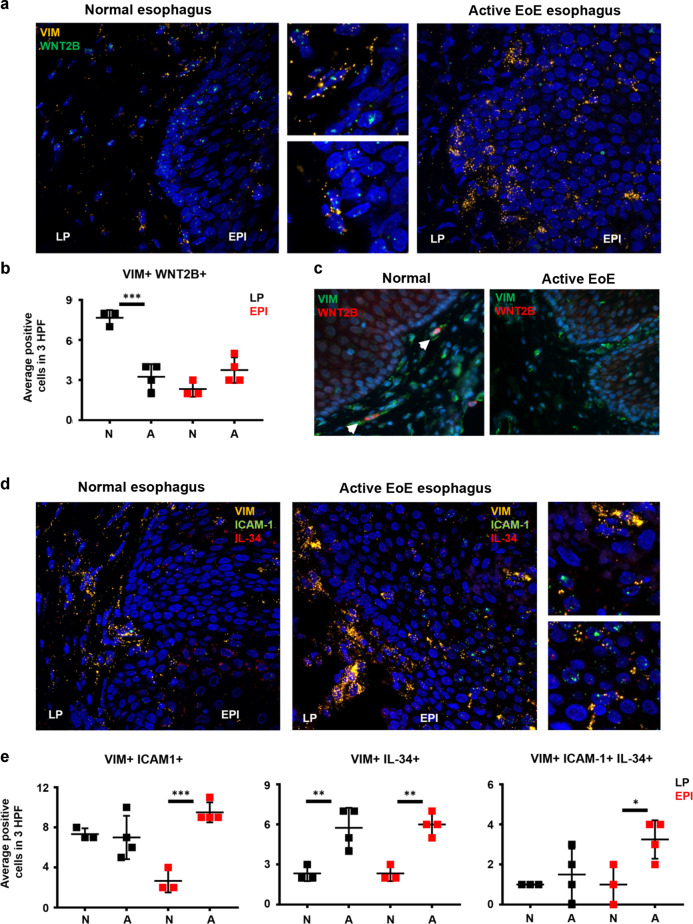
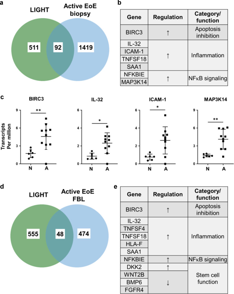
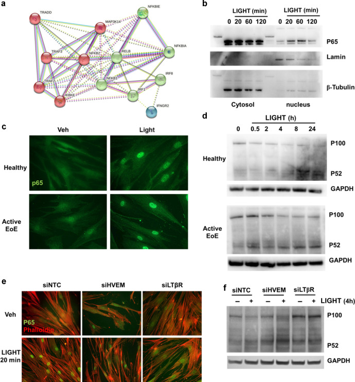
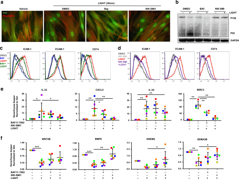
Fibroblasts mediate tissue remodeling in eosinophilic esophagitis (EoE), a chronic allergen-driven inflammatory pathology. Diverse fibroblast subtypes with homeostasis-regulating or inflammatory profiles have been recognized in various tissues, but which mediators induce these alternate differentiation states remain largely unknown. We recently identified that TNFSF14/LIGHT promotes an inflammatory esophageal fibroblast in vitro. Herein we used esophageal biopsies and primary fibroblasts to investigate the role of the LIGHT receptors, herpes virus entry mediator (HVEM) and lymphotoxin-beta receptor (LTβR), and their downstream activated pathways, in EoE. In addition to promoting inflammatory gene expression, LIGHT down-regulated homeostatic factors including WNTs, BMPs and type 3 semaphorins. In vivo, WNT2B+ fibroblasts were decreased while ICAM-1+ and IL-34+ fibroblasts were expanded in EoE, suggesting that a LIGHT-driven gene signature was imprinted in EoE versus normal esophageal fibroblasts. HVEM and LTβR overexpression and deficiency experiments demonstrated that HVEM regulates a limited subset of LIGHT targets, whereas LTβR controls all transcriptional effects. Pharmacologic blockade of the non-canonical NIK/p100/p52-mediated NF-κB pathway potently silenced LIGHT's transcriptional effects, with a lesser role found for p65 canonical NF-κB. Collectively, our results show that LIGHT promotes differentiation of esophageal fibroblasts toward an inflammatory phenotype and represses homeostatic gene expression via a LTβR-NIK-p52 NF-κB dominant pathway.


Conflict of interest statement

S.S.A. is a co-inventor of oral viscous budesonide for EoE patented by UCSD and licensed by Shire-Takeda. S.S.A. is a consultant for Regeneron, AImmune, Astellas, AstraZeneca, and Gossamer Bio. M.C has patents on TNFSF14/LIGHT. The remaining authors declare no competing interests.

Figures

Fig. 1
Fig. 1. HVEM and LTβR contribute to LIGHT-mediated responses in esophageal fibroblasts.
a Heat map of representative homeostatic and inflammatory mediators comparing vehicle or LIGHT treated esophageal fibroblasts from 4 different donors (>1.5 fold, p < 0.05). b Comparison of HVEM and LTβR expression (TPM) in normal esophagus (n = 6) and active EoE biopsies (n = 10) from Sherrill et al. 2014. c Representative plots showing the frequency of HVEM (red) and LTβR (green)-expressing cells compared to isotype control in active or inactive EoE fibroblasts (n = 6). qRT-PCR expression of inflammatory (d) or homeostatic (e) genes in cells transfected with non-targeting RNA (siNTC) or siRNAs against HVEM (siHVEM) or LTβR (siLTβR) in vehicle or LIGHT-treated (24 h) cells (each colored dot represents fibroblasts from a different donor, n ≥ 5). *p < 0.05 and ***p < 0.001.
Fig. 2
Fig. 2. Overexpression of HVEM partially modulates LIGHT-mediated gene expression in esophageal fibroblasts.
qRT-PCR of: HVEM and LTβR (a); ICAM-1 and IL-32 (b); IL-34 (c); and DKK1, WNT2B and BMP6 (d), in normal esophageal fibroblasts transfected with empty vector (pCMV6-XL4), HVEM-OE (pCMV6-XL4-HVEM) or a combination of HVEM-OE + siLTβR, and untreated or treated with LIGHT (n ≥ 3). Each colored dot represents fibroblasts from a different donor, *p < 0.05, **p < 0.01 and ***p < 0.001.
Fig. 3
Fig. 3. HVEM and LTβR-mediated mechanisms contribute to fibroblast-eosinophil tethering in co-culture.
a Representative haematoxylin/eosin stained co-cultures of fibroblasts and eosinophils in fibroblasts transfected with non-targeting control RNA (siNTC) or siRNAs against HVEM (siHVEM) or LTβR (siLTβR) in vehicle or LIGHT treated (24 h) cells followed by co-culture with eosinophils for 8 h in the absence of LIGHT (n = 3). Quantification of total eosinophils (b) or eosinophil clusters (c) in cells treated as in a (each dot represents fibroblasts from an independent donor). *p < 0.05.
Fig. 4
Fig. 4. Localization of VIM + cells expressing homeostatic and inflammatory markers reveals functional changes in EoE.
a Representative images of normal (n = 3) and active EoE esophagus (n = 4) hybridized with specific probes for VIM (yellow) and WNT2B (green, LP = lamina propria, EPI = epithelium). b Immunofluorescence staining of normal (n = 3) and active EoE esophagus (n = 4) for VIM (green) and WNT2B (red). c Quantification of VIM+ WNT2B+ cells in LP and EPI comparing normal and active EoE esophagus. d Representative images of normal (n = 3) and active EoE esophagus (n = 4) hybridized with specific probes for VIM (yellow), ICAM-1 (green) and IL-34 (red). e Quantification of VIM+ ICAM-1+ , VIM+ IL-34+ and VIM+ ICAM-1+ IL-34+ cells in LP and EPI comparing normal and active EoE esophagus. White arrows in c point positive cells. *p < 0.05, **p < 0.01 and ***p < 0.001.
Fig. 5
Fig. 5. Comparison of LIGHT-mediated and basal EoE transcriptomes reveals common regulated gene signatures.
Venn diagram (a) and list of selected commonly regulated genes (b) comparing genes differentially expressed in LIGHT-treated normal esophageal fibroblasts compared to vehicle (n = 4) and active EoE biopsies (n = 10) compared to normal esophagus (n = 6). c Normalized transcript per million analyses of BIRC3, IL-32, ICAM-1 and MAP3K14 in normal and active EoE esophageal biopsies. Venn diagram (d) and list of selected commonly regulated genes (e) comparing genes differentially expressed in LIGHT-treated normal esophageal fibroblasts compared to vehicle (n = 4) and basal expression in active EoE fibroblasts compared to normal (n = 4).
Fig. 6
Fig. 6. LIGHT activates canonical and alternative NF-κB signaling pathways in esophageal fibroblasts.
a STRING analysis of molecular signaling-related genes up-regulated by LIGHT in normal esophageal fibroblasts (>1.5 fold, p < 0.05). b Representative western blot (WB) comparing p65 translocation in nuclear vs cytosolic extracts from normal esophageal fibroblasts treated with LIGHT for the indicated times (n = 3). c Representative images of normal or active EoE esophageal fibroblast monolayers stained for p65 after 20 min of LIGHT stimulation (n = 3). d Representative WB comparing p100 cleavage into p52 in whole cell lysates from normal or active EoE esophageal fibroblasts treated with LIGHT for the indicated times (n = 3). Representative images of p65 translocation (e) and representative western blot of p100 cleavage (f) in cells transfected with scramble RNA (siNTC) or siRNAs against HVEM (siHVEM) or LTβR (siLTβR) and untreated or treated with LIGHT for the times indicated (n = 3).
Fig. 7
Fig. 7. NIK inhibition has a dominant suppressive role on LIGHT responses in esophageal fibroblasts.
Representative images of p65 translocation (a) and representative western blot of p100 cleavage (b) in cells pre-treated with BAY11-7082 or NIK-SMI1 for 1 h and then treated with LIGHT for the times indicated (n = 3). Representative histograms of flow cytometry of ICAM-1, VCAM-1 and CD74 in normal esophageal fibroblasts pre-treated with BAY11-7082 (c) or NIK-SMI1 (d) for 1 h and then treated with LIGHT for 24 h (n = 3). RT-PCR of inflammatory (e) and homeostatic (f) genes in esophageal fibroblasts pre-treated with BAY11-7082 or NIK-SMI1 for 1 h and then treated with LIGHT for 24 h (n = 6). Each colored dot represents fibroblasts from an independent donor. *p < 0.05, **p < 0.01 and ***p < 0.001.

References

    1. Croft AP, et al. Distinct fibroblast subsets drive inflammation and damage in arthritis. Nature. 2019;570:246–251. - PMC - PubMed
    1. Martin JC, et al. Single-cell analysis of Crohn’s disease lesions identifies a pathogenic cellular module associated with resistance to anti-TNF therapy. Cell. 2019;178:1493–1508. - PMC - PubMed
    1. Guerrero-Juarez C. F., et al. Single-cell analysis reveals fibroblast heterogeneity and myeloid-derived adipocyte progenitors in murine skin wounds. Nat. Commun.10 (2019). - PMC - PubMed
    1. Kinchen J, et al. Structural remodeling of the human colonic mesenchyme in inflammatory bowel disease. Cell. 2018;175:372. - PMC - PubMed
    1. Manresa MC, et al. Hydroxylase inhibition regulates inflammation-induced intestinal fibrosis through the suppression of ERK-mediated TGF-beta 1 signaling (Vol 311, Pg G1076, 2016) Am. J. Physiol. Gastr L. 2017;312:G405–G405. - PubMed

Publication types

MeSH terms

Substances