. 2022 Jan;21(1):e13528.
doi: 10.1111/acel.13528. Epub 2021 Dec 13.

Deletion of SA β-Gal+ cells using senolytics improves muscle regeneration in old mice

Affiliations

Deletion of SA β-Gal+ cells using senolytics improves muscle regeneration in old mice

Cory M Dungan et al. Aging Cell. 2022 Jan.

Abstract

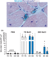
Systemic deletion of senescent cells leads to robust improvements in cognitive, cardiovascular, and whole-body metabolism, but their role in tissue reparative processes is incompletely understood. We hypothesized that senolytic drugs would enhance regeneration in aged skeletal muscle. Young (3 months) and old (20 months) male C57Bl/6J mice were administered the senolytics dasatinib (5 mg/kg) and quercetin (50 mg/kg) or vehicle bi-weekly for 4 months. Tibialis anterior (TA) was then injected with 1.2% BaCl2 or PBS 7- or 28 days prior to euthanization. Senescence-associated β-Galactosidase positive (SA β-Gal+) cell abundance was low in muscle from both young and old mice and increased similarly 7 days following injury in both age groups, with no effect of D+Q. Most SA β-Gal+ cells were also CD11b+ in young and old mice 7- and 14 days following injury, suggesting they are infiltrating immune cells. By 14 days, SA β-Gal+/CD11b+ cells from old mice expressed senescence genes, whereas those from young mice expressed higher levels of genes characteristic of anti-inflammatory macrophages. SA β-Gal+ cells remained elevated in old compared to young mice 28 days following injury, which were reduced by D+Q only in the old mice. In D+Q-treated old mice, muscle regenerated following injury to a greater extent compared to vehicle-treated old mice, having larger fiber cross-sectional area after 28 days. Conversely, D+Q blunted regeneration in young mice. In vitro experiments suggested D+Q directly improve myogenic progenitor cell proliferation. Enhanced physical function and improved muscle regeneration demonstrate that senolytics have beneficial effects only in old mice.

Keywords: regeneration; satellite cells; senescence; senolytics; skeletal muscle.


Conflict of interest statement

Z.J.T., Z.L., A.F., and M.F. are employees of Boehringer Ingelheim Pharmaceuticals, Inc. All other authors have no financial interests.

Figures
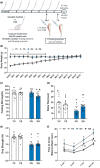
FIGURE 1
FIGURE 1
Senolytics lower blood glucose and improve physical function in old mice. (a) Study design schematic. (b) Weekly body weight for YV (white circles), YS (black and white circles), OV (blue squares), and OS (black and blue squares) mice. (c) Fasting blood glucose measurements in mg/dl. (d) Average time spent on the rotor rod in seconds. (e) Average forearm grip strength in Newtons. (f) Time to cross a 3 cm, 2 cm, 1 cm, and 0.5 cm wide balance beam for YV (white circles), YS (black and white circles), OV (blue squares), and OS (black and blue squares) mice. Error bars indicate −/+ the standard error of the mean. *p < 0.05 between young and old for an individual treatment group (YV vs. OV; YS vs. OS). ^p < 0.05 between vehicle and senolytics for a given age group (YV vs. YS; OV vs. OS). n = 17–20/group
FIGURE 2
FIGURE 2
Senolytics lower SA β‐Gal+ cell burden 28 days following BaCl2 injury. (a) Representative image of SA β‐Gal (blue staining outlined in black), hematoxylin (dark purple nuclei), and eosin (light purple cytoplasm). (b) SA β‐Gal+ cell abundance per area in YV, YS, OV, and OS mice. Young mice are labeled with white bars and old mice are labeled with blue bars. Error bars indicate −/+ the standard error of the mean. *p < 0.05 between young and old for an individual treatment group (YV vs. OV; YS vs. OS). ^p < 0.05 between vehicle and senolytics for a given age group (YV vs. YS; OV vs. OS). n = 12–14/group for PBS‐injected mice. n = 5–6/group for 7‐ and 28‐day BaCl2‐injected mice
FIGURE 3
FIGURE 3
Most C12FDG+ cells are CD11b+ suggesting they are infiltrating macrophages. Muscles from old mice 14 days following injury were dissociated and cells sorted as described in Figure S2. Representative images of isolated (a) C12FDG− and (b) C12FDG+ cells. Cells were immunoreacted with a CD11b antibody (red) shown in (c) C12FDG−/CD11b+, and (d) C12FDG+/CD11b+ cells. White circles in b and d indicate rare C12FDG+/CD11b− cells. Cells were also labeled with DAPI (blue) to visualize nuclei. n = 1 pooled sample of 4 TAs for each group. Scale bar equals 100 μm
FIGURE 4
FIGURE 4
Differential effects of senolytics on muscle fiber size during regeneration in young and old mice. Young mice are labeled with white bars and old mice are labeled with blue bars. (a) TA weight normalized to body weight. (b) Mean fiber CSA. (c–f) Representative images of immunohistochemical staining for eMyHC (red), laminin (green), and DAPI (blue) in PBS‐injected, (c’–f’) 7 days following BaCl2 injection, and (c’’–f’’) 28 days following BaCl2 injection. (g) Fiber size distribution for YV and YS groups. (h) Fiber size distribution for OV and OS groups. Error bars indicate −/+ the standard error of the mean. *p < 0.05 between young and old for an individual treatment group (YV vs. OV; YS vs. OS). ^p < 0.05 between vehicle and senolytics for a given age group (YV vs. YS; OV vs. OS). n = 12–14/group for PBS‐injected mice. n = 5–6/group for 7‐ and 28‐day BaCl2‐injected mice. Scale bar equals 50 μm
FIGURE 5
FIGURE 5
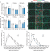
Senolytics increase satellite cell abundance in old mice. Young mice are labeled with white bars and old mice are labeled with blue bars. Representative images of immunohistochemical staining for Pax7 (pink), laminin (green), and DAPI (blue) from (a) PBS‐injected, (b) 7 days following BaCl2 injection, and (c) 28 days following BaCl2 injection. (d) Satellite cell abundance per area. Error bars indicate −/+ the standard error of the mean. Scale bar equals 50 μm. Representative images of EdU incorporation (red) into MPCs in vitro. Nuclei are visualized with DAPI (blue). (e) YV MPCs, (f) OV MPCs, (g) YS MPCs, and (h) OS MPCs. (i) Quantification of percent EdU+ MPCs. *p < 0.05 between young and old for an individual treatment group (YV vs. OV; YS vs. OS). ^p < 0.05 between vehicle and senolytics for a given age group (YV vs. YS; OV vs. OS). n = 12–14/group for PBS‐injected mice. n = 5–6/group for 7‐ and 28‐day BaCl2‐injected mice. For in vitro experiments, n = 3 technical replicates per group. Scale bar for Pax7 staining equals 50 μm. Scale bar for MPC experiments equals 20 μm
FIGURE 6
FIGURE 6
Inflammatory, metabolic, and extracellular matrix pathways are the most affected by the removal of senescent cells. Results of RNA‐seq of muscle from vehicle‐ and senolytic‐treated old mice 7 days following injury displayed by (a) Heat map and (b) Volcano plot. Upregulated (red) and downregulated (blue) genes in OS compared to OV mice are shown. (c) Pathway analysis of downregulated pathways in response to senolytics in old mice. (d) Pathway analysis of upregulated pathways in response to senolytics in old mice. n = 5/group

References

    1. Ademowo, O. S. , Dias, H. K. I. , Burton, D. G. A. , & Griffiths, H. R. (2017). Lipid (per) oxidation in mitochondria: An emerging target in the ageing process? Biogerontology, 18(6), 859–879. 10.1007/s10522-017-9710-z - DOI - PMC - PubMed
    1. Arneson‐Wissink, P. C. , Hogan, K. A. , Ducharme, A. M. , Samani, A. , Jatoi, A. , & Doles, J. D. (2020). The wasting‐associated metabolite succinate disrupts myogenesis and impairs skeletal muscle regeneration. JCSM Rapid Communications, 3(2), 56–69. 10.1002/rco2.14 - DOI - PMC - PubMed
    1. Baker, D. J. , Childs, B. G. , Durik, M. , Wijers, M. E. , Sieben, C. J. , Zhong, J. , A. Saltness, R. , Jeganathan, K. B. , Verzosa, G. C. , Pezeshki, A. , Khazaie, K. , Miller, J. D. , & van Deursen, J. M. (2016). Naturally occurring p16(Ink4a)‐positive cells shorten healthy lifespan. Nature, 530(7589), 184–189. 10.1038/nature16932 - DOI - PMC - PubMed
    1. Baker, D. J. , Wijshake, T. , Tchkonia, T. , LeBrasseur, N. K. , Childs, B. G. , van de Sluis, B. , Kirkland, J. L. , & van Deursen, J. M. (2011). Clearance of p16Ink4a‐positive senescent cells delays ageing‐associated disorders. Nature, 479(7372), 232–236. 10.1038/nature10600 - DOI - PMC - PubMed
    1. Basisty, N. , Kale, A. , Jeon, O. H. , Kuehnemann, C. , Payne, T. , Rao, C. , Holtz, A. , Shah, S. , Sharma, V. , Ferrucci, L. , Campisi, J. , & Schilling, B. (2020). A proteomic atlas of senescence‐associated secretomes for aging biomarker development. PLoS Biology, 18(1), e3000599. 10.1371/journal.pbio.3000599 - DOI - PMC - PubMed

Publication types

Substances