. 2022 Jan 11;50(1):522-535.
doi: 10.1093/nar/gkab1216.

Structural basis of dimerization and nucleic acid binding of human DBHS proteins NONO and PSPC1

Affiliations

Structural basis of dimerization and nucleic acid binding of human DBHS proteins NONO and PSPC1

Gavin J Knott et al. Nucleic Acids Res. .

Abstract

The Drosophila behaviour/human splicing (DBHS) proteins are a family of RNA/DNA binding cofactors liable for a range of cellular processes. DBHS proteins include the non-POU domain-containing octamer-binding protein (NONO) and paraspeckle protein component 1 (PSPC1), proteins capable of forming combinatorial dimers. Here, we describe the crystal structures of the human NONO and PSPC1 homodimers, representing uncharacterized DBHS dimerization states. The structures reveal a set of conserved contacts and structural plasticity within the dimerization interface that provide a rationale for dimer selectivity between DBHS paralogues. In addition, solution X-ray scattering and accompanying biochemical experiments describe a mechanism of cooperative RNA recognition by the NONO homodimer. Nucleic acid binding is reliant on RRM1, and appears to be affected by the orientation of RRM1, influenced by a newly identified 'β-clasp' structure. Our structures shed light on the molecular determinants for DBHS homo- and heterodimerization and provide a basis for understanding how DBHS proteins cooperatively recognize a broad spectrum of RNA targets.


Figures

Graphical Abstract
Graphical Abstract
Binding of nucleic acids by DBHS protein dimers involves RRM1 and RRM2 of each monomer. Although the two identical binding sites in a homodimer are not linked, independent rearrangement of RRM1 domains to accommodate nucleic acid requires the unfastening of the N-terminal β-clasp.
Figure 1.
Figure 1.
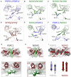
Overall structure of the human PSPC1 and NONO homodimers. (AB) The crystal structure of PSPC1-DBHS (61–320) and NONO-DBHS (53–312) homodimers, respectively. The domains crystallized are indicated schematically: β-clasp (green), RRM1 (red), RRM linker (orange), RRM2 (red), NOPS (blue) and CC (teal). (C) The six NONO dimers (chains AB, CD, EF, GH, IJ and KL) within the asymmetric unit superposed with an average RMSD of 0.46 Å between dimers (514/520 Cα-atoms). (D) The 12 NONO chains within the asymmetric unit overlaid. The variable positioning of the distal CC (teal) and the NOPS domain (blue) is indicated with ranges of motion. (E) The two conformers of W271, W1 (coloured) and W2 (transparent grey), overlaid and viewed from the dimerization interface between the NOPS domain (blue) and partnered RRM2′ (red) and CC (teal). All figures are coloured consistently throughout the manuscript.
Figure 2.
Figure 2.
Conformational plasticity of conserved residues at DBHS protein dimer interfaces, including a ‘β-clasp’ structure. (A–F) The core dimerization interface of crystallized DBHS dimers shown in a ribbon representation projected along the CC domain. Conserved residues are drawn in a stick representation. (GH) Side-by-side comparison of the NONO and PSPC1 β-clasp. (I) Superposition of the NONO and PSPC1 RRM1 domains relative to the 2-fold rotation axis centred at the β-clasp.
Figure 3.
Figure 3.
RRM1 is essential for nucleic binding and NONO and PSPC1 homodimers have different nucleic acid specificities. (A) Schematic representation of NONO/PSPC1 constructs used in this study. (B) MST binding curves for NONO/PSPC1 interacting with single-stranded homo-ribonucleic acids. Baseline-corrected normalized fluorescence (ΔFNORM) for polyG (blue) interacting with NONO is plotted on the right and ΔFNORM for polyU/C/A (green/grey/orange) and polyG interacting with PSPC1 on the left against concentration of NONO/PSPC1 in nM. The binding coefficient KD (μM) and Hill coefficient (nH) are summarized in a table. (C) Binding curves for NONO/PSPC1 interacting with 2′-modified ASOs. ΔFNORM is plotted against concentration of NONO/PSPC1 in nM. The binding coefficient KD (μM) and Hill coefficient (nH) are summarized in a table. (D) Binding curves for constructs of NONO interacting with IONIS742093, where ΔFNORM is plotted against concentration of NONO in nM. The binding coefficient KD (μM) and Hill coefficient (nH) are summarized in a table.
Figure 4.
Figure 4.
SAXS analysis of the NONO-DBHS homodimer, IONIS742093 and NONO:IONIS742093 complex. NONO data as published in (38). (A) Raw I(q) scattering data for NONO (green), IONIS742093 (blue) and NONO:IONIS742093 complex (black) plotted against the scattering angle q given in units of inverse Å (error bars, mean ± SD). The predicted scattering curves derived from models of NONO homodimer (5IFM) and bacterial group II intron (2M57) are overlaid with the NONO and IONIS742093 scattering data, respectively (solid black lines). (B) Guinier plot showing the reciprocal space derived radius of gyration (Rg) given in units of Å and the forward scattering vector, I(0). (C) Pairwise distribution [P(r)] profiles for the three samples plotted against r in units of Å. The real space derived Rg and DMAX are shown in a table. (D) Normalized Kratky plots for the three samples, where the intersection of qRg2 and qRg is denoted by a dashed grey line.
Figure 5.
Figure 5.
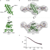
Solution structure of the NONO:IONIS742093 complex derived from SAXS data. (A) The X-ray crystal structure of a single NONO homodimer illustrated over two orthogonal perspectives highlighting the positions of RRM1 and RRM1′. (B) Ab initio reconstructions of the NONO:IONIS742093 complex calculated from SAXS data shown as a grey molecular envelope over two orthogonal perspectives. Superposed is a rigid body model for the NONO:IONIS742093 complex derived from SAXS data. The NONO homodimer is coloured green and the model for IONIS742093 (2M57) coloured blue and orange. The distances between the CCs, β-clasp and RRMs are shown to highlight the dimer compression upon binding. (C) Fit of refined model (red) to data (circles).

References

    1. Knott G.J., Bond C.S., Fox A.H.. The DBHS proteins SFPQ, NONO and PSPC1: a multipurpose molecular scaffold. Nucleic Acids Res. 2016; 44:3989–4004. - PMC - PubMed
    1. Yarosh C.A., Iacona J.R., Lutz C.S., Lynch K.W.. PSF: nuclear busy-body or nuclear facilitator?. Wiley Interdiscip. Rev. RNA. 2015; 6:351–367. - PMC - PubMed
    1. Liu P.Y., Erriquez D., Marshall G.M., Tee A.E., Polly P., Wong M., Liu B., Bell J.L., Zhang X.D., Milazzo G.et al. .. Effects of a novel long noncoding RNA, lncUSMycN, on N-Myc expression and neuroblastoma progression. J. Natl. Cancer Inst. 2014; 106:dju113. - PubMed
    1. Desai S.S., Kharade S.S., Parekh V.I., Iyer S., Agarwal S.K.. Pro-oncogenic roles of HLXB9 protein in insulinoma cells through interaction with Nono protein and down-regulation of the c-Met inhibitor Cblb (Casitas B-lineage lymphoma b). J. Biol. Chem. 2015; 290:25595–25608. - PMC - PubMed
    1. Zhu Z., Zhao X., Zhao L., Yang H., Liu L., Li J., Wu J., Yang F., Huang G., Liu J.. p54/NONO regulates lipid metabolism and breast cancer growth through SREBP-1A. Oncogene. 2016; 35:1399–1410. - PubMed

Publication types