. 2021 Dec 14;13(23):25440-25452.
doi: 10.18632/aging.203763. Epub 2021 Dec 14.

Titanium dioxide nanoparticles perturb the blood-testis barrier via disruption of actin-based cell adhesive function

Affiliations

Titanium dioxide nanoparticles perturb the blood-testis barrier via disruption of actin-based cell adhesive function

Dong-Qi Ni et al. Aging (Albany NY). .

Abstract

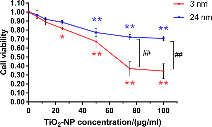
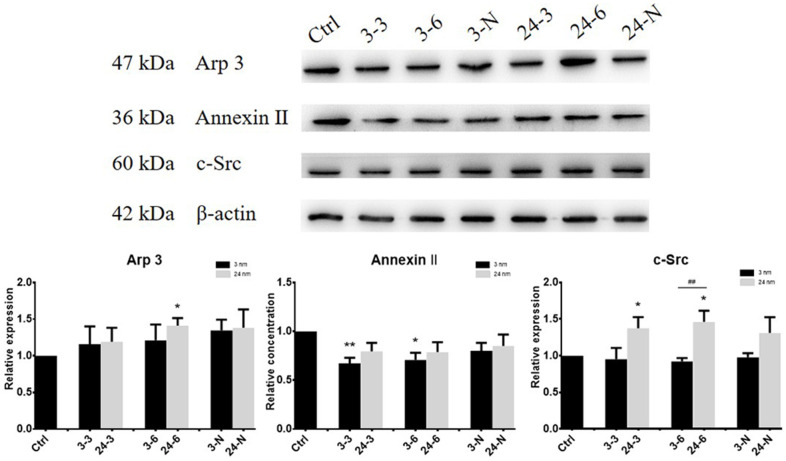
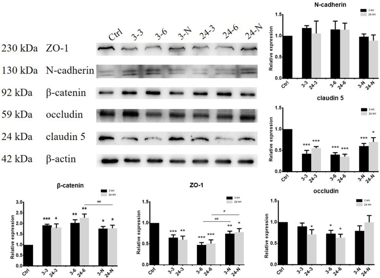
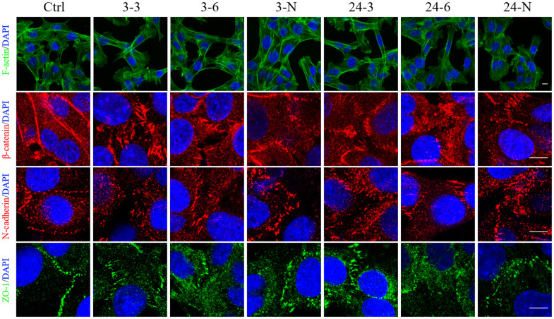
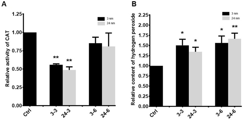
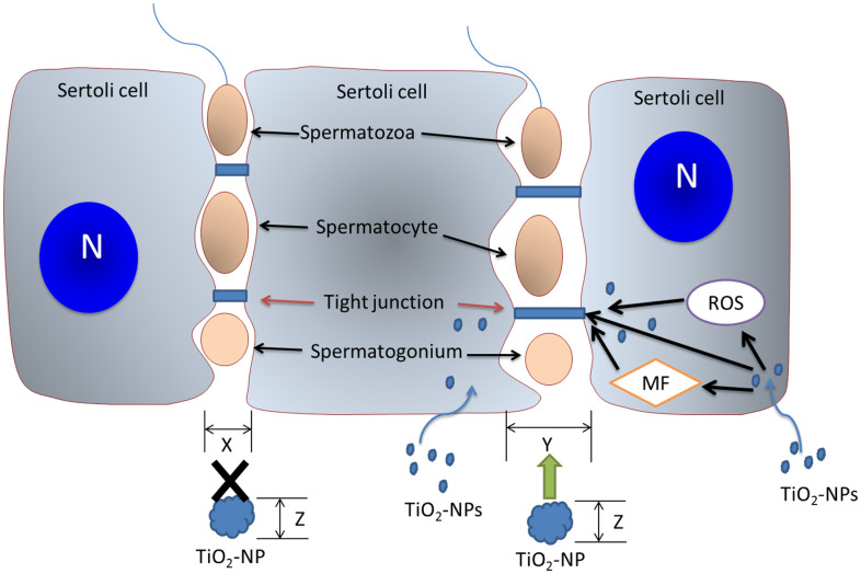
As one of the most commonly used nanoparticles, titanium dioxide nanoparticles (TiO2-NPs) are widely used as coating reagents in cosmetics, medicine and other industries. The increasing risk of exposure to TiO2-NPs raises concerns about their safety. In this study, we investigated the mechanism by which TiO2-NPs cross the blood-testis barrier (BTB). TM-4 cells were selected as an in vitro Sertoli cell model of BTB. Cell viability, cell morphological changes, apoptosis, oxidative damage, and the expression levels of actin regulatory and tight junction (TJ) proteins were assessed in TM-4 cells treated with 3-nm and 24-nm TiO2-NPs. Cells treated with 3-nm TiO2-NPs exhibited increased cytotoxicity and decreased Annexin II expression, whereas cells treated with 24-nm TiO2-NPs exhibited increased Arp 3 and c-Src expression. Both TiO2-NPs induced significant oxidative stress, decreased the expression of TJ proteins (occludin, ZO-1 and claudin 5), damaged the TJ structure, and exhibited enlarged gaps between TM-4 cells. Our results indicated that both TiO2-NPs crossed the BTB by disrupting actin-based adhesive junctions of TM-4 cells; however, apoptosis was not observed. Our results provide new insights into how TiO2-NPs cross the BTB.

Keywords: TM-4 cell; TiO2-NPs; actin; blood-testis barrier; tight junction.


Conflict of interest statement

CONFLICTS OF INTEREST: The authors declare that they have no conflicts of interest.

Figures

Figure 1
Figure 1
The effects of TiO2-NPs on TM-4 cell viability after exposure for 24 h. The results are expressed as the mean ± SEM. n=4, *p<0.05, **p<0.01 compared to the control; ##p<0.01 compared to the NPs.
Figure 2
Figure 2
The expression of actin regulatory proteins (Arp 3, Annexin II and c-Src) in TM-4 cells treated with two different concentrations of TiO2-NPs for 24 h. “3–3”: 30 μg/ml, “3–6”: 60 μg/ml, “3-N”: TM-4 cells were treated with 5 mM NAC for 2 h and then treated with 60 μg/ml 3-nm TiO2-NPs for 24 h. “24–3”: 30 μg/ml, “24–6”: 60 μg/ml, “24-N”: TM-4 cells were treated with 5 mM NAC for 2 h and then treated with 60 μg/ml of 24-nm TiO2-NPs for 24 h. n=5, *p<0.05, **p<0.01 compared to the control; ##p<0.01 compared to the NPs.
Figure 3
Figure 3
The expression of BTB-associated proteins in TM-4 cells upon treatment with two different concentrations of TiO2-NPs for 24 h. “3–3”: 30 μg/ml, “3–6”: 60 μg/ml, “3-N”: TM-4 cells were treated with 5 mM NAC for 2 h and then treated with 60 μg/ml 3-nm TiO2-NPs for 24 h. “24–3”: 30 μg/ml, “24–6”: 60 μg/ml, “24-N”: TM-4 cells were treated with 5 mM NAC for 2 h and then treated with 60 μg/ml of 24-nm TiO2-NPs for 24 h. n=5, *p<0.05, **p<0.01 compared to the control; #p<0.05, ##p<0.01 compared to the NPs.
Figure 4
Figure 4
The spatial distributions of F-actin and BTB-associated proteins in TM-4 cells treated with two different concentrations of TiO2-NPs for 24 h. The distribution of intracellular microfilaments (F-actin) and the distribution of BTB-associated proteins: ZO-1 (green), N-cadherin (red), and β-catenin (red). “3–3”: 30 μg/ml, “3–6”: 60 μg/ml, “3-N”: TM-4 cells were treated with 5 mM NAC for 2 h and then treated with 60 μg/ml 3-nm TiO2-NPs for 24 h. “24–3”: 30 μg/ml, “24–6”: 60 μg/ml, "24-N”: TM-4 cells were treated with 5 mM NAC for 2 h and then treated with 60 μg/ml of 24-nm TiO2-NPs for 24 h. Scale bar=10 μm.
Figure 5
Figure 5
The effect of TiO2-NPs on TM-4 cell apoptosis. (A) TUNEL results revealed no apoptosis signals (red fluorescence) in TM-4 cells treated with TiO2-NPs. (B) Caspase 3/8/9 activity in cells treated with TiO2-NPs showed no obvious changes. The results were expressed as the mean ± SEM. n=4, Scale bar=20 μm.
Figure 6
Figure 6
Oxidative stress in TM-4 cells treated with TiO2-NPs. (A) Intracellular CAT activity in TM-4 cells treated with TiO2-NPs for 24 h. (B) Intracellular hydrogen peroxide levels in TM-4 cells treated with TiO2-NPs for 24 h. The results are expressed as the mean ± SEM. n=7, *p<0.05, **p<0.01 compared to the control.
Figure 7
Figure 7
The mechanism of action of TiO2-NPs on BTB. TiO2-NPs damage the BTB via three mechanisms. TiO2-NPs interfere with BTB-associated proteins, induce cytoskeletal reorganization, and trigger oxidative stress to perturb TJ proteins. The BTB gap (size X) is expanded (size Y) by TiO2-NPs, and TiO2-NPs (particle size Z<Y) with particle sizes less than Y pass directly through the BTB gap and subsequently affect spermatogenesis. N: nucleus; MF: microfilament.

References

    1. Mascarenhas MN, Flaxman SR, Boerma T, Vanderpoel S, Stevens GA. National, regional, and global trends in infertility prevalence since 1990: a systematic analysis of 277 health surveys. PLoS Med. 2012; 9:e1001356. 10.1371/journal.pmed.1001356 - DOI - PMC - PubMed
    1. Carlsen E, Giwercman A, Keiding N, Skakkebaek NE. Evidence for decreasing quality of semen during past 50 years. BMJ. 1992; 305:609–13. 10.1136/bmj.305.6854.609 - DOI - PMC - PubMed
    1. Mruk DD, Cheng CY. Environmental contaminants: Is male reproductive health at risk? Spermatogenesis. 2011; 1:283–90. 10.4161/spmg.1.4.18328 - DOI - PMC - PubMed
    1. Ema M, Okuda H, Gamo M, Honda K. A review of reproductive and developmental toxicity of silver nanoparticles in laboratory animals. Reprod Toxicol. 2017; 67:149–64. 10.1016/j.reprotox.2017.01.005 - DOI - PubMed
    1. Liu L, He Y, Xiao Z, Tao W, Zhu J, Wang B, Liu Z, Wang M. Effects of Selenium Nanoparticles on Reproductive Performance of Male Sprague-Dawley Rats at Supranutritional and Nonlethal Levels. Biol Trace Elem Res. 2017; 180:81–9. 10.1007/s12011-017-0980-8 - DOI - PubMed

Publication types