

L-type amino acid transporter (LAT) 1 expression in 18F-FET-negative gliomas
- PMID: 34905134
- PMCID: PMC8671595
- DOI: 10.1186/s13550-021-00865-9
L-type amino acid transporter (LAT) 1 expression in 18F-FET-negative gliomas
Abstract
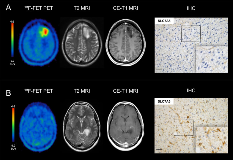
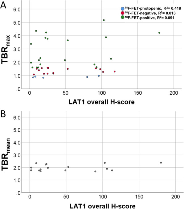
Background: O-(2-[18F]-fluoroethyl)-L-tyrosine (18F-FET) is a highly sensitive PET tracer for glioma imaging, and its uptake is suggested to be driven by an overexpression of the L-type amino-acid transporter 1 (LAT1). However, 30% of low- and 5% of high-grade gliomas do not present enhanced 18F-FET uptake at primary diagnosis ("18F-FET-negative gliomas") and the pathophysiologic basis for this phenomenon remains unclear. The aim of this study was to determine the expression of LAT1 in a homogeneous group of newly diagnosed 18F-FET-negative gliomas and to compare them to a matched group of 18F-FET-positive gliomas. Forty newly diagnosed IDH-mutant astrocytomas without 1p/19q codeletion were evaluated (n = 20 18F-FET-negative (tumour-to-background ratio (TBR) < 1.6), n = 20 18F-FET-positive gliomas (TBR > 1.6)). LAT1 immunohistochemistry (IHC) was performed using SLC7A5/LAT1 antibody. The percentage of LAT1-positive tumour cells (%) and the staining intensity (range 0-2) were multiplied to an overall score (H-score; range 0-200) and correlated to PET findings as well as progression-free survival (PFS).
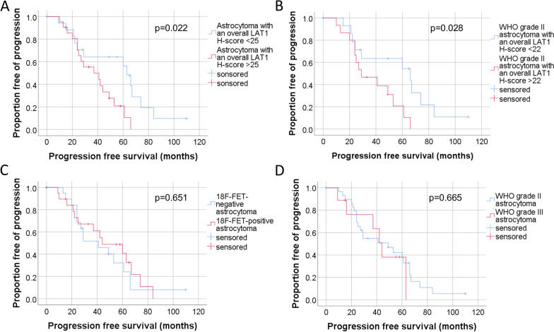
Results: IHC staining of LAT1 expression was positive in both, 18F-FET-positive as well as 18F-FET-negative gliomas. No differences were found between the 18F-FET-negative and 18F-FET-positive group with regard to percentage of LAT1-positive tumour cells, staining intensity or H-score. Interestingly, the LAT1 expression showed a significant negative correlation with the PFS (p = 0.031), whereas no significant correlation was found for TBRmax, neither in the overall group nor in the 18F-FET-positive group only (p = 0.651 and p = 0.140).
Conclusion: Although LAT1 is reported to mediate the uptake of 18F-FET into tumour cells, the levels of LAT1 expression do not correlate with the levels of 18F-FET uptake in IDH-mutant astrocytomas. In particular, the lack of tracer uptake in 18F-FET-negative gliomas cannot be explained by a reduced LAT1 expression. A higher LAT1 expression in IDH-mutant astrocytomas seems to be associated with a short PFS. Further studies regarding mechanisms influencing the uptake of 18F-FET are necessary.
Keywords: FET PET; Glioma; LAT1; Molecular imaging.
© 2021. The Author(s).
Conflict of interest statement
The authors declare that they have no competing interests.
Figures

References
-
- Lahoutte T, Caveliers V, Camargo SM, Franca R, Ramadan T, Veljkovic E, et al. SPECT and PET amino acid tracer influx via system L (h4F2hc-hLAT1) and its transstimulation. J Nucl Med. 2004;45(9):1591–1596. - PubMed
-
- Haase C, Bergmann R, Fuechtner F, Hoepping A, Pietzsch J. L-type amino acid transporters LAT1 and LAT4 in cancer: uptake of 3-O-methyl-6-18F-fluoro-L-dopa in human adenocarcinoma and squamous cell carcinoma in vitro and in vivo. J Nucl Med. 2007;48(12):2063–2071. - PubMed
LinkOut - more resources
Full Text Sources
Research Materials
