. 2022 Dec;60(1):56-64.
doi: 10.1080/13880209.2021.2010775.

Myristicin regulates proliferation and apoptosis in oxidized low-density lipoprotein-stimulated human vascular smooth muscle cells and human umbilical vein endothelial cells by regulating the PI3K/Akt/NF-κB signalling pathway

Affiliations

Myristicin regulates proliferation and apoptosis in oxidized low-density lipoprotein-stimulated human vascular smooth muscle cells and human umbilical vein endothelial cells by regulating the PI3K/Akt/NF-κB signalling pathway

Liang Luo et al. Pharm Biol. 2022 Dec.

Abstract

Context: Atherosclerosis (AS) is a chronic inflammatory disease. Human vascular smooth muscle cell (hVSMC) accumulation and human umbilical vein endothelial cell (HUVEC) dysfunction are associated with the pathogenesis of AS. This study explores whether myristicin plays a protective role in AS.

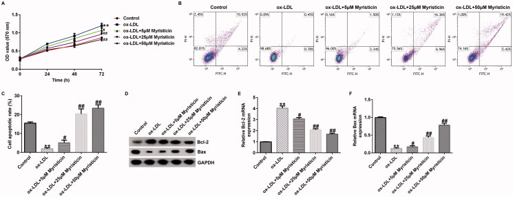
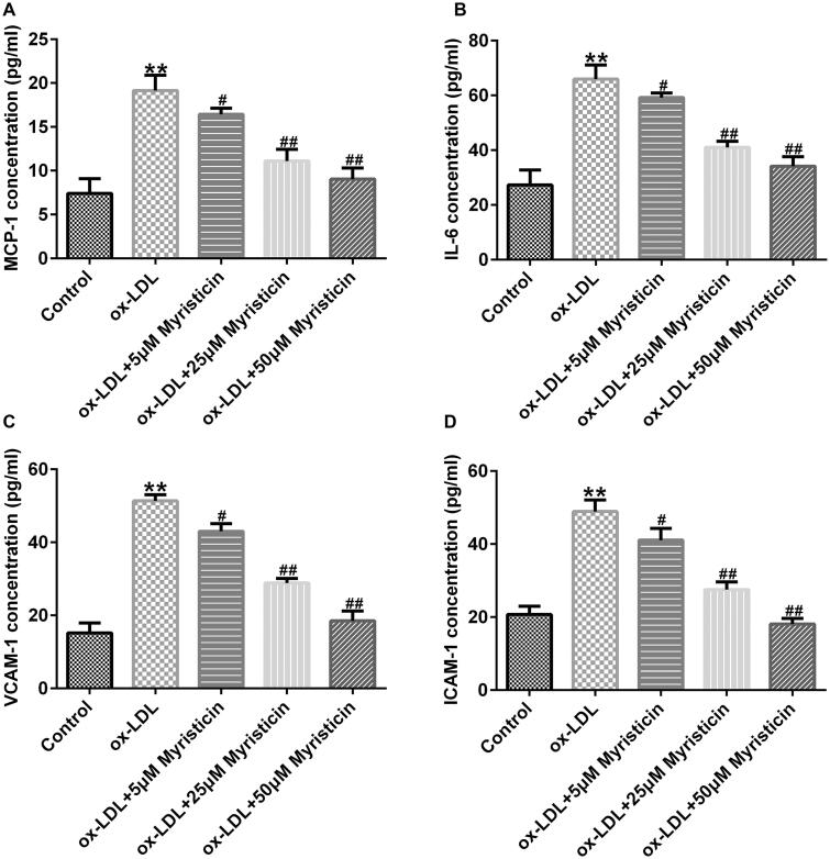
Materials and methods: hVSMCs and HUVECs were stimulated with 100 μg/mL oxidized low-density lipoprotein (ox-LDL) to establish a cellular model of AS. Cell viability, lactate dehydrogenase (LDH) release and cell apoptosis were evaluated using MTT, LDH and flow cytometry assays, respectively. Cell migration and inflammatory cytokine release were assessed using Transwell assay and ELISA.

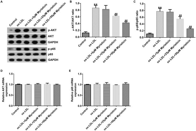
Results: Myristicin (5, 10, 25, and 50 μM) had no obvious effect on cell viability or the activity of LDH in hVSMCs, while 100 and 200 μM myristicin markedly suppressed hVSMCs viability and increased LDH release. Myristicin had no obvious effect on cell viability or the activity of LDH in HUVECs. Myristicin inhibited viability and increased apoptosis in ox-LDL-treated hVSMCs, but was associated with increased proliferation and inhibited apoptosis in HUVECs stimulated by ox-LDL. Additionally, myristicin markedly suppressed ox-LDL-induced hVSMCs migration and the release of inflammatory cytokines, including MCP-1, IL-6, VCAM-1 and ICAM-1, in HUVECs. Results also demonstrated that the promoting effects of ox-LDL on the PI3K/Akt and NF-κB signalling pathway in both hVSMCs and HUVECs were abolished by treatment with myristicin.

Discussion and conclusions: Myristicin regulated proliferation and apoptosis by regulating the PI3K/Akt/NF-κB signalling pathway in ox-LDL-stimulated hVSMCs and HUVECs. Thus, myristicin may be used as a new potential drug for AS treatment.

Keywords: Coronary heart disease; atherosclerosis; inflammation; migration.


Conflict of interest statement

No potential conflict of interest was reported by the author(s).

Figures

Figure 1.
Figure 1.
Myristicin had no distinct effects on hVSMC and HUVEC toxicity. A: Chemical formula of myristicin. Different concentrations of myristicin (0, 5, 10, 25, 50, 100, and 200 μM) were applied to induce hVSMCs and HUVECs for 24 h. (B and C) An MTT assay was performed to evaluate cell viability. (D and E) Detection of lactate dehydrogenase release. *p < 0.05, **p < 0.01 vs. control. HUVEC: human umbilical vein endothelial cell; hVSMC: human vascular smooth muscle cell.
Figure 2.
Figure 2.
Myristicin regulates ox-LDL-induced hVSMC proliferation and apoptosis. hVSMCs were stimulated with 100 μg/mL ox-LDL for 24 h and subsequently induced by myristicin (5, 25, and 50 μM) for 24 h. (A) Cell proliferation was assessed using an MTT assay. (B) Flow cytometry analysis of cell apoptosis. (C) Quantification of apoptotic cells. (D) Determination of Bcl-2 and Bax protein expression. (E and F) Reverse transcription-quantitative PCR analysis of Bcl-2 and Bax. **p < 0.01 vs. control; #p < 0.05, ##p < 0.01 vs. ox-LDL group. hVSMC: human vascular smooth muscle cell; ox-LDL: oxidized low-density lipoprotein.
Figure 3.
Figure 3.
Myristicin inhibits ox-LDL-induced hVSMC migration. hVSMCs were stimulated with 100 μg/mL ox-LDL for 24 h and subsequently induced by myristicin (5, 25, and 50 μM) for 24 h. (A) Migration of hVSMCs. (B) Quantification of migratory cells. (C) Determination of MMP-9 protein expression. (D) Reverse transcription-quantitative PCR analysis of MMP-9. **p < 0.01 vs. control; ##p < 0.01 vs. ox-LDL group. hVSMC: human vascular smooth muscle cell; ox-LDL: oxidized low-density lipoprotein.
Figure 4.
Figure 4.
Myristicin regulates ox-LDL-induced HUVEC proliferation and apoptosis. HUVECs were stimulated with 100 μg/mL ox-LDL for 24 h and subsequently induced by myristicin (5, 25, and 50 μM) for 24 h. (A) Cell proliferation was assessed using an MTT assay. (B) Flow cytometry analysis of cell apoptosis. (C) Quantification of apoptotic cells. (D) Determination of Bcl-2 and Bax protein expression. (E and F) Reverse transcription-quantitative PCR analysis of Bcl-2 and Bax. **p < 0.01 vs. control; #p < 0.05, ##p < 0.01 vs. ox-LDL group. HUVEC: human umbilical vein endothelial cell; ox-LDL: oxidized low-density lipoprotein.
Figure 5.
Figure 5.
Myristicin inhibits ox-LDL-induced inflammatory cytokine secretion in HUVECs. HUVECs were stimulated with 100 μg/mL ox-LDL for 24 h and subsequently induced by myristicin (5, 25, and 50 μM) for 24 h. The excretion of inflammatory cytokines, including (A) monocyte chemoattractant protein-1, (B) IL-6, (C) vascular cell adhesion molecule 1 and (D) intercellular adhesion molecule-1, was analyzed using ELISA. **p < 0.01 vs. control; #p < 0.05, ##p < 0.01 vs. ox-LDL group. HUVEC: human umbilical vein endothelial cell; ox-LDL: oxidized low-density lipoprotein.
Figure 6.
Figure 6.
Myristicin inactivates PI3K/AKT and NF-κB signalling pathway in hVSMCs. hVSMCs were stimulated with 100 μg/mL ox-LDL for 24 h and subsequently induced by myristicin (5, 25, and 50 μM) for 24 h. (A) Protein levels of p-p65 and p-AKT were evaluated by western blotting. (B and C) Ratio of p-AKT/AKT and p-p65/p65. (D and E) Reverse transcription-quantitative PCR analysis of AKT and p65 in different groups. **p < 0.01 vs. control; ##p < 0.01 vs. ox-LDL group. hVSMC: human vascular smooth muscle cell; ox-LDL: oxidized low-density lipoprotein; p-: phosphorylated.
Figure 7.
Figure 7.
Myristicin inactivates PI3K/AKT and NF-κB signalling pathway in HUVECs. HUVECs were stimulated with 100 μg/mL ox-LDL for 24 h and subsequently induced by myristicin (5, 25, and 50 μM) for 24 h. (A) Protein levels of p-p65 and p-AKT were evaluated by western blotting. (B and C) Ratio of p-AKT/AKT and p-p65/p65. (D and E) Reverse transcription-quantitative PCR analysis of AKT and p65 in different groups. **p < 0.01 vs. control; ##p < 0.01 vs. ox-LDL group. HUVEC: human umbilical vein endothelial cell; ox-LDL: oxidized low-density lipoprotein; p-: phosphorylated.

Similar articles

Cited by

References

    1. Berenji AS, Eftedal I, Pedersen M, Jeppesen PB, Norregaard R, Matchkov VV.. 2020. Endothelial dysfunction in small arteries and early signs of atherosclerosis in ApoE knockout rats. Sci Rep. 10(1):15296. - PMC - PubMed
    1. Bian W, Jing X, Yang Z, Shi Z, Chen R, Xu A, Wang N, Jiang J, Yang C, Zhang D, et al. . 2020. Downregulation of LncRNA NORAD promotes ox-LDL-induced vascular endothelial cell injury and atherosclerosis. Aging. 12(7):6385–6400. - PMC - PubMed
    1. Cao Z, Xia W, Zhang X, Yuan H, Guan D, Gao L.. 2020. Hepatotoxicity of nutmeg: a pilot study based on metabolomics. Biomed Pharmacother. 131:110780. - PubMed
    1. Colaiori I, Izzo R, Barbato E, Franco D, Di Gioia G, Rapacciuolo A, Bartunek J, Mancusi C, Losi MA, Strisciuglio T, et al. . 2019. Severity of coronary atherosclerosis and risk of diabetes mellitus. JCM. 8(7):1069. - PMC - PubMed
    1. Fashanu OE, Oyenuga AO, Zhao D, Tibuakuu M, Mora S, Otvos JD, Stein JH, Michos ED.. 2019. GlycA, a novel inflammatory marker and its association with peripheral arterial disease and carotid plaque: the multi-ethnic study of atherosclerosis. Angiology. 70(8):737–746. - PMC - PubMed

MeSH terms