. 2022 Feb 15;132(4):e153752.
doi: 10.1172/JCI153752.

Embryonic osteocalcin signaling determines lifelong adrenal steroidogenesis and homeostasis in the mouse

Affiliations

Embryonic osteocalcin signaling determines lifelong adrenal steroidogenesis and homeostasis in the mouse

Vijay K Yadav et al. J Clin Invest. .

Abstract

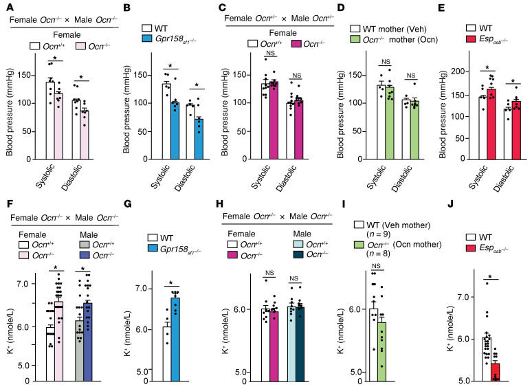
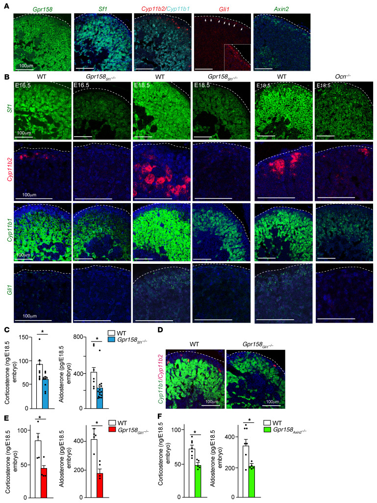
Through their ability to regulate gene expression in most organs, glucocorticoid (GC) hormones influence numerous physiological processes and are therefore key regulators of organismal homeostasis. In bone, GC hormones inhibit expression of the hormone Osteocalcin for poorly understood reasons. Here, we show that in a classical endocrine feedback loop, osteocalcin in return enhanced the biosynthesis of GC as well as mineralocorticoid hormones (adrenal steroidogenesis) in rodents and primates. Conversely, inactivation of osteocalcin signaling in adrenal glands significantly impaired adrenal growth and steroidogenesis in mice. Embryo-made osteocalcin was necessary for normal Sf1 expression in fetal adrenal cells and adrenal cell steroidogenic differentiation and therefore determined the number of steroidogenic cells present in the adrenal glands of adult animals. Embryonic, not postnatal, osteocalcin also governed adrenal growth, adrenal steroidogenesis, blood pressure, electrolyte equilibrium, and the rise in circulating corticosterone levels during the acute stress response in adult offspring. This osteocalcin-dependent regulation of adrenal development and steroidogenesis occurred even in the absence of a functional hypothalamus/pituitary/adrenal axis and explains why osteocalcin administration during pregnancy promoted adrenal growth and steroidogenesis and improved the survival of adrenocorticotropic hormone signaling-deficient animals. This study reveals that a bone-derived embryonic hormone influences lifelong adrenal functions and organismal homeostasis in the mouse.

Keywords: Bone Biology; Metabolism; Mouse models.


Conflict of interest statement

Conflict of interest: The authors have declared that no conflict of interest exists.

Figures

Figure 1
Figure 1. Osteocalcin increases circulating GCs and aldosterone in mice and monkeys.
(A) Circulating corticosterone levels 2 hours after injection of vehicle or recombinant osteocalcin (Ocn) from different sources (30 ng/g body weight) at 1200 hours in 2-month-old Sv129 male WT mice. (B and C) Circulating corticosterone levels in 2-month-old WT Sv129 males (B) and Sv129 females (C) at different time points after osteocalcin injection. (D) Circulating aldosterone levels 2 hours after vehicle or osteocalcin injection at 1200 hours in 2-month-old male WT Sv129 mice. (E and F) circulating aldosterone levels in Sv129 male (E) and Sv129 female (F) WT mice at different time points after osteocalcin injection. (GK) Circulating osteocalcin (G), corticosterone (H and J), and aldosterone (I and K) levels in WT, Esposb–/–, and Esposb–/– Ocn+/– mice at 1800 hours. (LN) Circulating osteocalcin (L), cortisol (M), and aldosterone (N) at different time points after vehicle or human osteocalcin injection at 1000 hours into rhesus monkeys. Statistical analyses were conducted using 1-way ANOVA followed by Tukey’s post hoc test (AF, G, K, M, and N) or a 2-tailed, unpaired t test (HJ and L). *P < 0.05. n = 6 or more each group for mice; n = 4 or more for rhesus monkeys.
Figure 2
Figure 2. Osteocalcin signaling through Gpr158 in adrenal glands is necessary for adrenal steroidogenesis.
(A and B) Expression of Gprc6a (A) and Gpr158 (B) in different tissues from WT mice (qRT-PCR). (C) ISH analysis of Gprc6a, Gpr158, Cyp11b1, and Cyp11b2 expression in WT adrenal glands. Scale bars: 100 μm. (DH) Gpr158 expression in adrenal glands (by qRT-PCR) (D), circulating corticosterone levels (E, 1-month-old and G, 3-month-old), and aldosterone levels in (F, 1-month-old and H, 3-month-old) female and male WT and Gpr158Sf1–/– mice. (I and J) Circulating corticosterone (I) and aldosterone (J) levels in 3-month-old WT and Gpr158Sf1–/– mice 2 hours after vehicle or osteocalcin injection. (K) Adrenal steroidogenic gene expression in WT and Gpr158Sf1–/– female mice. Statistical analyses were conducted using a 2-tailed, unpaired t test (DH and K) or 1-way ANOVA followed by Tukey’s post hoc test (I and J). *P < 0.05. n = 6 or more mice per group.
Figure 3
Figure 3. Neuronal deletion of Gpr158 through CamK2a-Cre does not affect adrenal steroidogenesis.
(A) β-Gal staining of a whole-mount mid-brain cross-section and adrenal gland from a 2-month-old Camk2a-Cre+ mouse crossed with a ROSA reporter mouse. (B) Eosin- and β-gal–stained section of an adrenal gland from a 2-month-old Camk2a-Cre+ mouse crossed with a ROSA reporter mouse. Scale bar: 100 μm. M, medulla. (C) Recombination analysis of genomic DNA in different tissues collected from Gpr158CamK2a–/– mice. Floxed (Fl) and deletion (Del) bands are indicated. (DF) Gpr158 expression in hypothalamus and adrenal glands (D) and circulating corticosterone (E) and aldosterone (F) levels in 3 month-old male WT and Gpr158Camk2a–/– mice. Statistical analyses were conducted using a 2-tailed, unpaired t test (DF). *P < 0.05. n = 5 or more in each group.
Figure 4
Figure 4. Embryonic osteocalcin promotes adrenal steroidogenesis and homeostasis in offspring.
(A and B) Circulating corticosterone (A) and aldosterone (B) levels at 1800 hours in 2-month-old female and male Ocn–/– mice born from Ocn+/– parents and in WT littermates. (C and D) Circulating corticosterone (C) and aldosterone (D) levels at 1800 hours in 3-month-old WT, Ocn+/–, Gpr158Sf1+/–, and Ocn+/– Gpr158Sf1+/– mice born from Ocn+/– Gpr158Sf1+/– parents. (E and F) Circulating corticosterone (E) and aldosterone (F) levels at 1800 hours in 8-week-old Ocn+/+ and Ocn–/– female and male mice born from Ocn+/+ or Ocn–/– isogenic parents. (GI) Adrenal steroidogenic gene expression (G), plasma ACTH levels (H), and plasma renin activity (I) in Ocn+/+ and Ocn–/– female mice born from Ocn+/+ or Ocn–/– isogenic parents. (J and K) Circulating corticosterone and aldosterone levels at 1800 hours in 2-month-old Ocn+/– and Ocn–/– female and male mice born from Ocn–/– (J) or Ocn+/– (K) mothers crossed with Ocn+/– or Ocn–/– fathers, respectively. (L) Circulating corticosterone and aldosterone levels in 2-month-old Ocn+/+ and Ocn–/– mice born from Ocn+/+ or Ocn–/– mothers that received either vehicle or osteocalcin (300 ng/day) from E14.5 until birth. In each panel, the parents are indicated on the top and progeny on the bottom. Statistical analyses were conducted using a 2-tailed, unpaired t test (A, B, and EK) or 1-way ANOVA followed by Tukey’s post hoc test (C, D, and L). *P < 0.05. n = 10 or more in each group except for GI: n = 5 or more.
Figure 5
Figure 5. Embryonic osteocalcin promotes homeostasis in offspring.
(AJ) Systolic and diastolic blood pressure and plasma K+ concentrations in 2-month-old Ocn+/+ and Ocn–/– mice born from Ocn+/+ or Ocn–/– isogenic parents (A and F); WT and Gpr158Sf1 Ocn–/– mice (B and G); Ocn+/+ and Ocn–/– mice born from Ocn+/– parents (C and H); Ocn+/+ and Ocn–/– offspring born from Ocn+/+ or Ocn–/– mothers that received either vehicle (Veh mother) or osteocalcin (Ocn mother, 300 ng/day) from E14.5 until birth (D and I); and WT and Esposb–/– mice (E and J). Statistical analyses were conducted using a 2-tailed, unpaired t test. *P < 0.05. n = 5 or more in each group.
Figure 6
Figure 6. Embryonic osteocalcin signaling in adrenal glands promotes cell proliferation during development.
(A) H&E-stained sections of adrenal glands of E14.5, E16.5, and E18.5 WT and Gpr158Sf1–/– embryos. Scale bars: 250 μm. (B and C) TUNEL staining showing apoptosis (B) and Ki67 staining showing proliferation (C) in E18.5 adrenal glands from WT and Gpr158Sf1–/– embryos. Scale bars: 100 μm. (D) Cyclin gene expression in adrenal glands from WT and Gpr158Sf1–/– newborn mice. (EK) Adrenal gland per body weight percentage (Adrenal wt/BW %) for mice of the indicated genotypes and crosses. In each panel, parents are indicated on the top and progeny on the bottom. Statistical analyses were conducted using a 2-tailed, unpaired t test (DI and K) or 1-way ANOVA followed by Tukey’s post hoc test (J). *P < 0.05. n = 10 or more in each group (EK); n = 5 or more in each group (AC).
Figure 7
Figure 7. Embryonic osteocalcin signaling in adrenal glands establishes the steroidogenic program during development.
(A) ISH analysis of adrenal Gpr158, Sf1, Cyp11b1, Cyp11b2, Gli1, and Axin2 expression in E18.5 WT embryos. Scale bars: 100 μm. (B) ISH analysis of adrenal Sf1, Cyp11b2, Cyp11b1, and Gli1 expression in E16.5 and E18.5 WT and Gpr158Sf1–/– embryos and E18.5 WT and Ocn–/– embryos. Scale bars: 100 μm. (C) Intra-adrenal content of corticosterone and aldosterone in E18.5 WT and Gpr158Sf1–/– embryos. (D) ISH analysis of Cyp11b2 and Cyp11b1 expression in E18.5 WT and Gpr158Gli1–/– embryos. Scale bars: 100 μm. (E) Intra-adrenal content of corticosterone and aldosterone in E18.5 WT and Gpr158Gli1–/– (E) and Gpr158Axin2–/– (F) embryos. Statistical analyses were conducted using a 2-tailed, unpaired t test (C, E, and F). *P < 0.05. n = 5 or more in each group of embryos or mice (C, E, and F); n = 3 or more for the ISH analysis (A, B, and D).
Figure 8
Figure 8. Osteocalcin induces adrenal steroidogenesis and growth in the absence of ACTH signaling.
(A) Circulating corticosterone levels following an acute ACTH challenge at 1000 hours in adult WT and Gpr158sf1–/– mice born from a Gpr158fl/fl female mouse crossed with a Gpr158fl/fl Sf1-Cre+ male mouse. (B) Gpr158 expression in adrenal glands of WT and Mc2r–/– newborn mice. (C) Adrenal Gpr158 expression in WT mice 2 hours after ACTH challenge. (D) Intra-adrenal content of corticosterone and aldosterone in P1 WT, Mc2r+/–, Gpr158Sf1+/–, and Mc2r+/– Gpr158Sf1+/– mice born from Mc2r+/– Gpr158Sf1+/– parents. (E and F) Adrenal Mc2r (E), Gpr158 (E), Cyp11b1, and Cyp11b2 (F) expression in WT and Mc2r+/– newborn mice. (G and H) H&E-stained sections of adrenal glands (G) and ISH analysis of adrenal Gli1, Cyp11b2, and Cyp11b1 expression (H) in E18.5 WT and Mc2r–/– embryos collected from Mc2r+/– mothers that received either vehicle or osteocalcin (300 ng/day) from E10.5 to E18.5. Scale bars: 250 μm (G) and 100 μm (H). (I and J) Intra-adrenal content of corticosterone (I) and aldosterone (J) in WT and Mc2r–/– newborn mice born from Mc2r+/– mothers that received vehicle or osteocalcin (300 ng/day) from E10.5 until birth. Statistical analyses were conducted using 1-way ANOVA followed by Tukey’s post hoc test (A, D, I, and J) or 2-tailed, unpaired t test (B, C, E, and F). *P < 0.05. n = 6 or more embryos or offspring in each group.
Figure 9
Figure 9. Model of the regulation of adrenal steroidogenesis and postnatal homeostasis by osteocalcin.
Embryonic osteocalcin signaling in the developing adrenal gland through Gpr158 is necessary for the differentiation of fetal, progenitor, and steroidogenic adrenal cells as well as for the proliferation of these cells. This affects lifelong adrenal growth and steroidogenesis and homeostasis in the offspring. Postnatally, exogenous osteocalcin can enhance steroidogenic functions in rodents and nonhuman primates.

Comment in

Similar articles

Cited by

References

    1. Xing Y, et al. Development of adrenal cortex zonation. Endocrinol Metab Clin North Am. 2015;44(2):243–274. doi: 10.1016/j.ecl.2015.02.001. - DOI - PMC - PubMed
    1. Pihlajoki M, et al. Adrenocortical zonation, renewal, and remodeling. Front Endocrinol (Lausanne) 2015;6:27. - PMC - PubMed
    1. Freedman BD, et al. Adrenocortical zonation results from lineage conversion of differentiated zona glomerulosa cells. Dev Cell. 2013;26(6):666–673. doi: 10.1016/j.devcel.2013.07.016. - DOI - PMC - PubMed
    1. Vidal V, et al. The adrenal capsule is a signaling center controlling cell renewal and zonation through Rspo3. Genes Dev. 2016;30(12):1389–1394. doi: 10.1101/gad.277756.116. - DOI - PMC - PubMed
    1. Lyraki R, Schedl A. Adrenal cortex renewal in health and disease. Nat Rev Endocrinol. 2021;17(7):421–434. doi: 10.1038/s41574-021-00491-4. - DOI - PubMed

Publication types