. 2022 Feb 1;132(3):e146926.
doi: 10.1172/JCI146926.

Smad7 effects on TGF-β and ErbB2 restrain myofibroblast activation and protect from postinfarction heart failure

Affiliations

Smad7 effects on TGF-β and ErbB2 restrain myofibroblast activation and protect from postinfarction heart failure

Claudio Humeres et al. J Clin Invest. .

Abstract

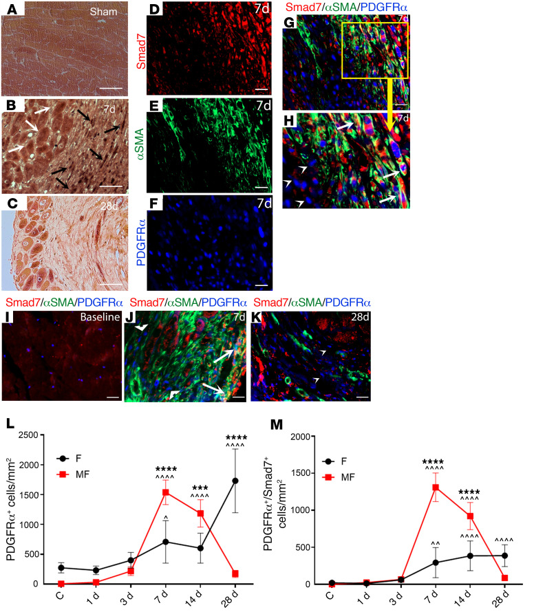
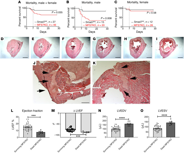
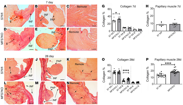
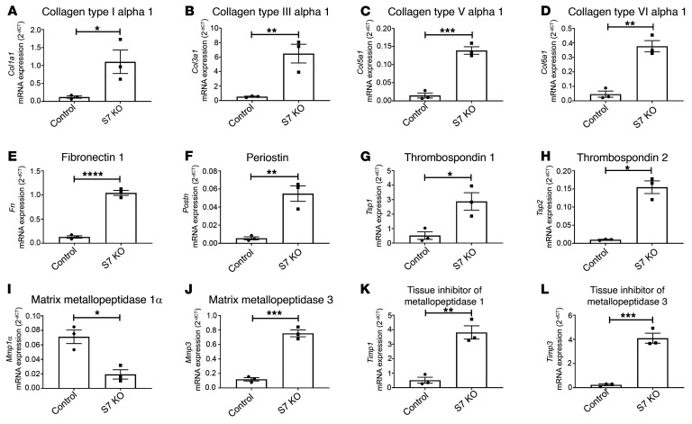
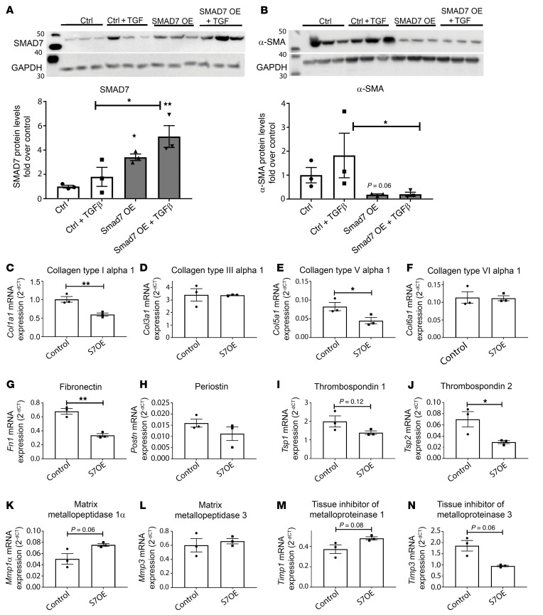
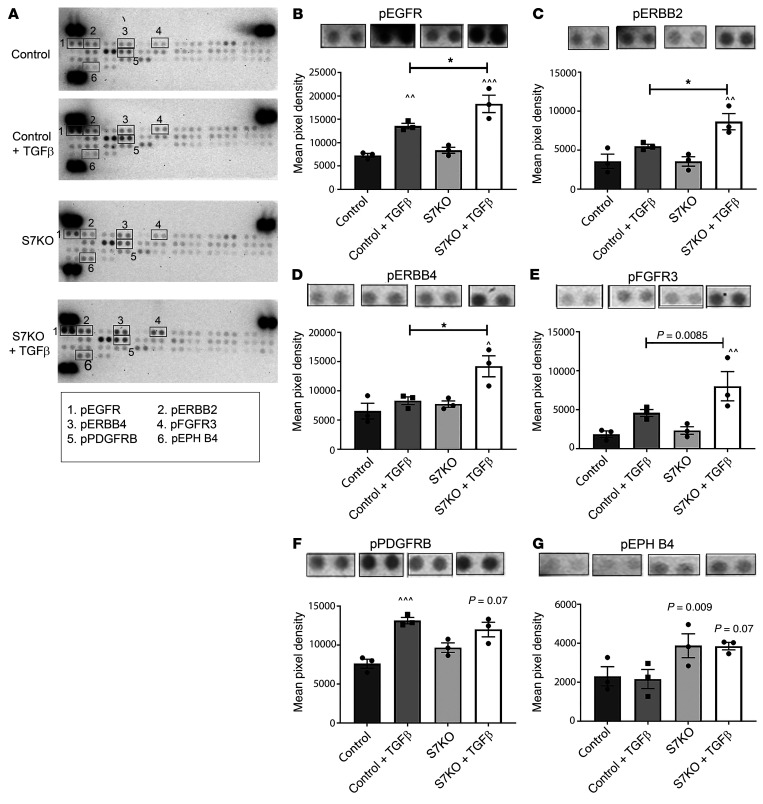
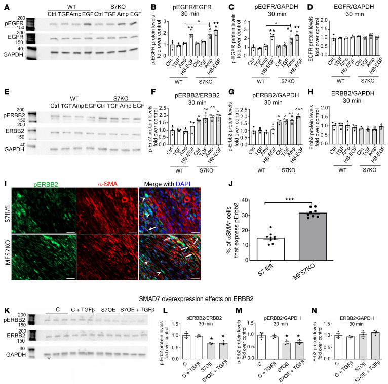
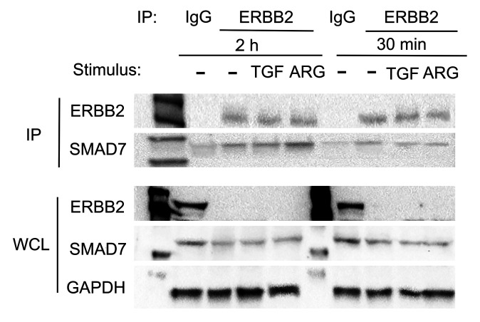
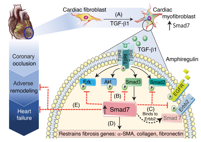
Repair of the infarcted heart requires TGF-β/Smad3 signaling in cardiac myofibroblasts. However, TGF-β-driven myofibroblast activation needs to be tightly regulated in order to prevent excessive fibrosis and adverse remodeling that may precipitate heart failure. We hypothesized that induction of the inhibitory Smad, Smad7, may restrain infarct myofibroblast activation, and we examined the molecular mechanisms of Smad7 actions. In a mouse model of nonreperfused infarction, Smad3 activation triggered Smad7 synthesis in α-SMA+ infarct myofibroblasts, but not in α-SMA-PDGFRα+ fibroblasts. Myofibroblast-specific Smad7 loss increased heart failure-related mortality, worsened dysfunction, and accentuated fibrosis in the infarct border zone and in the papillary muscles. Smad7 attenuated myofibroblast activation and reduced synthesis of structural and matricellular extracellular matrix proteins. Smad7 effects on TGF-β cascades involved deactivation of Smad2/3 and non-Smad pathways, without any effects on TGF-β receptor activity. Unbiased transcriptomic and proteomic analysis identified receptor tyrosine kinase signaling as a major target of Smad7. Smad7 interacted with ErbB2 in a TGF-β-independent manner and restrained ErbB1/ErbB2 activation, suppressing fibroblast expression of fibrogenic proteases, integrins, and CD44. Smad7 induction in myofibroblasts serves as an endogenous TGF-β-induced negative feedback mechanism that inhibits postinfarction fibrosis by restraining Smad-dependent and Smad-independent TGF-β responses, and by suppressing TGF-β-independent fibrogenic actions of ErbB2.

Keywords: Cardiology; Fibrosis; Growth factors; Heart failure; Immunology.


Conflict of interest statement

Conflict of interest: The authors have declared that no conflict of interest exists.

Figures

Figure 1
Figure 1. Smad7 expression is markedly upregulated in infarcted fibroblasts and is associated with fibroblast-to-myofibroblast conversion.
(A) Smad7 immunohistochemical staining shows low-level Smad7 immunoreactivity in sham mouse hearts. (B and C) Myocardial infarction is associated with a marked increase in Smad7 expression levels, peaking after 7 days of coronary occlusion. Smad7 immunoreactivity is localized in both interstitial cells (black arrows) and in border zone cardiomyocytes (white arrows). Smad7 immunoreactivity is reduced 28 days after coronary occlusion. (DK) In PDGFRα-GFP fibroblast reporter mice, triple immunofluorescent staining of Smad7, α-SMA, and PDGFRα-GFP was used to identify infarct fibroblasts (PDGFRα+α-SMA) and myofibroblasts (PDGFRα+α-SMA+) expressing Smad7. Individual (DF) and merged (G and H) channels for Smad7, α-SMA, and PDGFRα-GFP fluorescence show that Smad7 is predominantly expressed by α-SMA+ myofibroblasts (arrows) and not by α-SMA fibroblasts (arrowheads). The time course of Smad7 expression shows that (I) in sham hearts, myofibroblasts are absent and Smad7 levels are low. (J) Seven days after infarction, abundant myofibroblasts express high levels of cytosolic Smad7 (arrows), whereas fibroblasts have negligible Smad7 immunoreactivity (arrowheads). (K) Relatively few α-SMA–expressing myofibroblasts are noted 28 days after infarction. Abundant fibroblasts are present; the majority of these cells are Smad7 (arrow). (L and M) Quantitative analysis shows that at the 7- and 14-day time points, abundant myofibroblasts express Smad7, whereas in the mature scar (28 days after infarction), Smad7 is expressed by a fraction of fibroblasts. Statistical comparison (L and M) was performed using 1-way ANOVA followed by Tukey’s multiple comparison test (n = 6/group). ^P < 0.05, ^^P < 0.01, ^^^^P < 0.0001 vs. control (C); ***P < 0.001, ****P < 0.0001 between fibroblasts (F) and myofibroblasts (MF) at the same time point. Scale bars: 50 μm (AC) and 20 μm (DK).
Figure 2
Figure 2. Myofibroblast-specific Smad7 loss increases heart failure–related mortality in infarcted mice.
(AC) Comparison of survival curves between Smad7fl/fl and myofibroblast-specific Smad7-knockout (MFS7KO) mice after 28 days of permanent coronary occlusion. (A) When compared with Smad7fl/fl mice, MFS7KO mice have increased late mortality following nonreperfused infarction (Smad7fl/fl: 81% survival, n = 27; MFS7KO: 63% survival, n = 46; P = 0.0035). (B and C) Increased mortality is due to markedly accentuated death rates in male MFS7KO animals after 28 days of permanent occlusion when compared with sex-matched Smad7fl/fl mice (Smad7fl/fl: 73% survival, n = 15; MFS7KO: 35% survival, n = 20; P = 0.008), whereas for female MFS7KO mice, the difference in mortality does not reach statistical significance (Smad7fl/fl: 91% survival, n = 12; MFS7KO: 84% survival, n = 26; P = 0.58). (DI) To determine the cause of increased mortality in MFS7KO mice, systematic postmortem histological analysis was performed by sectioning the entire heart from base to apex into 300-μm segments (2 Smad7fl/fl and 11 MFS7KO hearts from dead mice after infarction). Original magnification, ×10. (J and K) Consecutive myocardial sections studied to identify rupture sites show that only 2 of 11 MFS7KO hearts had intramural rupture track (arrows). Thus, the excess mortality in MFS7KO mice was not due to rupture. Original magnification, ×100. (L and M) In order to further understand the basis for increased mortality in MFS7KO mice, we compared echocardiographic parameters measured at the 7-day time point between the MFS7KO mice that died between 7 and 28 days and the survivors that completed the protocol. MFS7KO mice that died between 7 and 28 days had much lower left ventricular ejection fraction (LVEF) and (N and O) increased left ventricular end-diastolic volume (LVEDV) and left ventricular end-systolic volume (LVESV), suggestive of heart failure–related mortality. Survival analysis was performed using the Kaplan-Meier method. Mortality was compared using the log-rank test (AC). Statistical comparison (LO) was performed using Student’s t test (MFS7KO survivors, n = 37; dead MFS7KO, n = 7). ***P < 0.001, ****P < 0.0001. Scale bars: 1 mm (DI) and 100 μm (J and K).
Figure 3
Figure 3. Myofibroblast-specific Smad7 loss increases left ventricular dysfunction and accentuates adverse postinfarction remodeling.
(A and B) Echocardiographic assessment of adverse remodeling after 7 to 28 days following infarction shows that MFS7KO mice have worse systolic dysfunction demonstrated by a lower ejection fraction at both time points (A), and significantly higher reduction in ejection fraction (ΔLVEF, B) in comparison with Smad7fl/fl (S7fl/fl) mice. (CF) MFS7KO mice have accentuated dilative remodeling evidenced by increased left ventricular end-diastolic volume (LVEDV, C), accentuated increases in LVEDV (ΔLVEDV, D), higher left ventricular end-systolic volume (LVESV, E), and accentuated ΔLVESV (F). (G) The E/E′ ratio, an indicator of diastolic dysfunction, is significantly increased in MFS7KO at the 7-day time point. Statistical comparison (AG) was performed using 1-way ANOVA followed by Tukey’s multiple comparison test (Smad7fl/fl, n = 30; MFS7KO, n = 44). *P < 0.05, **P < 0.01, ****P < 0.0001; ^^^P < 0.001, ^^^^P < 0.0001 versus baseline.
Figure 4
Figure 4. Myofibroblast-specific Smad7 loss accentuates postinfarction fibrosis in the border zone and in the papillary muscles.
Collagen staining was performed using picrosirius red, and the collagen-stained area was assessed in the papillary muscle (PAP), infarcted (INF), border (B), and remote remodeling areas of Smad7fl/fl (S7 fl/fl) and MFS7KO hearts at 7 (AF) and 28 days (IN) of coronary occlusion. (G and H) Quantitative analysis demonstrates increased collagen deposition in the infarct zone of MFS7KO hearts compared with Smad7fl/fl at 7 days after infarction. (O and P) Twenty-eight days after infarction, increased collagen deposition is noted in the infarct border zone (L) and in the papillary muscle (M) of MFS7KO hearts compared with Smad7fl/fl, with comparable collagen levels in the infarct zone and the remote myocardium. For comparisons between multiple groups (G and O), 1-way ANOVA was performed followed by Tukey’s multiple comparison test. For comparisons between 2 groups (H and P), unpaired 2-tailed Student’s t test with Welch’s correction for unequal variances was performed (7 days Smad7fl/fl, n = 6; MFS7KO, n = 7; 28 days Smad7fl/fl, n = 18; MFS7KO, n = 21). *P < 0.05; ****P < 0.0001. Scale bars: 100 μm.
Figure 5
Figure 5. Smad7 loss accentuates synthesis of structural and matricellular genes by cardiac fibroblasts and modulates expression of MMPs and TIMPs.
Expression of extracellular matrix genes was assessed using a PCR array and was compared between Smad7-KO fibroblasts (S7 KO, induced through overexpression of adeno-Cre in Smad7fl/fl cells) and control fibroblasts (Smad7fl/fl). (AH) Smad7 loss accentuates fibroblast expression of genes encoding structural matrix proteins, including collagen type I α1, collagen type III α1, collagen type V α1, collagen type VI α1, and fibronectin, and matricellular proteins, such as periostin, thrombospondin 1, and thrombospondin 2. (IL) Smad7 also modulates expression of genes associated with matrix remodeling, such as matrix metalloproteinases (MMPs) and tissue inhibitors of metalloproteinase (TIMPs). Smad7-KO cells have lower expression of Mmp1α, but markedly higher expression of Mmp3, Timp1, and Timp2. Comparisons between 2 groups (AL) was performed by unpaired, 2-tailed Student’s t test with Welch’s correction for unequal variances (n = 3/group). *P < 0.05, **P < 0.01, ***P < 0.001, ****P < 0.0001.
Figure 6
Figure 6. Smad7 overexpression restrains the TGF-β–induced increase in α-SMA and attenuates collagen I and fibronectin expression, without affecting collagen III levels.
(A) Efficiency of the Smad7 overexpression (OE) strategy was assessed by comparing Smad7 protein levels between cardiac fibroblasts transfected with a Smad7 cDNA plasmid (S7OE) and cells transfected with a control entry vector (Control). (B) Smad7 overexpression restrains myofibroblast conversion, markedly attenuating α-SMA protein levels in basal and TGF-β–induced conditions. (CN) A PCR array shows that Smad7 overexpression (S7OE) attenuates synthesis of Col1a1 (C), Fn (G), and Tsp2 (J) without affecting Col3a1 transcription (D). (KN) In contrast, expression of matrix remodeling genes, such as those encoding MMPs and TIMPs, is modestly affected by Smad7 overexpression. For comparisons between multiple groups (A and B), 1-way ANOVA was performed followed by Tukey’s post hoc test. For comparisons between 2 groups (CN), unpaired, 2-tailed Student’s t test was performed with Welch’s correction for unequal variances (n = 3 per group). *P < 0.05; **P < 0.01.
Figure 7
Figure 7. Smad7 acts downstream of the TGF-β receptors, restraining direct TGF-β–induced activation of Smad-dependent and Smad-independent signaling.
(A) Smad7 loss does not affect activity and expression (B) of the constitutively active TGF-βRII. Moreover, Smad7 loss has no effects on activity (C) and expression (D) of the TGF-β–activated TGF-βRI. In order to examine the effects of Smad7 loss on Smad-dependent and -independent pathways, we performed Western blotting for p-Smad3, Smad3, p-Smad2, Smad2, p-ERK1/2, ERK1/2, p-AKT, and AKT (EG). TGF-β–stimulated p-Smad3 (H) and p-Smad2 (J) activation was accentuated in Smad7-KO (S7KO) fibroblasts, in the absence of any effects on total Smad3 (I) or Smad2 levels (K). Smad-independent ERK1/2 (L) and AKT (N) activation, induced by TGF-β, was also accentuated in Smad7-KO fibroblasts without affecting total ERK (M) and AKT levels (O). Statistical comparison (AD and HO) was performed using 1-way ANOVA followed by Tukey’s multiple comparison test (n = 6 per group). **P < 0.01, ****P < 0.0001; ^^P < 0.01, ^^^^P < 0.0001 vs. unstimulated control.
Figure 8
Figure 8. Effects of Smad7 on RTK activation.
Because transcriptomic analysis identified receptor tyrosine kinase (RTK) signaling as the top-ranked pathway exhibiting differential gene expression in the absence of Smad7, we used a phospho-RTK proteomic array to identify specific RTKs modulated by Smad7. Smad7-KO (S7KO) fibroblasts were compared with control cells in the presence or absence of TGF-β1. (A) Representative array images for each condition are shown and phospho-RTKs with significant differences are highlighted in the numbered boxes. (BE) Quantitative analysis suggests that Smad7 loss is associated with accentuated activation of EGFR/ErbB1, ErbB2, ErbB4, and FGFR cascades in TGF-β1–stimulated fibroblasts. (F and G) Other phospho-RTKs such as PDGFRβ and EPH-4 are not affected by Smad7 loss. Intensity of signal was quantified in duplicate as mean pixel density. Statistical comparison (BG) was performed using 1-way ANOVA followed by Tukey’s multiple comparison test (n = 3). *P < 0.05; ^P < 0.05, ^^P < 0.01, ^^^P < 0.001 vs. unstimulated condition.
Figure 9
Figure 9. Smad7 restrains ErbB2 activation in a ligand-independent manner and limits amphiregulin-mediated EGFR/ErbB1 activation.
Representative blots and quantitative analysis of the effects of Smad7 on EGFR/ErbB1 (AD) and ErbB2 activation (EH) in the presence or absence of TGF-β1 and the ErbB activators amphiregulin and HB-EGF (30-minute stimulation). (AD) Smad7 loss does not affect EGFR/ErbB1 activity at baseline or after stimulation with TGF-β1 or HB-EGF. However, Smad7 loss accentuates amphiregulin-mediated EGFR activation. (EH) Markedly increased ErbB2 activation is observed in Smad7-KO (S7KO) fibroblasts both at baseline and upon stimulation with TGF-β1, amphiregulin, and HB-EGF. Total ErbB1 or ErbB2 levels are not affected. (I) Dual immunofluorescence combining α-SMA and p-ErbB2 staining was used to identify α-SMA+ myofibroblasts expressing p-ErbB2 (arrows) in the infarcted myocardium (7-day permanent occlusion) of Smad7fl/fl and MFS7KO mice. p-ErbB2 is also expressed in non-myofibroblasts (arrowheads). (J) Quantitative analysis shows that MFS7KO hearts have a significant increase in p-ErbB2+ infarct myofibroblasts, when compared with control floxed infarcted hearts. (KN) Smad7 overexpression (S7OE) attenuates ErbB2 activation in the presence or absence of TGF-β1, without affecting total ErbB2 levels (control, C). For comparisons between multiple groups (BD, FH, and LN), 1-way ANOVA was performed followed by Tukey’s multiple comparison test (n = 3). *P < 0.05, **P < 0.01 vs. corresponding unstimulated condition; ^P < 0.05, ^^P < 0.01, ^^^P < 0.001 vs. corresponding WT. For comparisons between 2 groups (J), unpaired, 2-tailed Student’s t test was performed (Smad7fl/fl, n = 8; MFS7KO, n = 9). ***P < 0.001. Scale bars: 20 μm.
Figure 10
Figure 10. The effects of Smad7 on fibroblast activity are mediated, at least in part, through an interaction with ErbB1/2.
Comparison of the effects of the dual ErbB1/2 inhibitor lapatinib (Inh) on extracellular matrix gene expression in Smad7-KO and WT fibroblasts, in the presence or absence of amphiregulin. (AJ) Unstimulated cardiac fibroblasts (first 4 bars of each graph): ErbB1/2 inhibition in unstimulated cardiac fibroblasts abrogates the effects of Smad7 loss on expression of Adamts1, Adamts2, Mmp12, and Mmp14 (proteases); Itga2, Itga3, and Itgb1 (integrins); Tsp3 (matricellular protein); and Cd44 and Vcam-1 (adhesion molecules), without exerting any effects on WT cells. Amphiregulin-stimulated cardiac fibroblasts (last 4 bars of each graph): In the presence of amphiregulin, the effects of Smad7 loss on Cd44, Itga2, Itgb1, and Mmp12 synthesis are accentuated. ErbB1/2 inhibition markedly attenuates the effects of Smad7 loss. Statistical comparison (AJ) was performed using 1-way ANOVA followed by Tukey’s multiple comparison test (n = 3). *P < 0.05, **P < 0.01, ***P < 0.001, ****P < 0.0001; ^P < 0.05, ^^P < 0.01, ^^^P < 0.001, ^^^^P < 0.0001 vs. corresponding WT; ##P < 0.01, ###P < 0.001, ####P < 0.0001 vs. unstimulated.
Figure 11
Figure 11. Smad7 binds to ErbB2.
Western blot analysis after immunoprecipitation by anti-ErbB2 magnetic beads to demonstrate the interaction between ErbB2 and Smad7 in cardiac fibroblasts treated with TGF-β1 (10 ng/mL) or amphiregulin (ARG, 1 ng/mL). Upper panel: Immunoprecipitated (IP) ErbB2 fractions from cell lysates (upper, first band), show Smad7 expression (upper, second band), demonstrating that Smad7 interacts with ErbB2 in unstimulated and stimulated cardiac fibroblasts. Validation of the ErbB2 IP technique is shown by both the absence of ErbB2 expression in the IgG-immunoprecipitated fraction, as well as the absence of ErbB2 protein in the whole-cell lysate (WCL) of ErbB2 pull-down cells (lower panel, first band). GAPDH levels in the WCL were used as loading controls. Representative blots are shown (n = 3).
Figure 12
Figure 12. Schematic illustration of the findings of the study.
Our study shows that (A) in healing infarcts, the inhibitory Smad, Smad7, is overexpressed in activated myofibroblasts through a Smad3-dependent pathway. (B) Smad7 induction restrains TGF-β–mediated Smad2/3, Erk, and Akt signaling without affecting TβR activation. (C) In addition to its inhibitory effects on TGF-β cascades, Smad7 directly interacts with the receptor tyrosine kinase ErbB2 and restrains EGFR/ErbB2 activation in a TGF-β–independent manner. (D) Inhibition of both TGF-β and ErbB1/2 cascades by Smad7 restrains synthesis of structural and matricellular matrix–associated genes and attenuates myofibroblast conversion. (E) Myofibroblast-specific Smad7-mediated effects protect the infarcted heart from adverse remodeling and reduce heart failure–related mortality. Considering the role of ErbB2 in mediating sustained actions of other ErbBs in fibrotic conditions, the Smad7-ErbB2 interaction may amplify the antifibrotic effects of Smad7. Our findings suggest that Smad7 should be viewed beyond its role as a negative regulator of the TGF-β superfamily.

References

    1. Tallquist MD, Molkentin JD. Redefining the identity of cardiac fibroblasts. Nat Rev Cardiol. 2017;14(8):484–491. doi: 10.1038/nrcardio.2017.57. - DOI - PMC - PubMed
    1. Pinto AR, et al. Revisiting cardiac cellular composition. Circ Res. 2016;118(3):400–409. doi: 10.1161/CIRCRESAHA.115.307778. - DOI - PMC - PubMed
    1. Humeres C, Frangogiannis NG. Fibroblasts in the infarcted, remodeling, and failing heart. JACC Basic Transl Sci. 2019;4(3):449–467. doi: 10.1016/j.jacbts.2019.02.006. - DOI - PMC - PubMed
    1. Kong P, et al. Opposing actions of fibroblast and cardiomyocyte Smad3 signaling in the infarcted myocardium. Circulation. 2018;137(7):707–724. doi: 10.1161/CIRCULATIONAHA.117.029622. - DOI - PMC - PubMed
    1. Fu X, et al. Specialized fibroblast differentiated states underlie scar formation in the infarcted mouse heart. J Clin Invest. 2018;128(5):2127–2143. doi: 10.1172/JCI98215. - DOI - PMC - PubMed

Publication types

MeSH terms