Clinical Trial
. 2022 Jan 25;7(2):e154633.
doi: 10.1172/jci.insight.154633.

Clinico-histopathologic and single-nuclei RNA-sequencing insights into cardiac injury and microthrombi in critical COVID-19

Affiliations
Clinical Trial

Clinico-histopathologic and single-nuclei RNA-sequencing insights into cardiac injury and microthrombi in critical COVID-19

Michael I Brener et al. JCI Insight. .

Abstract

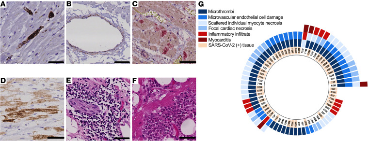
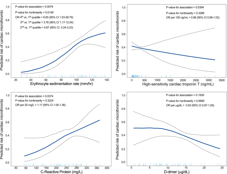
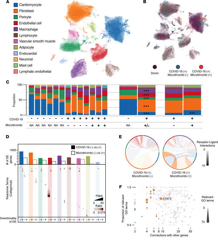
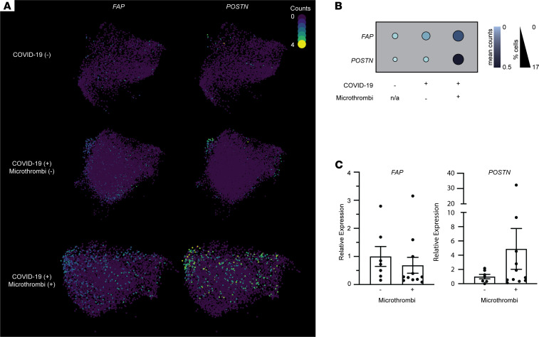
Acute cardiac injury is prevalent in critical COVID-19 and associated with increased mortality. Its etiology remains debated, as initially presumed causes - myocarditis and cardiac necrosis - have proved uncommon. To elucidate the pathophysiology of COVID-19-associated cardiac injury, we conducted a prospective study of the first 69 consecutive COVID-19 decedents at CUIMC in New York City. Of 6 acute cardiac histopathologic features, presence of microthrombi was the most commonly detected among our cohort. We tested associations of cardiac microthrombi with biomarkers of inflammation, cardiac injury, and fibrinolysis and with in-hospital antiplatelet therapy, therapeutic anticoagulation, and corticosteroid treatment, while adjusting for multiple clinical factors, including COVID-19 therapies. Higher peak erythrocyte sedimentation rate and C-reactive protein were independently associated with increased odds of microthrombi, supporting an immunothrombotic etiology. Using single-nuclei RNA-sequencing analysis on 3 patients with and 4 patients without cardiac microthrombi, we discovered an enrichment of prothrombotic/antifibrinolytic, extracellular matrix remodeling, and immune-potentiating signaling among cardiac fibroblasts in microthrombi-positive, relative to microthrombi-negative, COVID-19 hearts. Non-COVID-19, nonfailing hearts were used as reference controls. Our study identifies a specific transcriptomic signature in cardiac fibroblasts as a salient feature of microthrombi-positive COVID-19 hearts. Our findings warrant further mechanistic study as cardiac fibroblasts may represent a potential therapeutic target for COVID-19-associated cardiac microthrombi.

Keywords: Bioinformatics; COVID-19; Cardiology; Cardiovascular disease; Molecular pathology.


Figures

Figure 1
Figure 1. Acute cardiac histopathologic features in fatal COVID-19.
(A) Microthrombi, identified by intraluminal CD61 immunostaining (DAB chromogen, brown) of intramyocardial small vessels. (B) Small vascular endothelial cell damage, identified by C4d immunostaining (DAB chromogen, brown) of vascular endothelial cells. (C) Focal cardiac necrosis, identified by a confluence of C4d+ cardiomyocytes (DAB chromogen, brown) covering an area of at least 0.05 mm2 but less than or equal to 1 cm2. (D) Inflammatory infiltrates on H&E. (E) Scattered, isolated cardiomyocyte necrosis, identified by C4d+ staining (Alkaline Phosphatase chromogen, red) of cardiomyocytes. (F) Focal myocarditis on H&E. Scale bars: 50 μm. (G) Radial plot of acute cardiac histopathologic features detected in either ventricle of each COVID-19 decedent’s heart. Detection by quantitative reverse transcription polymerase chain reaction (RT-qPCR) of SARS-CoV-2 in cardiac tissue, where decedents’ age and sex are also shown.
Figure 2
Figure 2. Adjusted GAMs of the association between serum biomarker peak values and cardiac microthrombi.
We calculated a covariate balancing propensity score (CBPS) for each independent variable by regressing it on possible confounders; the resulting propensity score was used in the GAM as a single covariable. The covariates used to calculate CBPS were age, sex, race and ethnicity, BMI, duration of COVID-19 illness, outpatient ACE inhibitor/angiotensin receptor blocker (ARB) use, outpatient antiplatelet therapy, and inpatient administration of corticosteroids, remdesivir, interleukin-6 (IL-6) receptor antagonists, and therapeutic anticoagulation.
Figure 3
Figure 3. snRNA-Seq of right ventricular tissue from COVID-19 autopsies.
(A) Uniform manifold approximation and projection (UMAP) of 75,772 individual nuclei colored by cell type as determined from marker gene analysis. Colors observed within this legend are used throughout the figure. (B) UMAP colored by sample type of origin. (C) Compositional analysis for each cell type by individual sample, COVID-19 status, and microthrombi detection. *** indicates a credible increase or decrease in cell proportion when compared with the referent data set. (D) Bar plot displays the total number of DE genes at 0.25 LFC for COVID-19 versus non–COVID-19 control (filled bar) or microthrombi-positive versus microthrombi-negative in the COVID-19 (+) samples (unfilled bar). Dot plot indicates the Reactome terms associated with DE genes in a given comparison and direction. Size and shade of the dot represent the adjusted P value and ratio of Reactome genes present for a given term. (E) CellPhoneDB analysis of cell-cell communication through analysis of receptor-ligand pair expression. Size of each outer section corresponds to the proportion of that cell type in the tissue. Opacity of each connection indicates the number of connections between cell types (paracrine and autocrine). Color of the connection indicates the cell type producing the ligand. (F) Dot plot of GeneWalk analysis identifying regulator genes for the ontology observed within the microthrombi-positive versus microthrombi-negative comparison within the COVID-19 (+) samples. Color corresponds to the cell type, and size of the dot represents the number of significant Gene Ontology terms affiliated with that gene. Dots with high opacity represent those from the 0.5 LFC threshold for DE genes, while those with reduced opacity result from the more liberal 0.25 LFC threshold.
Figure 4
Figure 4. Analysis of fibroblasts’ activation in COVID-19 samples with versus without microthrombi.
(A) UMAPs of fibroblast cell populations. Colors correspond to the read counts for FAP and POSTN in each cell. (B) Dot plot displaying the expression of activated fibroblast markers FAP and POSTN in fibroblast clusters. Size and shade of the dot correspond to the percentage of cells with non-zero counts and the mean counts across all cells, respectively. (C) Scatter plot of relative expression of FAP and POSTN (corrected to GAPDH), as measured by probe-based RT-qPCR, in ventricular tissue samples from COVID-19 autopsies with PMI less than 8 hours (same PMI criteria as used for snRNA-Seq). Bars represent mean ± SEM. n = 7 for microthrombi-negative; n = 11 for microthrombi-positive COVID-19 groups. All RT-qPCR samples were run in duplicate. P = 0.96 for FAP, P = 0.20 for POSTN on Welch’s t test.

Update of

References

    1. Bangalore S, et al. ST-segment elevation in patients with Covid-19 — a case series. N Engl J Med. 2020;382(25):2478–2480. doi: 10.1056/NEJMc2009020. - DOI - PMC - PubMed
    1. Chung MK, et al. COVID-19 and cardiovascular disease: from bench to bedside. Circ Res. 2021;128(8):1214–1236. doi: 10.1161/CIRCRESAHA.121.317997. - DOI - PMC - PubMed
    1. Coyle J, et al. A recovered case of COVID-19 myocarditis and ARDS treated with corticosteroids, tocilizumab, and experimental AT-001. JACC Case Rep. 2020;2(9):1331–1336. doi: 10.1016/j.jaccas.2020.04.025. - DOI - PMC - PubMed
    1. Fried JA, et al. The variety of cardiovascular presentations of COVID-19. Circulation. 2020;141(23):1930–1936. doi: 10.1161/CIRCULATIONAHA.120.047164. - DOI - PMC - PubMed
    1. Inciardi RM, et al. Cardiac involvement in a patient with coronavirus disease 2019 (COVID-19) JAMA Cardiol. 2020;5(7):819–824. doi: 10.1001/jamacardio.2020.1096. - DOI - PMC - PubMed

Publication types

MeSH terms