. 2021 Dec 14;17(12):e1009962.
doi: 10.1371/journal.pgen.1009962. eCollection 2021 Dec.

TM2D genes regulate Notch signaling and neuronal function in Drosophila

Affiliations

TM2D genes regulate Notch signaling and neuronal function in Drosophila

Jose L Salazar et al. PLoS Genet. .

Abstract

TM2 domain containing (TM2D) proteins are conserved in metazoans and encoded by three separate genes in each model organism species that has been sequenced. Rare variants in TM2D3 are associated with Alzheimer's disease (AD) and its fly ortholog almondex is required for embryonic Notch signaling. However, the functions of this gene family remain elusive. We knocked-out all three TM2D genes (almondex, CG11103/amaretto, CG10795/biscotti) in Drosophila and found that they share the same maternal-effect neurogenic defect. Triple null animals are not phenotypically worse than single nulls, suggesting these genes function together. Overexpression of the most conserved region of the TM2D proteins acts as a potent inhibitor of Notch signaling at the γ-secretase cleavage step. Lastly, Almondex is detected in the brain and its loss causes shortened lifespan accompanied by progressive motor and electrophysiological defects. The functional links between all three TM2D genes are likely to be evolutionarily conserved, suggesting that this entire gene family may be involved in AD.


Conflict of interest statement

The authors have declared that no competing interests exist.

Figures

Fig 1
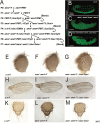
Fig 1. A clean null allele of TM2D3 fly ortholog almondex (amxΔ) behaves like the classic amx1 allele.
(A) Schematic diagram of almondex (amx) locus, amxΔ allele and genomic rescue constructs used in this study. (B) Predicted 2D-structure of Drosophila Amx protein. SS = signal sequence for membrane localization. Transmembrane 2 (TM2) domain is boxed in red. Hexagon denotes where 3xHA epitope is located in 3xHA::Amx protein. (C) Egg hatching assay shows that genomic rescue constructs can suppress embryonic lethality (amxΔ n = 857. amxΔ + amx n = 139. amxΔ + 3xHA::amx n = 1673. amxΔ + TM2D3 n = 257). Error bars show SEM. One-way ANOVA followed by Dunnett test. **** = p-value≤0.0001. (D-G) Embryonic nervous tissue (neuronal nuclei, Elav, green) of developing embryos. Embryos from homozygous amxΔ females (D) exhibit a neurogenic phenotype. This phenotype can be suppressed by wild-type amx (E), 3xHA::amx (F), or human TM2D3 (G) genomic rescue constructs. Scale bars = 100 μm in (D-G).
Fig 2
Fig 2. Null alleles of fly orthologs of TM2D2 (CG11103/amrt) and TM2D1 (CG10795/bisc) phenotypically mimics the loss of TM2D3 (amx).
(A) Protein alignment of human and Drosophila TM2D proteins. The TM2 domain (boxed in red) is composed of two transmembrane domains (TMD) and an intracellular DRF motif (denoted by black bars for TM2D3). (B) Schematic of amaretto (amrt) locus, amrt null allele (amrtΔ), and amrt genomic rescue plasmid construct generated for this study. (C) Embryos from homozygous amrtΔ females exhibit neurogenic phenotype, which can be suppressed by the amrt genomic rescue construct (D). (E) Egg hatching assay showing that amrt genomic rescue construct suppresses 68% of embryo lethality (amrtΔ n = 954. amrtΔ + amrt n = 1490). (F) Schematic of biscotti (bisc) locus, bisc null allele (biscΔ), and bisc::GFP genomic rescue fosmid construct generated for this study. (G) Embryos from homozygous biscΔ females exhibit neurogenic phenotype (n = 379) which can be suppressed by bisc::GFP genomic rescue construct (H). (I) Egg hatching assay showing that bisc::GFP suppresses 30% of embryonic lethality (biscΔ n = 379. biscΔ + bisc::GFP n = 436). t-test, **** = p-value<0.0001. Scale bars = 100 μm in (C, D, G, H).
Fig 3
Fig 3. Triple null mutant for all three TM2D fly genes is phenotypically similar to single null mutants.
(A) Crossing scheme used to generate TM2D triple null mutant flies. (B) A normal embryonic nervous system highlighted by neuronal nuclei marker Elav (green). (C-D) amxΔ amrtΔ double mutants (C) and amxΔ amrtΔ biscΔ triple mutants (D) exhibit a neurogenic phenotype. (E-K) TM2D double and triple null mutants exhibit no overt morphological phenotypes. Head structures of mutants (F, G) appear normal compared to y w control (E). Mutant wings (I, J) and thorax (L, M) also appear normal compared to control (H, K). Scale bars = 100 μm in (B-D).
Fig 4
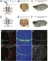
Fig 4. Amx that only possesses the highly conserved TM2 domain is a potent inhibitor of Notch signaling.
(A, D) Schematic of proteins generated from UAS-3xHA::amxFL and UAS-3xHA::amxΔECD transgenes. A 3xHA epitope (grey hexagon) was inserted after a predicted signal sequence (SS, blue box). The majority of the extracellular domain (ECD, light brown box) was removed to generate 3xHA::amxΔECD, consisting mostly of the TM2 domain (red box) tagged with the N-terminal 3xHA epitope. (B) Overexpression of 3xHA::AmxFL with pannier(pnr)-GAL4 has no effect on notum morphology. The absence of bristles along the midline is due to a very mild dorsal closure defect caused by the pnr-GAL4 insertion. (C) Expression of 3xHA::AmxFL in the posterior wing using engrailed (en)-GAL4 has no effect of the morphology of wings of adults raised at 29°C. (E) pnr-GAL4 driven overexpression of truncated Amx causes an increase in the number of micro- and macrochaete, indicative of loss of Notch mediated lateral inhibition. (F) en-GAL4 driven overexpression of 3xHA::AmxΔECD causes notching of the posterior wing margin. (G-H) Immunostaining of wing imaginal discs expressing full-length or truncated 3xHA::Amx. (G) 3xHA::AmxFL expression in the posterior imaginal wing disc using en-GAL4 has no effect on NRE (Notch response element)-GFP expression, a synthetic in vivo Notch signaling reporter (G’, rainbow) and on Cut (G”, white) expression, a downstream target of Notch activation in this context. The domain expressing GAL4 is marked by RFP (red). (H) Expression of 3xHA::AmxΔECD decreases NRE-GFP (rainbow) expression (H’) and reduces Cut expression (H”). Scale bars = 50 μm in (G-H).
Fig 5
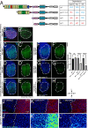
Fig 5. Genetic epistasis experiments place truncated Amx at the γ-secretase cleavage step of Notch activation.
(A) Schematics and characteristics of Notch proteins made from each UAS-Notch transgenes that were generated for this study. Full-length Notch (NFL) requires ligand binding and processing by Kuzbanian (Kuz) and γ-secretase (GS) for activation. Notch with EGF repeats and LNR domains removed (NΔEGF1-18.LNR) is not dependent on ligands but are dependent on both Kuz and GS for activation. Notch with an extracellular truncation (NEXT) is dependent only on GS for activation. The Notch intracellular domain (NICD) is constitutively active. (B) Cut (green) along the dorsoventral midline within the wing pouch [marked by nub-GAL4 UAS-mCherry (red), outlined by a white dash oval) is induced by Notch signaling. LacZ is expressed as a neutral protein. (C-E) Expression of Notch constructs leads to increase in Cut expression, quantified in (I). (F) Co-overexpression of 3xHA::AmxΔECD has no effect of NICD mediated increases in Cut expression (F’, I). (G-H) 3xHA::AmxΔECD expression suppresses the effects of NΔEGF1-18.LNR and NEXT on Cut expression (G’, H’, I). t-test. * = p<0.05. **** = p<0.0001. Error bars show SEM. (J-L) Overexpression of 3xHA::AmxΔECD causes an increase of Notch protein levels (K’) compared to overexpression of a neutral protein, LacZ (β-galactosidase) (J’). Knockdown of psn mediated by shRNA also results in mild increase Notch levels (L’), mimicking the effect of 3xHA::AmxΔECD. DAPI marks nuclei of wing disc cells whereas mCherry labels the dpp-GAL4 expression domain, the boundary of which is indicated by white dash lines, in J-L. Scale bars = 50 μm in (B-H). Scale bars = 5 μm in (J-L).
Fig 6
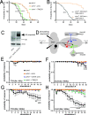
Fig 6. Loss of amx causes shortening of lifespan and age-dependent neurophysiological defects.
(A) Lifespan assay shows amxΔ animals (black, n = 247) have reduced lifespan compared to amxΔ + amx controls (orange, n = 224). amxΔ + human TM2D3 flies (green, n = 234) have significantly longer lifespan than amxΔ animals, but shorter than control. Animals were reared at 25°C; Log-rank test (Mantel-Cox), **** = p<0.0001. (B) Restoring Amx expression specifically in neuronal cells rescues the reduced lifespan in amx null animals. LacZ overexpression was used as a control. Animals were reared at 25°C; Log-rank test (Mantel-Cox), **** = p<0.0001. (C) Western blot of amxΔ; 3xHA::amx brains shows that 3xHA::Amx (predicted 35 kDa size) is expressed in the adult nervous system (arrow). Protein isolate from five brains was loaded per lane and Actin was probed as a loading control. (D) Schematization of the giant fiber electrophysiological recordings. Stimulating electrodes are inserted into the brain and recording electrodes record responses from the TTM and DLM muscles. (E-F) TTM muscles of 5d old amxΔ mutants (black) have a response similar to amxΔ + amx controls (orange) while DLM muscles have small but significant decrease in response probability. 3xHA::amx (blue) flies also perform as well as controls. (G-H) TTM and DLM response in 25d old amxΔ mutants is significantly reduced. DLM response of 25d old amxΔ + human TM2D3 (green) flies is reduced compared to controls (I). Multiple unpaired t-tests with Holm-Šídák correction for multiple comparisons. * = p<0.05. ** = p≤0.01. *** = p<0.001, **** = p<0.0001. Error bars show SEM. Additional data can be found in S12–S14 Figs.

Similar articles

Cited by

References

    1. Long JM, Holtzman DM. Alzheimer Disease: An Update on Pathobiology and Treatment Strategies. Cell. Cell Press; 2019. pp. 312–339. doi: 10.1016/j.cell.2019.09.001 - DOI - PMC - PubMed
    1. Sala Frigerio C, De Strooper B. Alzheimer’s Disease Mechanisms and Emerging Roads to Novel Therapeutics. Annu Rev Neurosci. 2016;39: 57–79. doi: 10.1146/annurev-neuro-070815-014015 - DOI - PubMed
    1. Bellenguez C, Grenier-Boley B, Lambert JC. Genetics of Alzheimer’s disease: where we are, and where we are going. Current Opinion in Neurobiology. Elsevier Ltd; 2020. pp. 40–48. doi: 10.1016/j.conb.2019.11.024 - DOI - PubMed
    1. Karran E, Mercken M, Strooper B De. The amyloid cascade hypothesis for Alzheimer’s disease: An appraisal for the development of therapeutics. Nature Reviews Drug Discovery. Nature Publishing Group; 2011. pp. 698–712. doi: 10.1038/nrd3505 - DOI - PubMed
    1. Zhang S, Zhang M, Cai F, Song W. Biological function of Presenilin and its role in AD pathogenesis. Translational Neurodegeneration. BioMed Central; 2013. p. 15. doi: 10.1186/2047-9158-2-15 - DOI - PMC - PubMed

Publication types