. 2022 Mar 3;139(9):1374-1388.
doi: 10.1182/blood.2021012537.

Hypofibrinogenemia with preserved hemostasis and protection from thrombosis in mice with an Fga truncation mutation

Affiliations

Hypofibrinogenemia with preserved hemostasis and protection from thrombosis in mice with an Fga truncation mutation

Woosuk S Hur et al. Blood. .

Abstract

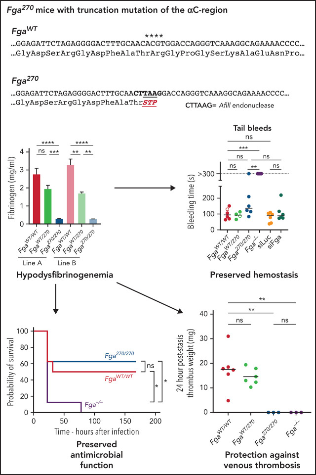
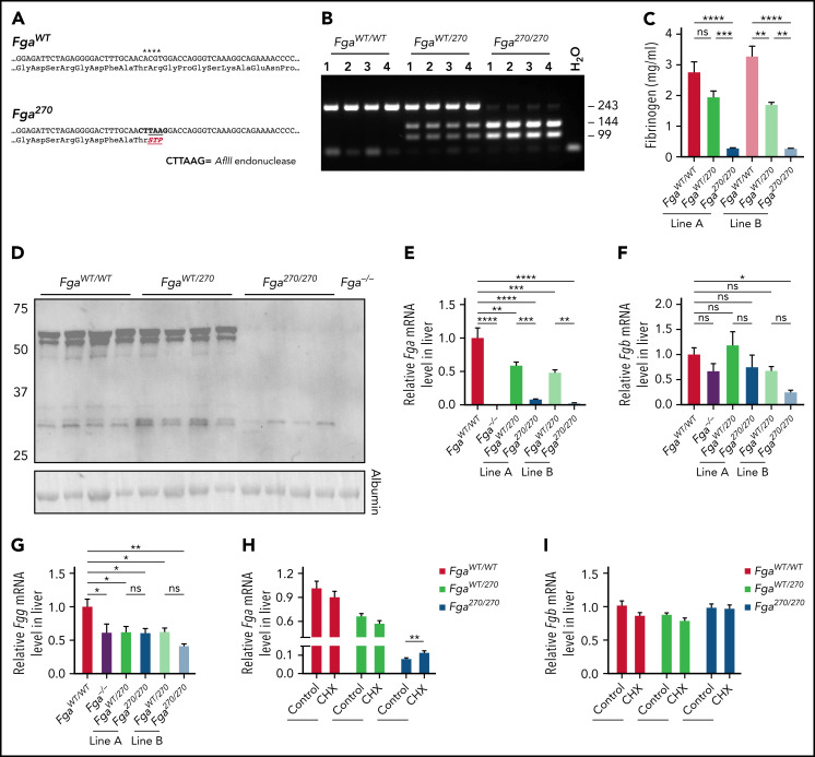
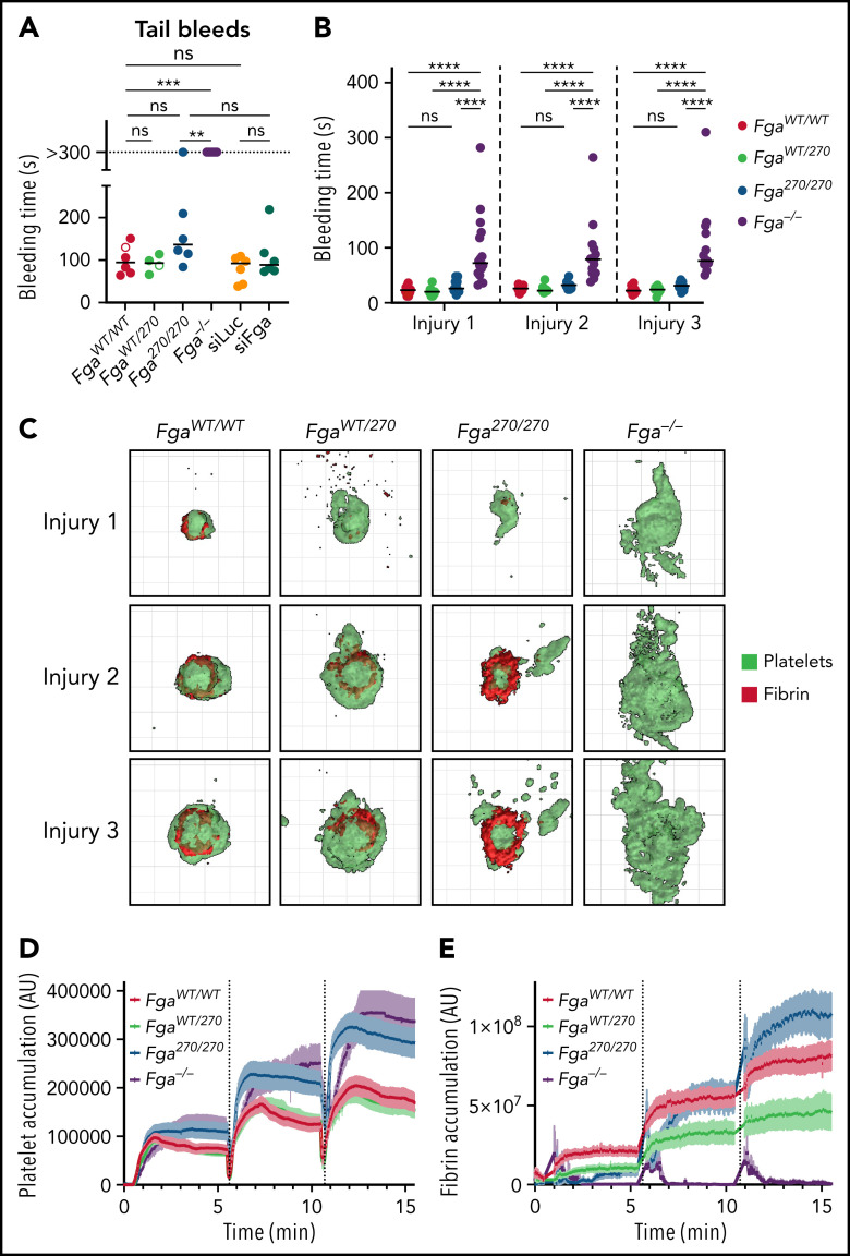
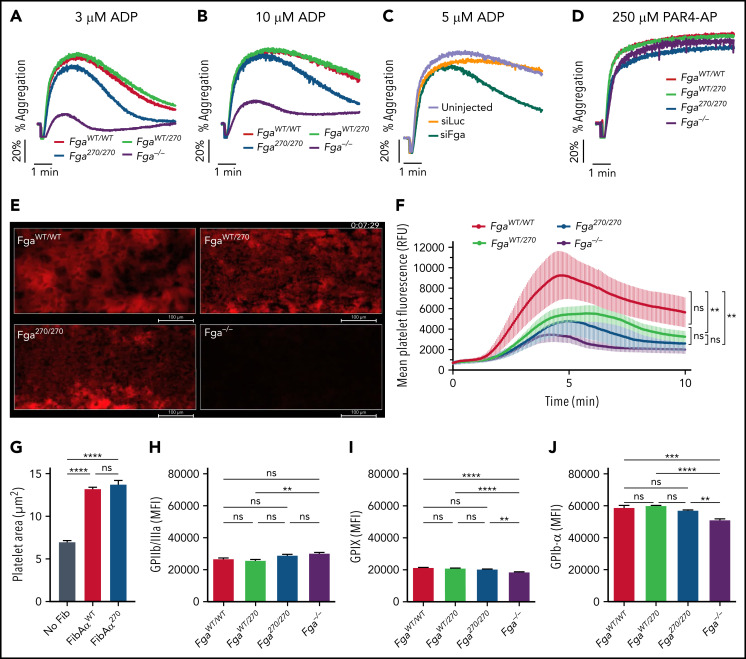
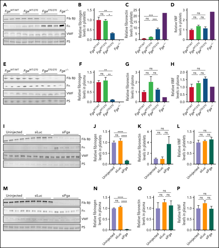
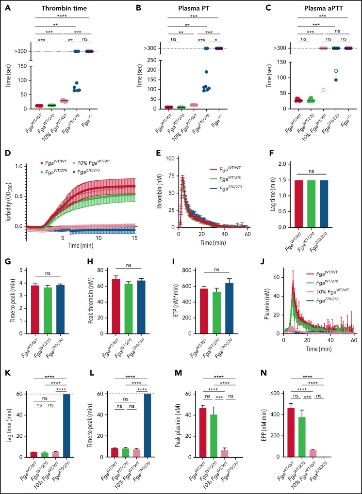
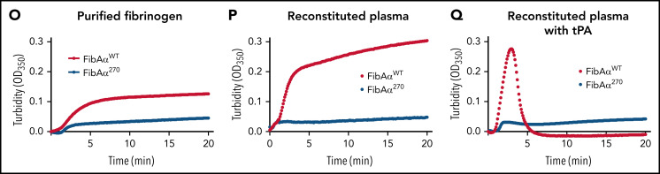
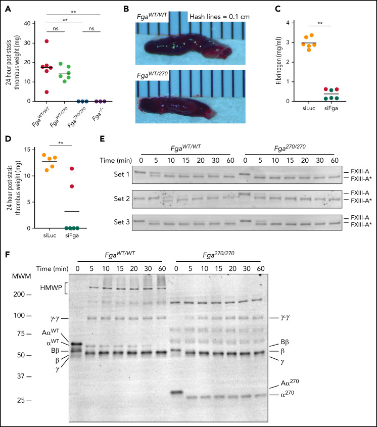
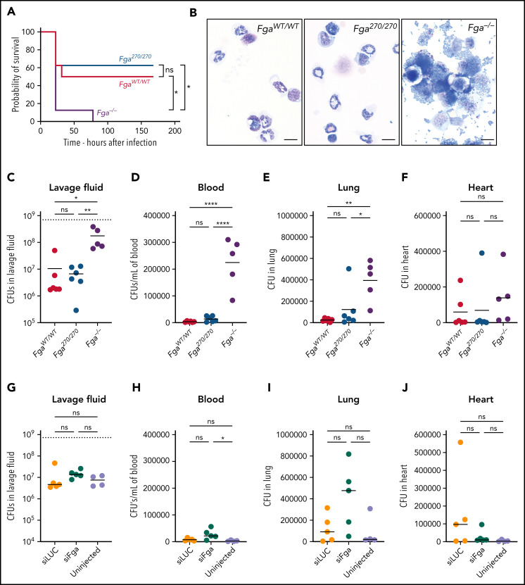
Genetic variants within the fibrinogen Aα chain encoding the αC-region commonly result in hypodysfibrinogenemia in patients. However, the (patho)physiological consequences and underlying mechanisms of such mutations remain undefined. Here, we generated Fga270 mice carrying a premature termination codon within the Fga gene at residue 271. The Fga270 mutation was compatible with Mendelian inheritance for offspring of heterozygous crosses. Adult Fga270/270 mice were hypofibrinogenemic with ∼10% plasma fibrinogen levels relative to FgaWT/WT mice, linked to 90% reduction in hepatic Fga messenger RNA (mRNA) because of nonsense-mediated decay of the mutant mRNA. Fga270/270 mice had preserved hemostatic potential in vitro and in vivo in models of tail bleeding and laser-induced saphenous vein injury, whereas Fga-/- mice had continuous bleeding. Platelets from FgaWT/WT and Fga270/270 mice displayed comparable initial aggregation following adenosine 5'-diphosphate stimulation, but Fga270/270 platelets quickly disaggregated. Despite ∼10% plasma fibrinogen, the fibrinogen level in Fga270/270 platelets was ∼30% of FgaWT/WT platelets with a compensatory increase in fibronectin. Notably, Fga270/270 mice showed complete protection from thrombosis in the inferior vena cava stasis model. In a model of Staphylococcus aureus peritonitis, Fga270/270 mice supported local, fibrinogen-mediated bacterial clearance and host survival comparable to FgaWT/WT, unlike Fga-/- mice. Decreasing the normal fibrinogen levels to ∼10% with small interfering RNA in mice also provided significant protection from venous thrombosis without compromising hemostatic potential and antimicrobial function. These findings both reveal novel molecular mechanisms underpinning fibrinogen αC-region truncation mutations and highlight the concept that selective fibrinogen reduction may be efficacious for limiting thrombosis while preserving hemostatic and immune protective functions.


Figures

None
Graphical abstract
Figure 1.
Figure 1.
Generation of Fga270 mice that express low levels of fibrinogen with a truncated form of Aα chain after residue 270. (A) Summary of the nucleic acid substitutions introduced by Crispr-Cas9 gene editing and resulting amino acid changes for the mutated fibrinogen Aα-chain gene of the Fga270 mice. Asterisks highlight positions of the nucleotide substitutions. (B) Representative PCR analysis to establish animal genotypes of FgaWT/WT, FgaWT/270, and Fga270/270 mice. (C) ELISA measurement of plasma fibrinogen levels from FgaWT/WT, FgaWT/270, and Fga270/270 mice from 2 independent lines (n = 3-4 per genotype). One-way ANOVA was used to determine statistical significance. (D) Western blot analysis of plasma (reducing conditions) from FgaWT/WT, FgaWT/270, Fga270/270, and Fga−/− mice using antibodies directed against the Aα chain of fibrinogen. Analysis of hepatic mRNA levels by quantitative reverse transcriptase PCR for (E) Fga, (F) Fgb, and (G) Fgg in Fga−/− mice and 2 independent lines of Fga270 mice (n = 4 per genotype). One-way ANOVA test was used to determine statistical significance. Analysis of fibrinogen (H) Fga and (I) Fgb gene expression from primary mouse hepatocytes isolated from FgaWT/WT, FgaWT/270, and Fga270/270 mice treated with or without 100 μM cycloheximide (CHX) for 3 hours (n = 3-4 per treatment group). Two-tailed Student t test was used to determine statistical significance. *P < .05, **P < .01, ***P < .001, ****P < .0001. ns, no statistical significance.
Figure 2.
Figure 2.
Fga270/270 and siFga-treated mice achieve hemostasis following vascular injury. (A) Time to cessation of bleeding (sustained >30 seconds) of FgaWT/WT, FgaWT/270, Fga270/270, and Fga−/− mice as well as siLuc- and siFga-treated mice following 3-mm excision of the distal portion of the tail. Horizontal bars indicate mean times for each group. Note that 1 of 6 Fga270/270 mice and 6 of 6 Fga−/− mice did not stop bleeding during the 5-minute evaluation period. Open dots represent mice in which rebleeding episodes occurred and the values indicate total bleeding time. Kaplan-Meier analysis was used to determine statistical significance. (B) Time to cessation of bleeding of FgaWT/WT, FgaWT/270, Fga270/270, and Fga−/− mice following 3 consecutive laser-induced saphenous injuries (n = 14-16 per genotype). Two-way ANOVA was used to determine statistical significance. (C) Representative 3-dimensional reconstruction of injury sites depicting the intravascular view 5 minutes following each injury. Each grid box = 50 μm × 50 μm. Quantification of (D) platelet and (E) fibrin accumulation at the site of injury over the course of 3 injuries. **P < .01, ***P < .001, ****P < .0001. ns, no statistical significance.
Figure 3.
Figure 3.
Fga270/270 platelets form weaker aggregates in response to ADP stimulation. Representative aggregation traces of platelet-rich plasma collected from FgaWT/WT, FgaWT/270, Fga270/270, and Fga−/− mice following stimulation with (A) 3 μM ADP or (B) 10 μM ADP (n = 4 per genotype). (C) Representative aggregation traces of platelet-rich plasma collected from untreated or siLuc- or siFga- treated mice following stimulation with 5 μM ADP (n = 4-5 per group). (D) Representative aggregation traces of platelet-rich plasma collected from FgaWT/WT, FgaWT/270, Fga270/270, and Fga−/− mice following stimulation with 250 μM of protease-activated receptor 4 activating peptide (PAR4-AP) (n = 4 per genotype). (E) Representative images and (F) quantification of platelet adhesion (indicated in red) to collagen coated surface at venous (400 seconds−1) shear rate using heparinized whole blood from FgaWT/WT, FgaWT/270, Fga270/270, and Fga−/− mice (n = 4 per genotype). (G) Adhesion and spreading of Fga−/− platelets on uncoated or FibAαWT- or FibAα270- coated coverslips following stimulation with 50 μM of ADP for 30 minutes (n = 3 per fibrinogen). One-way ANOVA was used to determine statistical significance. Expression of (H) GPIIb/IIIa, (I) GPIX, and (J) GPIbα on platelet membrane of FgaWT/WT, FgaWT/270, Fga270/270, and Fga−/− mice (n = 5 per genotype). One-way ANOVA was used to determine statistical significance. **P < .01, ****P < .0001. ns, no statistical significance.
Figure 4.
Figure 4.
Fga270/270 platelets contain higher levels of fibrinogen relative to fibrinogen levels in plasma. (A-D) Platelet lysates and (E-H) plasma harvested from FgaWT/WT, FgaWT/270, Fga270/270, and Fga−/− mice using antibodies against fibrinogen Bβ-chain (Fib Bβ), fibronectin (Fn), and VWF. Ponceau S staining of albumin (PS) was used as a loading control (n = 3 per genotype). (I-L) Platelet lysates and (M-P) plasma harvested from untreated or siLuc- or siFga-treated mice using antibodies against fibrinogen Bβ-chain, fibronectin, and VWF. Ponceau S staining of albumin was used as a loading control (n = 4-5 per treatment). One-way ANOVA test was used to determine statistical significance. *P < .05, **P < .01, ****P < .0001. ns, no statistical significance.
Figure 5.
Figure 5.
Altered clotting with Fga270/270 plasma and FibAα270 protein. (A) TT, (B) PT, and (C) APTT of FgaWT/WT, FgaWT/270, 10% FgaWT/WT, Fga270/270, and Fga−/− mice (n = 4-6 per genotype). Kaplan-Meier analysis was used to determine statistical significance. (D) Turbidity analysis of FgaWT/WT, FgaWT/270, 10% FgaWT/WT, and Fga270/270 plasma (n = 3-6 per genotype). (E) Representative thrombin generation curves and (F-I) associated parameters, and (J) representative plasmin generation curves and (K-N) associated parameters (n = 3-4 per genotype). One-way ANOVA was used to determine statistical significance. Turbidity analysis using (O) 2 mg/mL of purified FibAαWT and FibAα270 fibrinogen in buffered system and 1 mg/mL of FibAαWT and FibAα270 fibrinogen reconstituted in Fga−/− plasma in the (P) absence or (Q) presence of tPA (n = 1 per group). *P < .05, **P < .01, ***P < .001, ****P < .0001. ns, no statistical significance.
Figure 5.
Figure 5.
Altered clotting with Fga270/270 plasma and FibAα270 protein. (A) TT, (B) PT, and (C) APTT of FgaWT/WT, FgaWT/270, 10% FgaWT/WT, Fga270/270, and Fga−/− mice (n = 4-6 per genotype). Kaplan-Meier analysis was used to determine statistical significance. (D) Turbidity analysis of FgaWT/WT, FgaWT/270, 10% FgaWT/WT, and Fga270/270 plasma (n = 3-6 per genotype). (E) Representative thrombin generation curves and (F-I) associated parameters, and (J) representative plasmin generation curves and (K-N) associated parameters (n = 3-4 per genotype). One-way ANOVA was used to determine statistical significance. Turbidity analysis using (O) 2 mg/mL of purified FibAαWT and FibAα270 fibrinogen in buffered system and 1 mg/mL of FibAαWT and FibAα270 fibrinogen reconstituted in Fga−/− plasma in the (P) absence or (Q) presence of tPA (n = 1 per group). *P < .05, **P < .01, ***P < .001, ****P < .0001. ns, no statistical significance.
Figure 6.
Figure 6.
Fga270/270 and siFga-treated mice are protected from venous thrombosis. (A) Thrombus weights from FgaWT/WT, FgaWT/270, Fga270/270, and Fga−/− mice 24 hours after IVC ligation (stasis model). Kruskal-Wallis test was used to determine statistical significance. (B) Representative images of thrombi isolated 24 hours after IVC ligation. (C) Circulating fibrinogen levels of siLuc- and siFga-treated mice immediately before the IVC ligation, measured by ELISA. Red dots indicate mice that later developed occlusive thrombi. (D) Thrombus weights from siLuc- and siFga-treated mice 24 hours after ligation. Mantel-Cox test was used to determine statistical significance. Time course of (E) FXIII activation and (F) fibrin crosslinking in FgaWT/WT and Fga270/270 plasma. HMWP, high-molecular-weight polymer; MWM, molecular weight marker. **P < .01. ns, no statistical significance.
Figure 7.
Figure 7.
Fga270/270 and siFga-treated mice have preserved fibrinogen-dependent antimicrobial function. (A) Kaplan-Meier analysis of survival of FgaWT/WT, Fga270/270, and Fga−/− mice after intraperitoneal infection with 0.7 × 10⁹ CFUs of S aureus (n = 8 per genotype). (B) Representative photomicrographs of cytospin preparations of peritoneal lavage fluid 1 hour after intraperitoneal infection with 0.7 × 10⁹ CFUs of S aureus from FgaWT/WT, Fga270/270, and Fga−/− mice (n = 6 per genotype). Note the cell-associated and free bacteria in samples from Fga−/− mice that are absent in samples from FgaWT/WT and Fga270/270 mice. Scale bar: 10 μm. CFUs within (C) lavage fluid, (D) blood, (E) lung homogenates, and (F) heart homogenates after intraperitoneal infection of S aureus in FgaWT/WT, Fga270/270, and Fga−/− mice. CFUs within (G) lavage fluid, (H) blood, (I) lung homogenates, and (J) heart homogenates after intraperitoneal infection with S aureus in untreated, siLuc- and siFga-treated mice. The dashed lines in panels C and G indicate the initial inoculum dose in CFUs. One-way ANOVA test was used to determine statistical significance. *P < .05, **P < .01, ****P < .0001. ns, no statistical significance.

Comment in

Similar articles

Cited by

References

    1. Soria J, Mirshahi S, Mirshahi SQ, et al. . Fibrinogen αC domain: its importance in physiopathology. Res Pract Thromb Haemost. 2019;3(2):173-183. - PMC - PubMed
    1. Ridgway HJ, Brennan SO, Faed JM, George PM. Fibrinogen Otago: a major alpha chain truncation associated with severe hypofibrinogenaemia and recurrent miscarriage. Br J Haematol. 1997;98(3): 632-639. - PubMed
    1. Koopman J, Haverkate F, Grimbergen J, Egbring R, Lord ST. Fibrinogen Marburg: a homozygous case of dysfibrinogenemia, lacking amino acids A alpha 461-610 (Lys 461 AAA-->stop TAA). Blood. 1992;80(8):1972-1979. - PubMed
    1. Tsurupa G, Medved L. Identification and characterization of novel tPA- and plasminogen-binding sites within fibrin(ogen) alpha C-domains. Biochemistry. 2001;40(3):801-808. - PubMed
    1. Collet JP, Moen JL, Veklich YI, et al. . The alphaC domains of fibrinogen affect the structure of the fibrin clot, its physical properties, and its susceptibility to fibrinolysis. Blood. 2005;106(12):3824-3830. - PMC - PubMed

Publication types