. 2022 Jan;21(1):e13531.
doi: 10.1111/acel.13531. Epub 2021 Dec 14.

Regulatory role of cathepsin L in induction of nuclear laminopathy in Alzheimer's disease

Affiliations

Regulatory role of cathepsin L in induction of nuclear laminopathy in Alzheimer's disease

Md Imamul Islam et al. Aging Cell. 2022 Jan.

Abstract

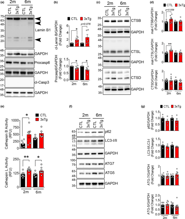
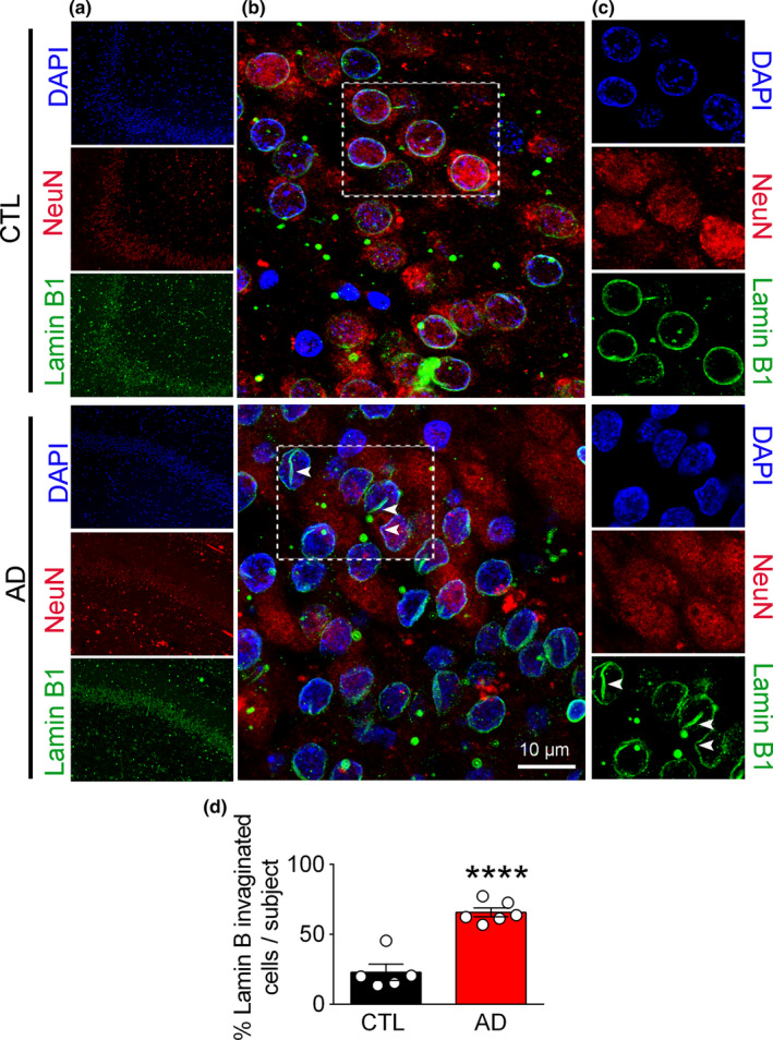
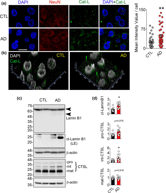
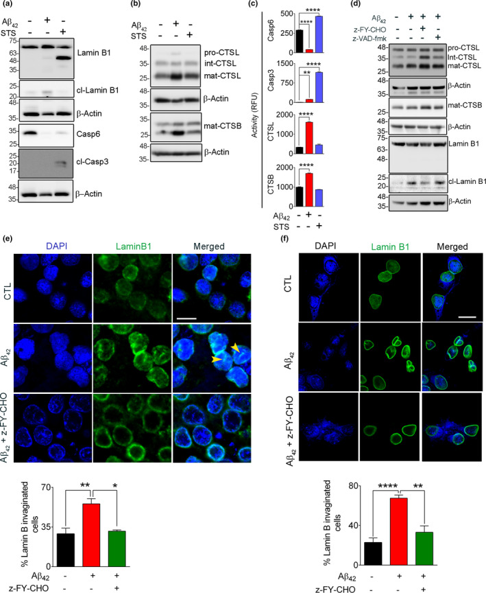
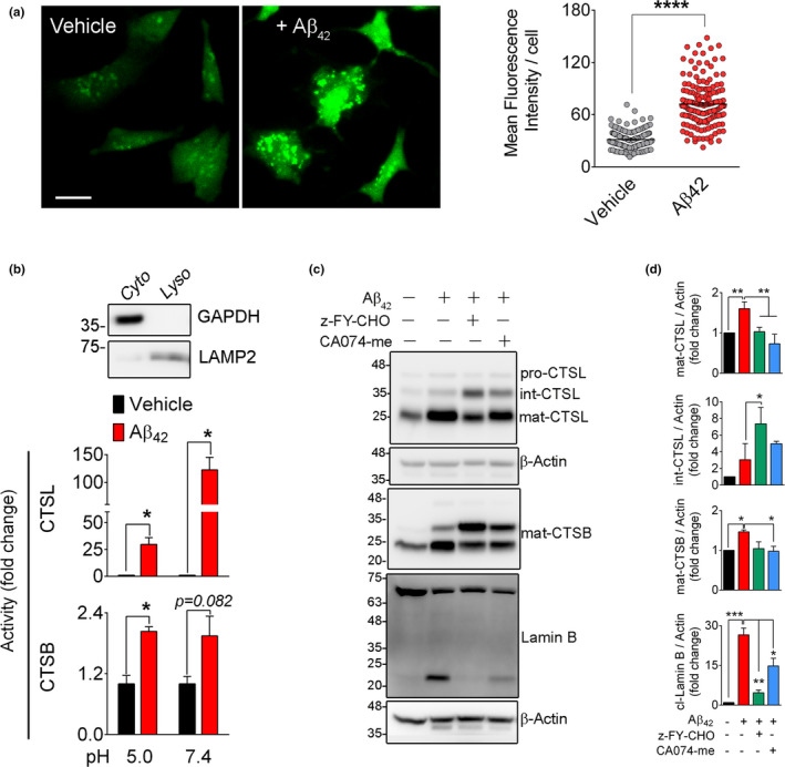
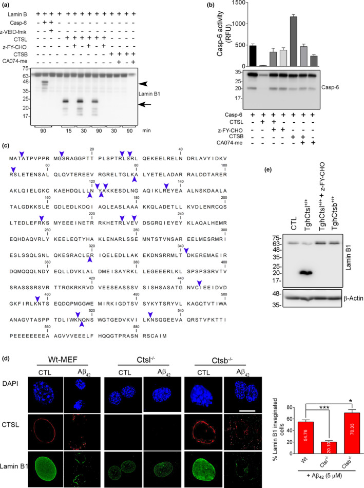
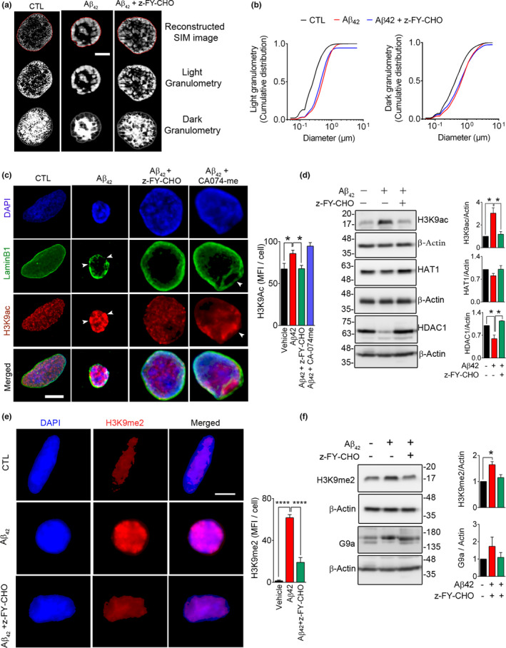
Experimental and clinical therapies in the field of Alzheimer's disease (AD) have focused on elimination of extracellular amyloid beta aggregates or prevention of cytoplasmic neuronal fibrillary tangles formation, yet these approaches have been generally ineffective. Interruption of nuclear lamina integrity, or laminopathy, is a newly identified concept in AD pathophysiology. Unraveling the molecular players in the induction of nuclear lamina damage may lead to identification of new therapies. Here, using 3xTg and APP/PS1 mouse models of AD, and in vitro model of amyloid beta42 (Aβ42) toxicity in primary neuronal cultures and SH-SY5Y neuroblastoma cells, we have uncovered a key role for cathepsin L in the induction of nuclear lamina damage. The applicability of our findings to AD pathophysiology was validated in brain autopsy samples from patients. We report that upregulation of cathepsin L is an important process in the induction of nuclear lamina damage, shown by lamin B1 cleavage, and is associated with epigenetic modifications in AD pathophysiology. More importantly, pharmacological targeting and genetic knock out of cathepsin L mitigated Aβ42 induced lamin B1 degradation and downstream structural and molecular changes. Affirming these findings, overexpression of cathepsin L alone was sufficient to induce lamin B1 cleavage. The proteolytic activity of cathepsin L on lamin B1 was confirmed using mass spectrometry. Our research identifies cathepsin L as a newly identified lamin B1 protease and mediator of laminopathy observed in AD. These results uncover a new aspect in the pathophysiology of AD that can be pharmacologically prevented, raising hope for potential therapeutic interventions.

Keywords: acetylation; amyloid beta; chromatin; histone; lysosomal membrane permeabilization; methylation; nuclear lamina; super-resolution microscopy.


Conflict of interest statement

All the authors declare no competing of interests.

Figures

FIGURE 1
FIGURE 1
Lamin B1 degradation and upregulation of lysosomal cathepsins in 3xTg mouse hippocampus tissue (a) Representative Western blots showing the expression of lamin B1, pro‐casp6, and cl‐Casp3 in hippocampal lysates from CTL and age‐matched 3xTg mouse. (b) Quantification of cleaved lamin B1 (21 kDa) and pro‐casp6 is shown in 2 months (2m) and 6 months (6m) old mice. (n = 5–6 mice/group), data shown as ±SEM, *p < 0.05. Increased cleaved lamin B1 in 3xTg mice is detected by appearance of a 46 kDa and a 21 kDa (arrowhead). As reported previously, a significant decrease in pro‐casp6 protein was confirmed in 3xTg mouse. We did not detect any indication of apoptosis in these mice as assessed by lack of cl‐Casp3. (c) Representative Western blots showing lysosomal cathepsins in 3xTg mouse hippocampus were quantified using densitometry. (d) Bar graph shows increased levels of CTSB and CTSL in 3xTg mouse, although CTSD levels remained unchanged. (e) Enzymatic activity of CTSB and CTSL activity in CTL and age‐matched 3xTg hippocampal lysates. (f) Markers of autophagy progression were assessed using immunoblotting. (g) No significant change in autophagy progression was observed in this model
FIGURE 2
FIGURE 2
Nuclear lamina damage is detected by invagination and focal loss of Lamin B1 in human AD brain. (a) Confocal microscopic micrographs depicting histological examination of hippocampal and medial temporal cortex from autopsy samples: LB1 (green), NeuN (red), and DAPI (blue). (b and c) Higher magnifications of the selected regions from A are shown (scale bar = 10 µm). (d) Quantification of damaged neuronal nuclei, as identified with their coffee‐bean nuclei immunolabeled with LB1 is shown. Cell counting was performed using ImageJ. A minimum of 109 cells were counted for each sample and the number of invaginated cells were expressed as % of total cells. (CTL = 5 and AD = 6 samples). Data reported as mean ±SEM. **** represents p < 0.0001
FIGURE 3
FIGURE 3
Cathepsin L (CTSL) mediated lamin B1 cleavage is observed in human AD samples. (a) Immunohistochemical labeling of hippocampal and medial temporal sections of human samples for NeuN (red), CTSL (green), and DAPI (blue), scale bar = 10 µm. Mean fluorescence intensity for CTSL was quantified and shown as mean ± SEM, **p < 0.01. (CTL = 5 and AD = 6). (b) Graphs representing distribution and intensity of CTSL in CTL and AD are shown. Higher intensity and closer apposition of CTSL‐positive bodies to the nucleus were observed in AD tissues in comparison with the CTL. (c) Representative Western blotting showing the expression of LB1 and CTSL in human hippocampus samples. (d) Densitometric quantification of the blots is shown as mean ± SEM, CTL (n = 7), and AD (n = 10), *p < 0.05
FIGURE 4
FIGURE 4
Aβ42‐induced lamin B1 cleavage is independent of apoptosis. To differentiate the contribution of apoptosis and autophagy to lamin B1 cleavage, SH‐SY5Y cells were exposed to Aβ42 (5 µM, 16 h) and STS (0.5 µM, 6 h). (a) Western blots showing the appearance of a lamin B1 46 kDa fragment that was only observed in STS‐treated cells. This was associated with a significant loss of pro‐casp6 protein and presence of cleaved Casp3, indicating the involvement of caspases. The 46 kDa fragment of lamin B1 and pro‐casp6 were not observed in Aβ42‐treated cells and no cleaved Casp3 was detected in these cells. (b) Representative immunoblots examining the changes in lysosomal cathepsin protein level. (c) Enzymatic activity assays for Casp6, Casp3, CTSL, and CTSB indicated the induction of caspases in STS‐treated cells and preferential activation of cathepsins in Aβ42‐treated cells. **p < 0.01 and **** p < 0.0001. (one‐way ANOVA/Tukey's post hoc). (d) Western blot confirming the involvement of CTSL in lamin B1 damage in rat primary hippocampal neurons when exposed to Aβ42 (5 µM, 16 h). Overnight pre‐treatment of neurons with CTSL inhibitor alleviated lamin B1 damage, but a pan‐caspase inhibitor z‐VAD‐fmk (20 µM) did not have any effect. (e) Confocal micrographs depicting cultured rat hippocampal neurons labeled for lamin B1 status in Aβ42 toxicity. LB1 invagination was observed in this model but was prevented significantly by CTSL inhibitor. The bar graph represents the percentage of invaginated/nicked neuronal LB1. A minimum of 130 cells/condition were analyzed. (f) The sensitivity of neuronal LB1 damage and NL invagination was also confirmed in mouse primary cortical neurons. This was preventable by pre‐treatment with CTSL inhibitor. An average of 56 cells were examined/experimental group. Results are mean ±SEM, *p < 0.05, **p < 0.01 and ****p < 0.0001, one‐way ANOVA/Tukey's post hoc, scale bar = 10 µm
FIGURE 5
FIGURE 5
Lamin B1 damage in Aβ42 toxicity is associated with lysosomal membrane permeabilization. (a) Representative acridine orange staining confirmed the induction of LMP in Aβ42‐treated SH‐SY5Y cells. Scale bar = 20 µm. LMP was quantified using green fluorescence intensity/cell. A minimum of 100 cells/condition were examined. ****p < 0.0001. (t test) (b) LMP was further assessed by measuring CTSL and CTSB enzymatic activities in cytosolic fractions at pH 5.0 and pH 7.4. Data shown here represents mean ±SEM of n = 3 independent experiments. *p < 0.05. The purity of cytosolic fraction was confirmed by a Western blot against β‐actin (cytosol) and LAMP2 (lysosome). (c) Pharmacological inhibition of CTSL prevents Aβ42‐induced lamin B1 cleavage. SH‐SY5Y cells were pre‐treated with indicated inhibitors (20 µM each) overnight. Medium was changed, and cells were exposed to Aβ42 (5 µM) for 16 h. Cathepsin and lamin B were assessed by Western blotting in whole‐cell lysates. (d) Quantification of Western blot results in c, data presented as mean ± SEM, n = 3 independent experiments, *p < 0.05, **p < 0.01, and ***p < 0.001, respectively (one‐way ANOVA/Tukey's post hoc). Upregulation of cathepsin B and L coincided with appearance of the 21 kDa lamin B1 product in Aβ42‐treated group. This was robustly prevented by z‐FY‐CHO and partially by CA074‐me
FIGURE 6
FIGURE 6
Cathepsin L (CTSL) is the main protease in degradation of lamin B1. (a) Recombinant human (rh) LB1 (1 µg) was incubated with equal amount of (50 ng) rhCasp6, rhCTSL, and rhCTSB at pH 7.4 for the indicated time. Western blot comparing the protease activity of CTSL, CTSB, and CASP6 in processing of LB1, in a cell‐free condition. The 21 kDa fragment of LB1, similar to the one seen in Aβ42 treatment and AD mouse and human samples was only produced by rhCTSL and inhibited by its specific inhibitor. Data are representative of 4 independent experiments. CASP6 produced a ~46 kDa fragment that was inhibited by z‐VEID‐FMK. rhCTSB did not have any effect on rhLB1. (b) Enzymatic activity and Western blotting confirming that CASP6 is degraded by CTSL. rhCasp6 (1 µg) was incubated with rhCTSL and rhCTSB (50 ng each) without/with their specific inhibitors for 1 h at 37°C. Only rhCTSL degraded rhCasp6. These results were further validated using rhCasp6 enzymatic activity using Ac‐VEID‐amc as substrate (bar graph). (c) In vitro reaction mixture of rhLB1 and rhCTSL was subjected to mass spectrometry, and CTSL‐mediated cleavage sites on LB1 were determined by analyzing the small peptides produced in mass spectrometry. The abundance of the peptides was established by considering peptide spectrum match (PSM) 7 or more. The cleavage sites are indicated with blue arrow heads. Peptides are listed in Table S1. (d) Genetic inhibition of CTSL prevents invagination of LB1. Representative 3D confocal microscopy depicting the effect of Aβ42 on LB1 was assessed in WT, Ctsl−/− , and Ctsb−/− MEF cells after treatment with 5 µM of Aβ42 for 16 h. Depletion of CTSL−/− effectively prevented LB1 damage. n = 3 independent experiments, *p < 0.05, and ***p < 0.001, respectively (one‐way ANOVA/Tukey's post hoc), Scale bar: 10 µm. (e) Western blotting confirmed that overexpression of CTSL (TghCTSL+/+) is sufficient to induce LB1 cleavage in normal conditions, however, pre‐treatment with z‐FY‐CHO effectively prevented LB1 damage. WT, Ctsl−/− , and Ctsb−/− indicate wilt type, CTSL, and CTSB knock out, respectively
FIGURE 7
FIGURE 7
Lamin B1 damage in Aβ42 toxicity affects nuclear architecture and chromatin remodeling. To examine the effect of LB1 invagination observed in Aβ42 toxicity, we exposed the SH‐SY5Y cells to Aβ42 (5 µM, 16 h), with/without pre‐treatment with CTSL inhibitor (z‐FY‐CHO, 20 µM). (a) Micrographs representing 3D‐Struture Illumination Microscopy (3D‐SIM) of DAPI‐stained cells. (Scale bar = 1 µm) (b) Granulometry analysis of images in a is shown by graph (cumulative distribution vs size in µm). A minimum of 39 cells/condition were analyzed. Data are representative of two independent experiments and reported as mean ± SEM. Statistical analysis (one‐way ANOVA/Tukey's post hoc) showed that administration of Aβ42 significantly increased DNA compaction, as shown by light granulometry: (p = 1.1744E‐138) that was prevented in cells pre‐treated with z‐FY‐CHO (p = 0.700E‐04). DNA‐free space, assessed by dark granulometry, was also increased after Aβ42 administration (CTL vs. Aβ42, p = 5.7328E‐17), although it did not fully recover after inhibition of CTSL (Aβ42 + z‐FY‐CHO: p = 0.5810). (c) Representative 3D confocal microscopy depicting epigenetic changes (H3K9ac) associated with lamin B1 damage. While inhibition of CTSL (z‐FY‐CHO, 20 µM) robustly alleviated these changes, inhibition of CTSB (CA074‐me, 20 µM) was not equally effective. Quantitative analysis of H3K9ac mean fluorescence intensity (MFI)/cells (≥ 46 cells/treatment group) is shown as a bar graph. Data are representative of three independent experiments and reported as mean ± SEM, *p < 0.05 (one‐way ANOVA/Tukey's post hoc), Scale bar = 10 µm. (d) To gain a mechanistic view of molecular changes in acetylation, Western blotting was performed, showing that the increase in H3K9ac was due to decreased HDAC1 levels in Aβ42 treated cells and was normalized by inhibition of CTSL. n = 2 independent experiments, data are mean ± SEM. *p < 0.05 (one‐way ANOVA/Tukey's post hoc). (e) Confocal micrographs depicting changes in H3K9me2 in this experimental model. Mean fluorescence intensity for H3K9me2 was analyzed (n ≥ 41 cells/conditions) and reported as mean ±SEM. ****p < 0.0001 (one‐way ANOVA/Tukey's post hoc), Scale bar: 10 µm. (f) H3K9me2 and G9a were probed for Western blotting after treating SH‐SY5Y cells as stated in e. Quantitative bar graphs of the indicated protein levels. Data reported as mean ± SEM. *p < 0.05 (one‐way ANOVA/Tukey's post hoc)

Similar articles

Cited by

References

    1. Arranz, A. M. , & De Strooper, B. (2019). The role of astroglia in Alzheimer's disease: Pathophysiology and clinical implications. The Lancet Neurology, 18(4), 406–414. 10.1016/S1474-4422(18)30490-3 - DOI - PubMed
    1. Bertero, A. , Fields, P. A. , Smith, A. S. T. , Leonard, A. , Beussman, K. , Sniadecki, N. J. , Kim, D.‐H. , Tse, H.‐F. , Pabon, L. , Shendure, J. , Noble, W. S. , & Murry, C. E. (2019). Chromatin compartment dynamics in a haploinsufficient model of cardiac laminopathy. Journal of Cell Biology, 218(9), 2919–2944. 10.1083/jcb.201902117 - DOI - PMC - PubMed
    1. Boya, P. , & Kroemer, G. (2008). Lysosomal membrane permeabilization in cell death. Oncogene, 27(50), 6434–6451. 10.1038/onc.2008.310 - DOI - PubMed
    1. Broers, J. L. , Ramaekers, F. C. , Bonne, G. , Yaou, R. B. , & Hutchison, C. J. (2006). Nuclear lamins: Laminopathies and their role in premature ageing. Physiological Reviews, 86(3), 967–1008. 10.1152/physrev.00047.2005 - DOI - PubMed
    1. Butin‐Israeli, V. , Adam, S. A. , Jain, N. , Otte, G. L. , Neems, D. , Wiesmüller, L. , Berger, S. L. , & Goldman, R. D. (2015). Role of lamin b1 in chromatin instability. Molecular and Cellular Biology, 35(5), 884–898. 10.1128/MCB.01145-14 - DOI - PMC - PubMed

Publication types

MeSH terms