. 2021 Dec 28;93(51):16995-17002.
doi: 10.1021/acs.analchem.1c02546. Epub 2021 Dec 14.

Tear-Based Vibrational Spectroscopy Applied to Amyotrophic Lateral Sclerosis

Affiliations

Tear-Based Vibrational Spectroscopy Applied to Amyotrophic Lateral Sclerosis

Diletta Ami et al. Anal Chem. .

Abstract

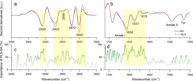
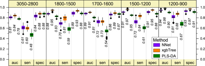
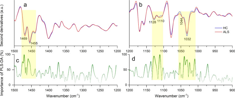
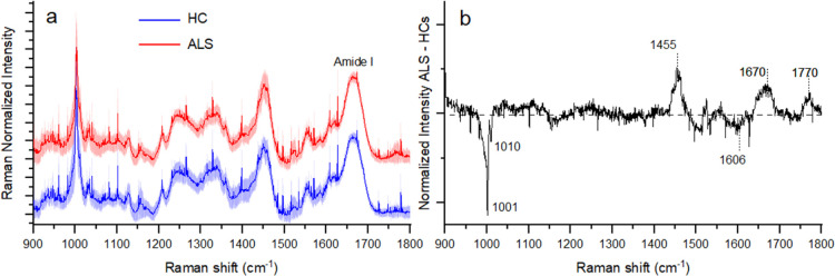
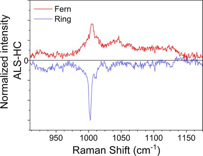
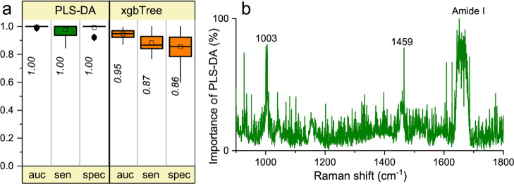
Biofluid analysis by optical spectroscopy techniques is attracting considerable interest due to its potential to revolutionize diagnostics and precision medicine, particularly for neurodegenerative diseases. However, the lack of effective biomarkers combined with the unaccomplished identification of convenient biofluids has drastically hampered optical advancements in clinical diagnosis and monitoring of neurodegenerative disorders. Here, we show that vibrational spectroscopy applied to human tears opens a new route, offering a non-invasive, label-free identification of a devastating disease such as amyotrophic lateral sclerosis (ALS). Our proposed approach has been validated using two widespread techniques, namely, Fourier transform infrared (FTIR) and Raman microspectroscopies. In conjunction with multivariate analysis, this vibrational approach made it possible to discriminate between tears from ALS patients and healthy controls (HCs) with high specificity (∼97% and ∼100% for FTIR and Raman spectroscopy, respectively) and sensitivity (∼88% and ∼100% for FTIR and Raman spectroscopy, respectively). Additionally, the investigation of tears allowed us to disclose ALS spectroscopic markers related to protein and lipid alterations, as well as to a reduction of the phenylalanine level, in comparison with HCs. Our findings show that vibrational spectroscopy is a new potential ALS diagnostic approach and indicate that tears are a reliable and non-invasive source of ALS biomarkers.


Conflict of interest statement

The authors declare no competing financial interest.

Figures

Figure 1
Figure 1
FTIR analysis of fern-like morphologies. Average second-derivative spectra of ALS-positive samples and HCs in the CHx stretching range (a) and in the amide I and amide II bands (b). Wavenumber importance (domain 0–100) for the PLS-DA method in the CHx stretching range (c) and the amide I and amide II bands (d).
Figure 2
Figure 2
Multivariate analysis of the FTIR second-derivative spectra of fern-like morphologies: overall performance of the NNet, xgbTree, and PLS-DA methods in all the analyzed spectral ranges. In particular, the 5-fold cross-validation resampled area under the curve (auc), sensitivity (sen.), and specificity (spec.) are reported. The black horizontal line within the box is the median, the square within the box is the mean, the box ends show the first (Q1) and third (Q3) quartiles, the lower whisker is computed as the maximum value between the absolute minimum and Q1 – 1.5 × IQR, and the upper whisker is the minimum between the absolute maximum and Q3 + 1.5 × IQR. Here, IQR is the interquartile range computed as Q3–Q1. The values beyond whiskers (outliers) are shown as black diamonds. The median values are also reported.
Figure 3
Figure 3
FTIR analysis of fern-like morphologies. Average second-derivative spectra of ALS-positive samples and HCs in the 1500–1200 cm–1 (a) and 1200–900 cm–1 (b) spectral ranges. Wavenumber importance (domain 0–100) for the PLS-DA method in the 1500–1200 cm–1 (c) and 1200–900 cm–1 (d) ranges.
Figure 4
Figure 4
Comparison of the mean Raman spectra obtained by considering all the measured tears from ALS patients and HCs (a). The shadowed area refers to the standard deviation of the data. (b) Spectrally resolved differential average Raman spectra of the two investigated groups.
Figure 5
Figure 5
Differential average Raman spectra of ALS patients and HCs measured in the fern-like patterns at the center of the dried drop (red line) and in the coffee ring (blue line).
Figure 6
Figure 6
Multivariate analysis of Raman spectra. (A) Overall performances of PLS-DA and xgbTree methods in the 900–1800 cm–1 spectral range. In particular, the resampled area under the curve (auc), sensitivity (sens.), and specificity (spec.) are reported as in Figure 2. (B) Wavenumber importance (domain 0–100) for PLS-DA method in the 900–1800 cm–1 spectral range.

Similar articles

Cited by

References

    1. Chiti F.; Dobson C. M. Protein Misfolding, Amyloid Formation, and Human Disease: A Summary of Progress Over the Last Decade. Annu. Rev. Biochem. 2017, 86, 27–68. 10.1146/annurev-biochem-061516-045115. - DOI - PubMed
    1. Deng H.-X.; Chen W.; Hong S.-T.; Boycott K. M.; Gorrie G. H.; Siddique N.; Yang Y.; Fecto F.; Shi Y.; Zhai H.; Jiang H.; Hirano M.; Rampersaud E.; Jansen G. H.; Donkervoort S.; Bigio E. H.; Brooks B. R.; Ajroud K.; Sufit R. L.; Haines J. L.; Mugnaini E.; Pericak-Vance M. A.; Siddique T. Mutations in UBQLN2 Cause Dominant X-Linked Juvenile and Adult-Onset ALS and ALS/Dementia. Nature 2011, 477, 211–215. 10.1038/nature10353. - DOI - PMC - PubMed
    1. Strong M. J. Revisiting the Concept of Amyotrophic Lateral Sclerosis as a Multisystems Disorder of Limited Phenotypic Expression. Curr. Opin. Neurol. 2017, 30, 599–607. 10.1097/WCO.0000000000000488. - DOI - PubMed
    1. Kadena K.; Vlamos P. Amyotrophic Lateral Sclerosis: Current Status in Diagnostic Biomarkers. Adv. Exp. Med. Biol. 2020, 1195, 179–187. 10.1007/978-3-030-32633-3_26. - DOI - PubMed
    1. Pampalakis G.; Mitropoulos K.; Xiromerisiou G.; Dardiotis E.; Deretzi G.; Anagnostouli M.; Katsila T.; Rentzos M.; Patrinos G. P. New Molecular Diagnostic Trends and Biomarkers for Amyotrophic Lateral Sclerosis. Hum. Mutat. 2019, 40, 361–373. 10.1002/humu.23697. - DOI - PubMed

Publication types