

Molecular characterization of equine thymidine kinase 1 and preliminary evaluation of its suitability as a serum biomarker for equine lymphoma
- PMID: 34906077
- PMCID: PMC8670147
- DOI: 10.1186/s12860-021-00399-x
Molecular characterization of equine thymidine kinase 1 and preliminary evaluation of its suitability as a serum biomarker for equine lymphoma
Abstract
Background: Thymidine kinase 1 (TK1) plays a key role in the synthesis of deoxythymidine triphosphate (dTTP) and is thus important for DNA replication and cell proliferation. The expression of TK1 is highest during S-phase, and it is rapidly degraded after mitosis. In cancer cells, TK1 is upregulated, resulting in leakage of excess TK1 into the blood. Consequently, serum TK1 has been used as a diagnostic and prognostic cancer biomarker, mainly in human medicine. The aims of this work were to characterize equine TK1 and to evaluate its suitability as a serum biomarker for equine lymphoma.
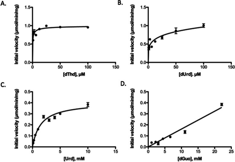
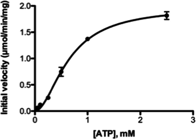
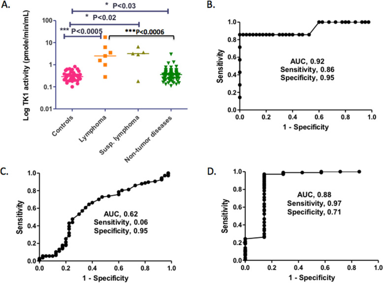
Results: Equine TK1 was cloned, expressed in E. coli and affinity purified. The purified recombinant horse TK1 showed broad substrate specificity, phosphorylating pyrimidine deoxyribo- and ribonucleosides and, to some extent, purine deoxynucleosides, including anticancer and antiviral nucleoside analogues. ATP was the preferred phosphate donor. Serum TK1 activity was measured in samples collected from horses with confirmed or suspected lymphoma and control horses with and without concurrent diseases. Serum TK1 activity levels were significantly higher in horses with lymphoma (p < 0.0005) and suspected lymphoma (p < 0.02) and in tumour-free groups with diverse diseases (p < 0.03) than in controls without concurrent diseases. There was a significant difference between the lymphoma group and the tumour-free group with diverse diseases (p < 0.0006). Furthermore, receiver operating characteristic analysis revealed a sensitivity of 0.86, a specificity of 0.95 and an AUC (area under the curve) of 0.92 compared to the controls without concurrent diseases, with a sensitivity of 0.97, a specificity of 0.71 and an AUC of 0.88 when compared with the tumour-free group with diverse diseases.
Conclusion: Equine TK1 showed high specific activity and broader substrate specificity than human TK1. Anticancer and antiviral thymidine analogues were efficiently phosphorylated by horse TK1, suggesting that these analogues might be good candidates for chemotherapy in horses. Serum TK1 activity was significantly higher in horses with lymphoma than in controls. ROC analysis indicated that serum TK1 could serve as a promising cancer biomarker in horses.
Keywords: Enzyme kinetics; Equine lymphoma; Equine thymidine kinase 1; Nucleoside analogues; Serum biomarker; cancer.
© 2021. The Author(s).
Conflict of interest statement
HS is employed by Alertix Veterinary Diagnostic AB; SE is a consultant and cofounder, and HR is member of the scientific advisory board. SE, HR, HS, and LW own shares of the same company. The other authors declare no conflicts of interest.
Figures
References
-
- Taintor J, Schleis S. Equine lymphoma. Equine Vet Educ. 2011;23(4):205–213. doi: 10.1111/j.2042-3292.2010.00200.x. - DOI
