. 2021 Dec 14;22(1):312.
doi: 10.1186/s12931-021-01908-4.

TRB3 mediates vascular remodeling by activating the MAPK signaling pathway in hypoxic pulmonary hypertension

Affiliations

TRB3 mediates vascular remodeling by activating the MAPK signaling pathway in hypoxic pulmonary hypertension

Xiaopei Cao et al. Respir Res. .

Abstract

Background: Hypoxic pulmonary hypertension (PH) is a refractory pulmonary vascular remodeling disease, and the efficiency of current PH treatment strategies is unsatisfactory. Tribbles homolog 3 (TRB3), a member of the pseudokinase family, is upregulated in diverse types of cellular stresses and functions as either a pro-proliferative or pro-apoptotic factor depending on the specific microenvironment. The regulatory mechanisms of TRB3 in hypoxic PH are poorly understood.

Methods: We performed studies using TRB3-specific silencing and overexpressing lentiviral vectors to investigate the potential roles of TRB3 on hypoxic pulmonary artery smooth muscle cells (PASMCs). Adeno-associated virus type 1(AVV1) vectors encoding short-hairpin RNAs against rat TRB3 were used to assess the role of TRB3 on hypoxic PH. TRB3 protein expression in PH patients was explored in clinical samples by western blot analysis.

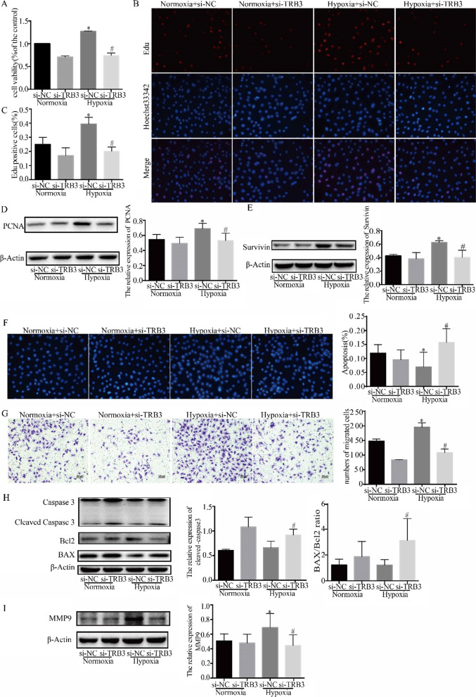
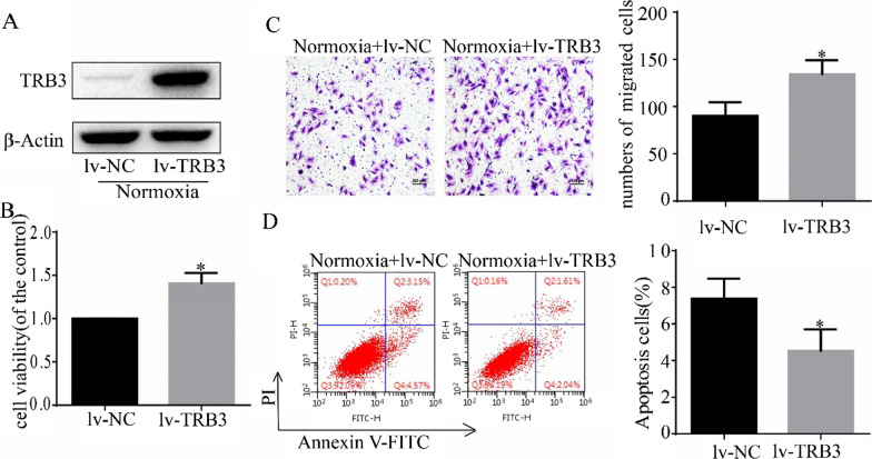
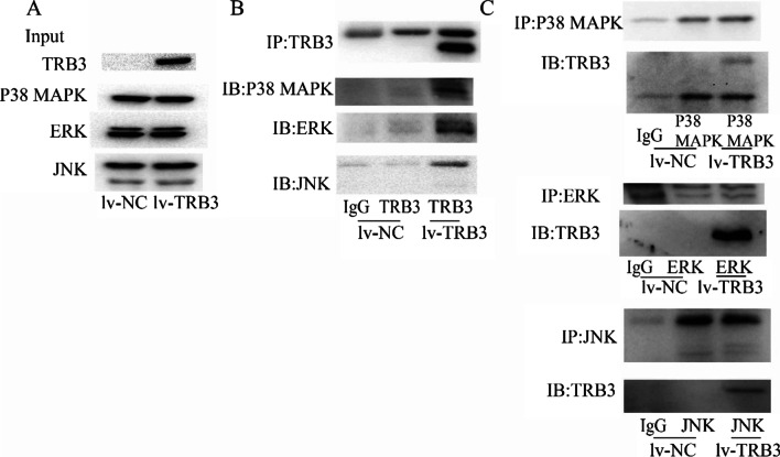
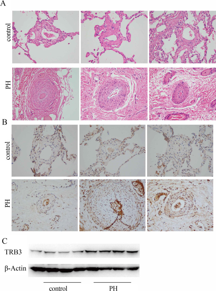
Results: The results of whole-rat genome oligo microarrays showed that the expression of TRB3 and endoplasmic reticulum stress (ERS)-related genes was upregulated in hypoxic PASMCs. TRB3 protein expression was significantly upregulated by hypoxia and thapsigargin. In addition, 4-PBA and 4μ8C, both inhibitors of ERS, decreased the expression of TRB3. TRB3 knockdown promoted apoptosis and damaged the proliferative and migratory abilities of hypoxic PASMCs as well as inhibited activation of the MAPK signaling pathway. TRB3 overexpression stimulated the proliferation and migration of PASMCs but decreased the apoptosis of PASMCs, which was partly reversed by specific inhibitors of ERK, JNK and p38 MAPK. The Co-IP results revealed that TRB3 directly interacts with ERK, JNK, and p38 MAPK. Knockdown of TRB3 in rat lung tissue reduced the right ventricular systolic pressure and decreased pulmonary medial wall thickness in hypoxic PH model rats. Further, the expression of TRB3 in lung tissues was higher in patients with PH compared with those who have normal pulmonary artery pressure.

Conclusions: TRB3 was upregulated in hypoxic PASMCs and was affected by ERS. TRB3 plays a key role in the pathogenesis of hypoxia-induced PH by binding and activating the ERK, JNK, and p38 MAPK pathways. Thus, TRB3 might be a promising target for the treatment of hypoxic PH.

Keywords: Hypoxia; MAPK; PASMCs; PH; TRB3.


Conflict of interest statement

The authors declare that they have no competing interests.

Figures

Fig. 1
Fig. 1
The expression of TRB3 was regulated by hypoxia and ERS. A A heat map of the hierarchical clustering of gene expression profiling of rat PASMCs for ERS from the Agilent Whole Rat Genome Oligo Microarrays. Each gene is depicted by one row, in which the red color denotes an increase in gene expression and the green color denotes a decrease in gene expression compared to the other group. Brighter colors represent higher gene expression levels. B TRB3 protein expression in PASMCs treated with hypoxia for different times. C The rats in the hypoxia group were exposed to 10% O2 for 8 h a day, and after 4 weeks, lung tissues were stained with hematoxylin–eosin (HE). D TRB3 protein expression in lung tissues and pulmonary arteries. EG TRB3 protein expression in PASMCs treated with 4-PBA, TG, and 4μ8C. Data represent the mean ± SEM (n = 4). *P < 0.05 and **P < 0.01 compared to the group exposed to hypoxia for 0 h. #P < 0.05 and ##P < 0.01 compared to PASMCs exposed to hypoxia for 48 h
Fig. 2
Fig. 2
Effect of TRB3 knockdown on proliferation, migration, and apoptosis of hypoxic PASMCs. PASMCs were transfected with lentiviral vectors encoding short hairpin RNAs targeting against rat TRB3(si-TRB3) or negative control(si-NC) for further analysis. A CCK-8 assays were performed to test cell viability after TRB3 knockdown in hypoxic PASMCs. B The EdU proliferation assay was used to measure cell proliferation. All images are × 400 magnification. C EdU‐positive cells were analyzed. D and E The expression of PCNA and Survivin was examined by western blot, and the gray value was quantified by ImageJ software. F Cell apoptosis was evaluated by Hoechst 33342 staining, and the percentage of apoptosis was quantified. G Crystal violet staining of PASMCs is presented at × 200 magnification for the Transwell assay, and the migrated cells were counted manually. H Apoptosis-related protein expression was evaluated and quantification of the analysis is shown. I The expression of MMP9 was evaluated. Data represent the mean ± SEM (n = 4). *P < 0.05 and **P < 0.01 compared to normoxic PASMCs transfected with si-NC. #P < 0.05 and ##P < 0.01 compared to hypoxic PASMCs transfected with si-TRB3. si-NC, lentiviral vectors encoding the negative control sequence; si-TRB3, lentiviral vectors encoding the short-hairpin RNAs against rat TRB3; PCNA, proliferating cell nuclear antigen; TRB3, Tribbles homolog 3; BAX, BCL2 associated X; Bcl2, B cell leukemia/lymphoma 2; MMP9, matrix metallopeptidase 9
Fig. 3
Fig. 3
TRB3 overexpression in PASMCs increased cell proliferation and migration but impeded cell apoptosis. A TRB3 overexpression was achieved by lentiviral transfection, and the efficiency was determined by Western blot analysis. B The CCK8 test was used to determine cell proliferation. C Crystal violet staining is presented at × 200 magnification for the Transwell assay, and the migrated cells were counted. D Cell apoptosis induced by TRB3 overexpression was detected using flow cytometry and the percentage of early apoptotic (Annexin V+/PI−) and late apoptotic (Annexin V+/PI+) cells was determined. Data represent the mean ± SEM (n = 4). *P < 0.05 compared to the normoxic PASMCs transfected with lv-NC. lv-TRB3, PASMCs transfected with a lentiviral vector containing TRB3 CDS; lv-NC (negative control), PASMCs transfected with an empty lentiviral vector
Fig. 4
Fig. 4
The connection of TRB3 and MAPK signaling in PASMCs was explored by western blot analysis. A and C Western blot analysis of the phosphorylation of ERK, JNK, and p38 MAPK in PASMCs after TRB3 knockdown and overexpression. B Effect of ERK, JNK, and p38 MAPK inhibitors on TRB3 in hypoxic PASMCs by Western blot analysis. D Effect of ERK, JNK, and p38 MAPK inhibitors on TRB3 in PASMCs transfected with a lentiviral vector expressing TRB3 (lv-TRB3) by Western blot analysis. Data represent the mean ± SEM (n = 4). *P < 0.05 and **P < 0.01. ERK, extracellular signal‐regulated kinase; JNK, c‐Jun N‐terminal kinase; p38 MAPK, p38 mitogen-activated protein kinase (MAPK)
Fig. 5
Fig. 5
Inhibitors of ERK, JNK, and p38 MAPK reversed the effect of TRB3 overexpression on PASMC biological behavior. A The CCK-8 assay was used to evaluated the proliferation of TRB3-overexpressing cells after treating with U0126, SP600125, and SB203580. BD Western blot analysis of PCNA expression in TRB3 overexpressing cells incubated with U0126, SP600125, or SB203580 for 12 h. E Cell apoptosis induced by U0126, SP600125 and SB203580 in TRB3-overexpressing cells was evaluated using flow cytometry, and the percentage of early apoptotic (Annexin V+/PI−) and late apoptotic (Annexin V+ /PI+) cells was analyzed. FH Western blot analysis of the protein expression of PARP, BAX, and Bcl2 in TRB3-overexpressing cells incubated with U0126, SP600125, and SB203580 for 12 h. I Crystal violet staining is presented at × 200 magnification for the Transwell assay and the migrated cells were counted and analyzed. JL Western blot analysis of MMP9 expression in TRB3-overexpressing cells incubated with U0126, SP600125, and SB203580 for 12 h. Data represent the mean ± SEM (n = 4). *P < 0.05 compared to normoxic PASMCs transfected with lv-NC. #P < 0.05 and ##P < 0.01 compared to PASMCs transfected with lv-TRB3. U0126, an ERK signaling inhibitor; SP600125, an JNK signaling inhibitor; SB203580, an p38 MAPK signaling inhibitor
Fig. 6
Fig. 6
Co-immunoprecipitation (IP) analysis was performed to examine the association between TRB3 and the MAPK signaling pathway. A The protein expression of TRB3, ERK, JNK and p38 MAPK in whole cell extracts was evaluated in PASMCs transfected with a negative control vector(lv-NC) or lentiviral vector containing TRB3 CDS (lv-TRB3). B Total cell extracts were immunoprecipitated with an anti‐TRB3 antibody followed by blotting with anti‐ERK, anti‐JNK, and anti-p38 MAPK antibodies. C Whole‐cell extracts were separately immunoprecipitated with anti‐ERK, anti‐JNK, and anti-p38 MAPK antibodies followed by blotting with anti‐TRB3
Fig. 7
Fig. 7
TRB3 knockdown reversed and prevented the progression of hypoxia -induced PH. A The right ventricular systolic pressure(RVSP) was measured by a 1.2-Fr pressure catheter (GuGeShengWu, Wuhan, China) inserted via the right jugular vein and positioned in the right ventricle. B The values of RVSP were obtained using the ADInstruments Powerlab data acquisition system. C Right ventricular hypertrophy was evaluated by the ratio of the RV/(LV + S). The right ventricle (RV) was peeled off from the left ventricle (LV) and the septum (S), and the dry weight of the two components was measured. D Systemic blood pressure was measured by a 1.2-Fr pressure catheter inserted via the left carotid artery. E Lung tissue sections were stained for hematoxylin–eosin (HE) (magnification, × 200) and α-SMA (magnification, × 400). F Pulmonary vascular remodeling was evaluated by the percentage of medial wall thickness of distal pulmonary vessels, which was expressed using the following equation: [(external diameter − internal diameter)/external diameter]. G The whole pulmonary artery of each rat from different groups was extracted, and TRB3 protein expression in each group was measured by western blot analysis. H and I PCNA protein expression in pulmonary arteries was examined by immunohistochemical staining and western blot analysis. J MMP9 protein expression in pulmonary arteries was examined by western blot analysis. K The phosphorylated forms of ERK, JNK, and p38 MAPK in pulmonary arteries in each group were evaluated by western blot analysis. Data represent the mean ± SEM (n = 4). *P < 0.05 and **P < 0.01
Fig. 8
Fig. 8
TRB3 protein expression was expressed at a higher level in PH patients. A Lung sections stained for hematoxylin–eosin (HE) (magnification, × 400). B TRB3 expression in pulmonary arteries in lung sections of the two groups stained for TRB3 (magnification, × 400). C TRB3 protein expression in pulmonary tissues of PH patients and controls was evaluated by western blot analysis

References

    1. Spiekerkoetter E, Kawut SM, de Jesus Perez VA. New and emerging therapies for pulmonary arterial hypertension. Annu Rev Med. 2018;70:45–59. doi: 10.1146/annurev-med-041717-085955. - DOI - PMC - PubMed
    1. Tajsic T, Morrell NW. Smooth muscle cell hypertrophy, proliferation, migration and apoptosis in pulmonary hypertension. Compr Physiol. 2011;1:295–317. - PubMed
    1. Morrell NW, Adnot S, Archer SL, Dupuis J, Jones PL, MacLean MR, McMurtry IF, Stenmark KR, Thistlethwaite PA, Weissmann N, et al. Cellular and molecular basis of pulmonary arterial hypertension. J Am Coll Cardiol. 2009;54:S20–31. doi: 10.1016/j.jacc.2009.04.018. - DOI - PMC - PubMed
    1. Hetz C. The unfolded protein response: controlling cell fate decisions under ER stress and beyond. Nat Rev Mol Cell Biol. 2012;13:89–102. doi: 10.1038/nrm3270. - DOI - PubMed
    1. Cao X, He Y, Li X, Xu Y, Liu X. The IRE1alpha-XBP1 pathway function in hypoxia-induced pulmonary vascular remodeling, is upregulated by quercetin, inhibits apoptosis and partially reverses the effect of quercetin in PASMCs. Am J Transl Res. 2019;11:641–654. - PMC - PubMed

MeSH terms

Substances