. 2022 Feb;24(2):384-397.
doi: 10.1016/j.gim.2021.09.019. Epub 2021 Nov 30.

Pathogenic variants in RNPC3 are associated with hypopituitarism and primary ovarian insufficiency

Affiliations

Pathogenic variants in RNPC3 are associated with hypopituitarism and primary ovarian insufficiency

Leyla Akin et al. Genet Med. 2022 Feb.

Abstract

Purpose: We aimed to investigate the molecular basis underlying a novel phenotype including hypopituitarism associated with primary ovarian insufficiency.

Methods: We used next-generation sequencing to identify variants in all pedigrees. Expression of Rnpc3/RNPC3 was analyzed by in situ hybridization on murine/human embryonic sections. CRISPR/Cas9 was used to generate mice carrying the p.Leu483Phe pathogenic variant in the conserved murine Rnpc3 RRM2 domain.

Results: We described 15 patients from 9 pedigrees with biallelic pathogenic variants in RNPC3, encoding a specific protein component of the minor spliceosome, which is associated with a hypopituitary phenotype, including severe growth hormone (GH) deficiency, hypoprolactinemia, variable thyrotropin (also known as thyroid-stimulating hormone) deficiency, and anterior pituitary hypoplasia. Primary ovarian insufficiency was diagnosed in 8 of 9 affected females, whereas males had normal gonadal function. In addition, 2 affected males displayed normal growth when off GH treatment despite severe biochemical GH deficiency. In both mouse and human embryos, Rnpc3/RNPC3 was expressed in the developing forebrain, including the hypothalamus and Rathke's pouch. Female Rnpc3 mutant mice displayed a reduction in pituitary GH content but with no reproductive impairment in young mice. Male mice exhibited no obvious phenotype.

Conclusion: Our findings suggest novel insights into the role of RNPC3 in female-specific gonadal function and emphasize a critical role for the minor spliceosome in pituitary and ovarian development and function.

Keywords: Growth hormone deficiency; Hypopituitarism; Minor spliceosome; Primary ovarian insufficiency; U12-type spliceosome.


Conflict of interest statement

Conflict of Interest All authors declare that they have no conflicts of interest.

Figures

Figure 1
Figure 1
A) Pedigrees 1-9 harbouring the RNPC3 variants. All affected patients are homozygous in pedigrees 1-5 and compound heterozygotes in pedigrees 6-9 for the RNPC3 variants shown under each symbol while the unaffected parents are heterozygotes for the relevant variants. Shaded squares and circles indicate affected family members, squares for males, circles for females. Heterozygote members are indicated with a dot in each symbol. Shapes joined by thick lines indicate consanguinity between those individuals. Pedigree numbers are given at the top of each pedigree, and each affected subject is marked with the relevant patient IDs relating to the text and tables. B) The pathogenic variants found in RNPC3 in pedigrees 1-9. C) The structure of the C-Terminal RNA recognition motif of the U11/U12 65K protein (PDB-ID: 3EGN) displayed in dark grey cartoon and transparent surface. Residues P474 and L483, discussed in the text, are displayed in red sticks. D) The conservation of substitutions, RNPC3 (p.L483F), (p.P474T), (p.R502X), (p.R205X) and (p.P474LfsX10) across multiple species. (HSA: Homo sapiens; PTR: Pan troglodytes; MMU: Macaca mulatta; CAN: Canis lupus; BTA: Bos taurus; MM: Mus musculus; RN: Rattus norvegicus; GG: Gallus gallus; DR: Danio rerio; AT: Arabidopsis thaliana)
Figure 2
Figure 2. Murine data including the methods and the results.
A) Alignment of the mouse and human RNPC3 protein sequences. The leucine residue at position 483 that is mutated in patients and the surrounding region are conserved between mouse and human. B) Two single guide RNA were designed where the leucine residue (ttg) was mutated into phenylalanine (ttt). C) Chromatogram of a mouse heterozygous Rnpc3 mutant. Sanger sequencing showing mutated nucleotides giving rise to the desired mutation (L to F) and the introduction of two silent mutations to avoid subsequent cutting by the sgRNA. D) RIA performed on female pituitaries comparing homozygous mutants and wild-type littermate controls. A decrease in GH levels was observed in both sgRNA1 and 5 strains, but it was only statistically significant in sgRNA1 (64.28±4.6 SEM in controls versus 43.4±3.5 SEM in mutants, p=0.016).
Figure 3
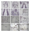
Figure 3. RNPC3 expression in the developing hypothalamic-pituitary axis during human embryogenesis.
In situ hybridization using the antisense probe against the human RNPC3 mRNA transcript (hRNPC3) on human sections from different developmental stages during embryogenesis. (A-D) CS19: high hRNPC3 expression is seen in the telencephalon, diencephalon, trigeminal ganglia and Rathke’s pouch. (E) CS19: expression can be seen in the spinal cord and spinal ganglia. (F-I) 9 post conception week (pcw): hRNPC3 mRNA transcripts are present in the mesonephros; the ducts that will develop into the kidney, indicated by the labelled arrows. The boxes in F correspond to G and H respectively. (I-J) 9 pcw: expression is noted in the presumptive developing fallopian tube. The box in I corresponds to J. (K-L) 10 pcw: hRNPC3 expression is seen in the vertebrae and lamina of the vertebrae, indicated by the labelled arrows. T, telencephalon; D, diencephalon; TG, trigeminal ganglia; RP, Rathke’s \pouch; Hyp, hypothalamus; S, spinal cord; SG, spinal ganglia, M, mesonephros; FT, fallopian tube; V, vertebrae; LV, lamina of the vertebrae.

References

    1. Le Quesne Stabej P, Williams HJ, James C, et al. STAG3 truncating variant as the cause of primary ovarian insufficiency. Eur J Hum Genet. 2016;24:135–138. - PMC - PubMed
    1. Jiao X, Ke H, Qin Y, Chen ZJ. Molecular Genetics of Premature Ovarian Insufficiency. Trends Endocrinol Metab. 2018;29:795–07. - PubMed
    1. Beysen D, De Paepe A, De Baere E. FOXL2 mutations and genomic rearrangements in BPES. HumMutat. 2009;30:158–169. - PubMed
    1. Jenkinson EM, Rehman AU, Walsh T, et al. Perrault syndrome is caused by recessive mutations in CLPP, encoding a mitochondrial ATP-dependent chambered protease. Am J Hum Genet. 2013;92:605–613. - PMC - PubMed
    1. Caburet S, Arboleda VA, Llano E, et al. Mutant cohesin in premature ovarian failure. N Engl J Med. 2014;370:943–949. - PMC - PubMed

Publication types