

Differences in clinical and imaging presentation of maxillary sinus fungus ball with and without intralesional hyperdensity
- PMID: 34907314
- PMCID: PMC8671531
- DOI: 10.1038/s41598-021-03507-1
Differences in clinical and imaging presentation of maxillary sinus fungus ball with and without intralesional hyperdensity
Abstract
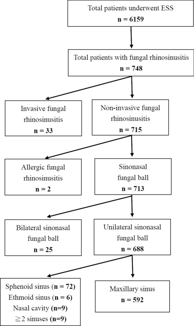
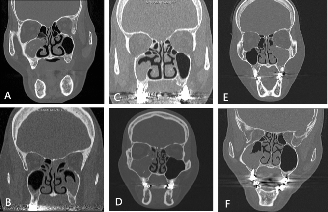
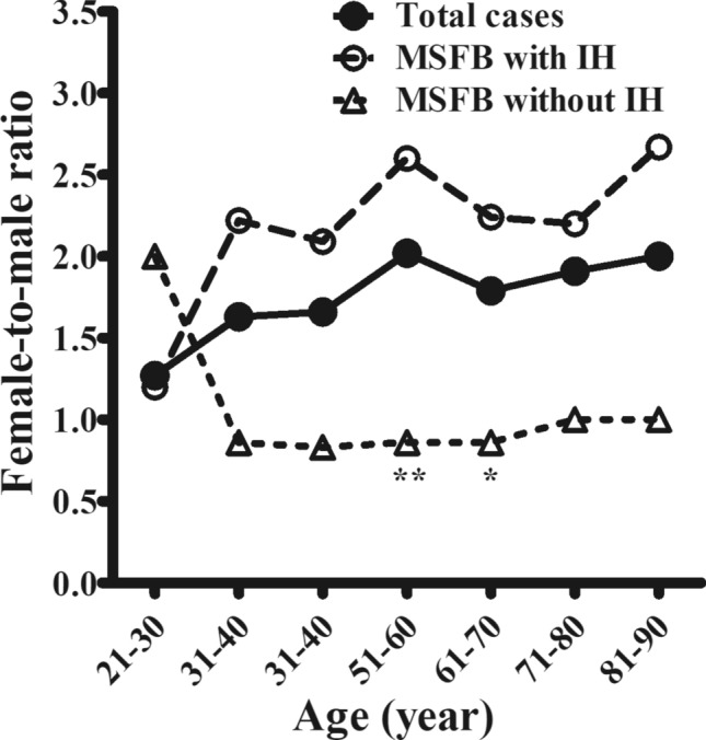
Maxillary sinus fungal balls (MSFBs) mostly occur in older individuals and demonstrate female predominance. Early diagnosis is important to avoid treatment delays. Intralesional hyperdensity (IH) indicates the presence of heavy metal deposition within fungal hyphae and has been the most specific characteristic of MSFB on computed tomography (CT). For those without IH on CT, the diagnosis of MSFB remains challenging. This study aimed to characterize clinical presentation of MSFB with and without IH and to study factors contributing to MSFB with no IH formation. We retrospectively identified 588 patients with MSFB. The clinical characteristics and CT findings were reviewed. Patients with unilateral MSFB had a mean age of 57.4 years and demonstrated female predominance (64.63%). The female-to-male ratio was highest at 51-60 years (2.02) and rose to 2.60 in MSFB with IH only. Compared to those with IH, MSFB without IH was significantly more common in males (OR = 2.49), in those with diabetes mellitus (DM) (OR = 1.87), adjacent maxillary odontogenic pathology (OR = 1.75). Complete opacification on CT was less common in MSFB without IH (OR = 0.60). Patients with MSFB without IH were more likely to have DM, no female predominance, adjacent maxillary odontogenic pathology, and partial opacification of the sinus, compared to those with IH. These may be helpful in better understanding of the formation of MSFBs without IH, early identification of them and prevention of post-operative recurrence.
© 2021. The Author(s).
Conflict of interest statement
The authors declare no competing interests.
Figures
Similar articles
-
Predicting the Probability of the Incidence of Maxillary Sinus Fungus Ball in Patients Using Nomogram Models.Diagnostics (Basel). 2023 Oct 9;13(19):3156. doi: 10.3390/diagnostics13193156. Diagnostics (Basel). 2023. PMID: 37835900 Free PMC article.
-
Diagnosis of a maxillary sinus fungus ball without intralesional hyperdensity on computed tomography.Laryngoscope. 2019 May;129(5):1041-1045. doi: 10.1002/lary.27670. Epub 2018 Dec 24. Laryngoscope. 2019. PMID: 30582161
-
Analysis of clinical characteristics of fungus ball based on the number of involved sinuses.Rhinology. 2025 Apr 1;63(2):230-236. doi: 10.4193/Rhin23.501. Rhinology. 2025. PMID: 39982171
-
Anatomical Variations Associated With Maxillary Sinus Fungal Ball.Ear Nose Throat J. 2023 Nov;102(11):727-732. doi: 10.1177/01455613211028470. Epub 2021 Jun 28. Ear Nose Throat J. 2023. PMID: 34182819 Review.
-
Paranasal sinus fungus ball: diagnosis and management.Mycoses. 2007 Nov;50(6):451-6. doi: 10.1111/j.1439-0507.2007.01416.x. Mycoses. 2007. PMID: 17944705 Review.
Cited by
-
Predicting the Probability of the Incidence of Maxillary Sinus Fungus Ball in Patients Using Nomogram Models.Diagnostics (Basel). 2023 Oct 9;13(19):3156. doi: 10.3390/diagnostics13193156. Diagnostics (Basel). 2023. PMID: 37835900 Free PMC article.
-
Acremonium strictum Sphenoid Sinusitis in an Immunocompetent Patient.Am J Case Rep. 2025 Apr 8;26:e946501. doi: 10.12659/AJCR.946501. Am J Case Rep. 2025. PMID: 40195081 Free PMC article.
-
A clinical comparative study of an outpatient treatment group and an endoscopic sinus surgery group for maxillary sinus fungus ball.Eur Arch Otorhinolaryngol. 2025 Jan;282(1):225-233. doi: 10.1007/s00405-024-08994-2. Epub 2024 Oct 2. Eur Arch Otorhinolaryngol. 2025. PMID: 39356354
-
Unprecedented Finding of Isolated Sphenoid Fungal Ball in a Child: A Case Report.Case Rep Otolaryngol. 2025 Jul 14;2025:4404155. doi: 10.1155/crot/4404155. eCollection 2025. Case Rep Otolaryngol. 2025. PMID: 40692643 Free PMC article.
-
Identifying the Risk Factors for Orbital Complications in Isolated Sphenoid Rhinosinusitis.Medicina (Kaunas). 2024 Jan 10;60(1):128. doi: 10.3390/medicina60010128. Medicina (Kaunas). 2024. PMID: 38256389 Free PMC article.
