. 2022 Mar 31;171(4):399-410.
doi: 10.1093/jb/mvab141.

MAB21L4 regulates the TGF-β-induced expression of target genes in epidermal keratinocytes

Affiliations

MAB21L4 regulates the TGF-β-induced expression of target genes in epidermal keratinocytes

Tomohiro Ogami et al. J Biochem. .

Erratum in

  • Erratum.
    [No authors listed] [No authors listed] J Biochem. 2022 Mar 31;171(4):469. doi: 10.1093/jb/mvac016. J Biochem. 2022. PMID: 35181785 Free PMC article. No abstract available.

Abstract

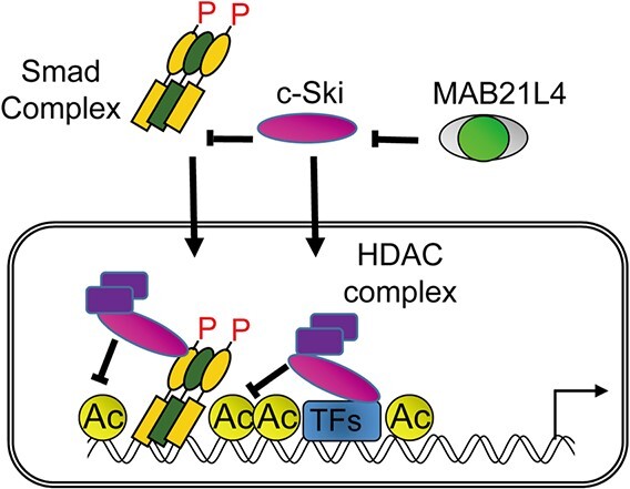
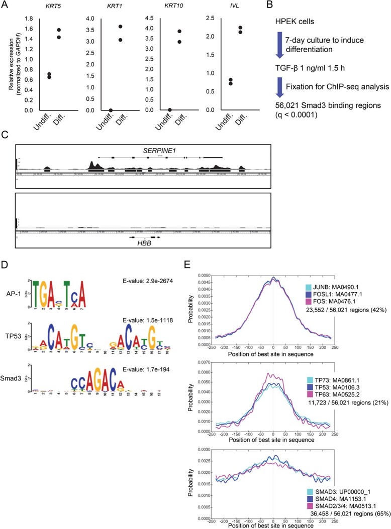
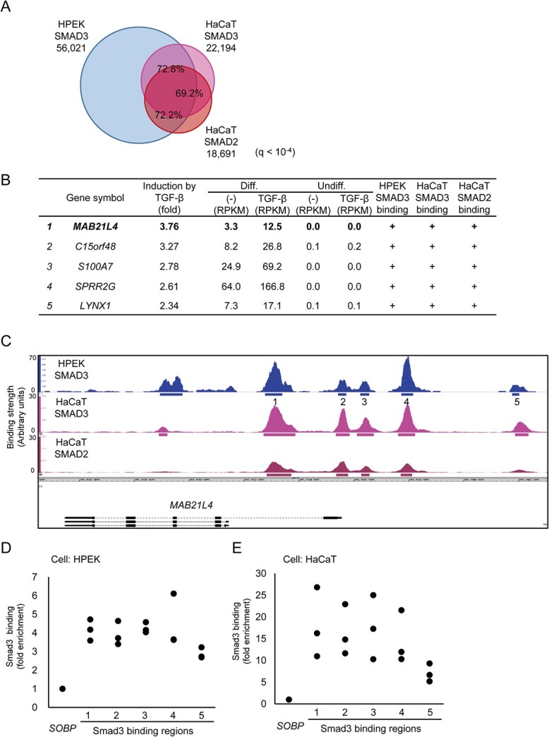
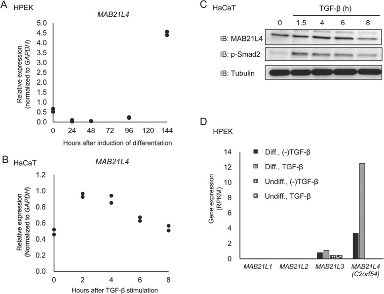
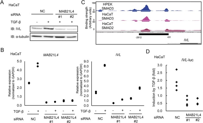
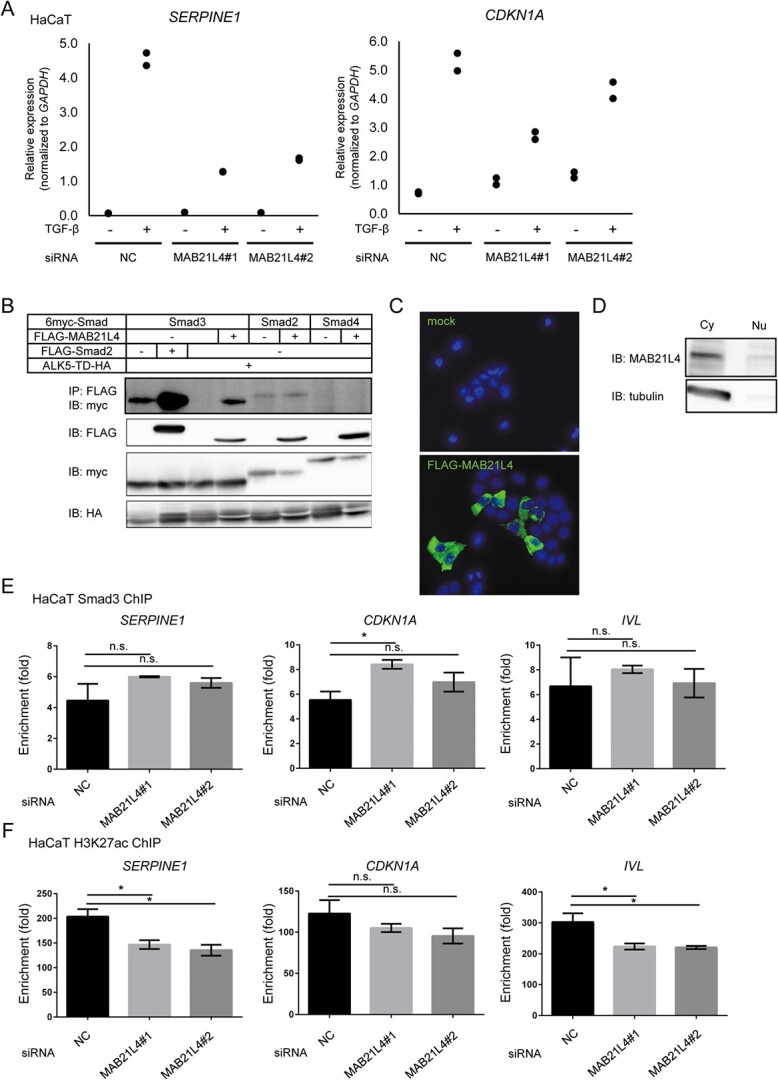
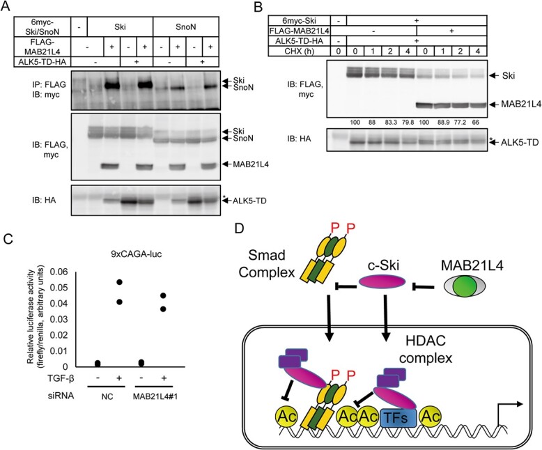
Smad proteins transduce signals downstream of transforming growth factor-β (TGF-β) and are one of the factors that regulate the expression of genes related to diseases affecting the skin. In the present study, we identified MAB21L4, also known as male abnormal 21 like 4 or C2orf54, as the most up-regulated targets of TGF-β and Smad3 in differentiated human progenitor epidermal keratinocytes using chromatin immunoprecipitation sequencing (ChIP-seq) and RNA sequencing (RNA-seq). We found that TGF-β induced expression of the barrier protein involucrin (encoded by the IVL gene). Transcriptional activity of the IVL promoter induced by TGF-β was inhibited by MAB21L4 siRNAs. Further analysis revealed that MAB21L4 siRNAs also down-regulated the expression of several target genes of TGF-β. MAB21L4 protein was located mainly in the cytosol, where it was physically bound to Smad3 and a transcriptional corepressor c-Ski. siRNAs for MAB21L4 did not inhibit the binding of Smad3 to their target genomic regions but down-regulated the acetylation of histone H3 lys 27 (H3K27ac), an active histone mark, near the Smad3 binding regions. These findings suggest that TGF-β-induced MAB21L4 up-regulates the gene expression induced by TGF-β, possibly through the inhibition of c-Ski via physical interaction in the cytosol.

Keywords: ChIP-seq; Smad3; TGF-β; c-Ski; epidermal keratinocytes.


Figures

Graphical Abstract
Graphical Abstract
Fig. 1
Fig. 1
Smad3 binding regions in epidermal keratinocytes. (A) Expression of epidermal differentiation markers in HPEK cells. Total RNA was isolated after the induction of differentiation of HPEK cells for 6 days, and the expression of keratinocyte differentiation markers was compared to that of undifferentiated progenitor cells using qRT-PCR. Data were obtained from two biological replicates. (B) Experimental design of Smad3 ChIP-seq analysis of differentiated HPEK cells. Cells were cultured in differentiation medium for 7 days and then stimulated with TGF-β for 1.5 h before fixation. Smad3 binding regions were determined by ChIP and ChIP-seq. (C) Smad3 binding signals at the known target of TGF-β. Peak signals at the SERPINE1 locus show enrichment of Smad3 binding signals, and black bars show significant binding regions at q < 0.0001. The HBB locus is shown as a negative control. (D) The top enriched motif groups, AP-1 and TP53, according to CentriMo. Smad binding motifs were also identified. (E) Relative position of the enriched motifs alongside the peak summit of the Smad3 binding regions. The top 3 motifs included in each motif group are shown. Percentages show the frequency of motifs in the Smad3 binding regions. Undiff., undifferentiated cells; Diff., differentiated cells.
Fig. 2
Fig. 2
Identification of MAB21L4 as a target of Smad3. (A) Comparison of the Smad3 and Smad2 binding regions in HPEK and HaCaT cells. ChIP-seq data of HaCaT cells stimulated with 1 ng/ml of TGF-β3 for 1.5 h were obtained for comparison to the data obtained in Fig. 1. Significant binding regions were determined at q < 10−4 and overlap analysis was performed by using bedtools (https://bedtools.readthedocs.io/en/latest/). (B) MAB21L4 was identified as the most up-regulated target gene of TGF-β and Smad3 in differentiated HPEK cells. Genes with expression levels of at least 10 FPKM in any of the differentiated HPEK samples were included in the analysis. The data were then sorted by the order of induction by TGF-β. Genes exclusively expressed in the differentiated HPEK cells (FPKM < 1 in undifferentiated HPEK cells) are shown. ‘Smad3 binding’ is defined as significant Smad3 binding within 100 kb from the gene. (C) Smad3 and Smad2 binding regions at the MAB21L4 locus are shown. There are three variants (a longer form and lower two shorter forms) of MAB21L4. Numbers show the regions analysed by ChIP-qPCR in (D) and (E). (D) ChIP-qPCR analysis of Smad3 binding at the MAB21L4 locus. Differentiated HPEK cells were treated with TGF-β for 1.5 h and fixed. The SOBP locus was used as the negative control region (9). Data were obtained from three biological replicates. (E) ChIP-qPCR analysis of Smad3 binding in an epidermal keratinocyte cell line HaCaT. ChIP was performed as in (D). Data were obtained from three biological replicates.
Fig. 3
Fig. 3
Regulation of MAB21L4 expression. (A) MAB21L4 is expressed after a delay following the induction of differentiation in HPEK cells. Total RNA was serially isolated after the induction of differentiation of HPEK cells as indicated, and the expression of MAB21L4 was determined by qRT-PCR. Data were obtained from two biological replicates. (B) Up-regulation of MAB21L4 mRNA by TGF-β in HaCaT cells. Cells were stimulated with TGF-β as indicated. Data were obtained from two biological replicates. (C) The change in the amount of MAB21L4 protein after TGF-β stimulation in HaCaT cells. The experiment was repeated with the similar results, and the representative data are shown. (D) Expression of MAB21-family mRNAs in HaCaT cells. RPKM values of each mRNA were obtained from the RNA-seq data used in Supplementary Fig. S1. IB, immunoblotting.
Fig. 4
Fig. 4
MAB21L4 up-regulates involucrin expression. (A) Up-regulation of involucrin protein induced by TGF-β was abrogated by MAB21L4 siRNA. HaCaT cells were transfected with two different MAB21L4 siRNAs and stimulated with TGF-β for 24 h. The experiment was repeated with the similar results, and the representative data are shown. (B) The effect of MAB21L4 siRNA on the expression of MAB21L4 mRNA in HaCaT cells. HaCaT cells were transfected with MAB21L4 siRNAs and stimulated with TGF-β for 24 h. Data were obtained from two biological replicates. (C) Smad3 and Smad2 binding regions at the promoter of the IVL locus. Significant binding regions are represented as in Fig. 2C. The region (−2812 to +81 from the TSS) used in the promoter-reporter analysis shown in (D) is also indicated in the bottom panel. (D) MAB21L4 siRNAs down-regulated TGF-β-induced transcriptional activity of the IVL promoter. HaCaT cells were transfected with luciferase reporter plasmids and siRNA as indicated. Cells were then stimulated with TGF-β or left untreated. Data were obtained from four biological replicates.
Fig. 5
Fig. 5
MAB21L4 up-regulates TGF-β target genes. (A) The effect of MAB21L4 siRNA on the TGF-β-induced expression of SERPINE1 and CDKN1A in HaCaT cells. The same samples shown in Fig. 4B were used for this evaluation. (B) MAB21L4 protein physically binds to Smad proteins; 293T cells were transfected with the expression plasmids as indicated and lysed for co-immunoprecipitation analysis. The experiment was repeated with the similar results and the representative data are shown. (C) Cytoplasmic localization of FLAG-MAB21L4 protein in transfected HaCaT cells. Cells were transfected with the plasmids encoding FLAG-tagged MAB21L4 or empty vector. Data were obtained from the two microscopic fields, and the representative images are shown. (D) Cell fractionation assay showing the localization of endogenous MAB21L4 protein. HaCaT cells were lysed with NE-PER (Thermo Fisher Scientific) to obtain the cytoplasmic and nuclear fractions. The experiment was repeated with similar results, and the representative data are shown. (E) Effect of knockdown of MAB21L4 on the Smad3 binding to the target genomic regions. HaCaT cells transfected with siRNA as indicated were stimulated with TGF-β for 1.5 h and harvested for ChIP-qPCR analysis. Data represents the mean ± standard deviation of two biological replicates. (F) Down-regulation of H3K27ac by MAB21L4 siRNAs. HaCaT cells were transfected with the siRNAs as indicated and were stimulated with TGF-β for 1.5 h. Data represents the mean ± standard deviation of two biological replicates. Note that the most enriched genomic positions of Smad3 and H3K27ac appeared different at the IVL locus and the different primers were used between Fig. 5E and F, as shown in Supplementary Table S2. NC, control siRNA; IP, immunoprecipitation; IB, immunoblotting; ALK5-TD, constitutively active ALK5; n.s.: not significant. *P < 0.05 by Dunnett test.
Fig. 6
Fig. 6
MAB21L4 physically interacts with c-Ski and SnoN proteins. (A) Co-immunoprecipitation analysis showing the physical interaction of c-Ski and SnoN with MAB21L4 protein; 293T cells were transfected as indicated. 6myc-tagged c-Ski and SnoN proteins co-immunoprecipitated with anti-FLAG antibody was detected in the top panel. The blotted membrane was serially used for detection of different antigens and thus anti-HA image shows residual anti-FLAG and anti-myc bands shown as *. (B) Effect of co-expression of MAB21L4 protein on the turnover of c-Ski protein; 293T cells were transfected with the expression plasmids as indicated. Cells were then treated with CHX at 100 μg/ml for the indicated times before cell lysis. Amounts of the c-Ski protein relative to the samples without CHX treatment (%) are shown below the top panel. (C) Effect of MAB21L4 siRNA on Smad-specific transcriptional activity. HaCaT cells were transfected with a luciferase reporter plasmid and siRNA. Cells were then treated with TGF-β. Data were obtained from two biological replicates. (D) A model of mechanism of enhanced target gene expression by MAB21L4 (also shown in Graphical Abstract). MAB21L4 protein physically binds to c-Ski in the cytosol to inhibit the known repressor function of c-Ski, either inhibition of the nuclear translocation of Smad complex or recruitment of HDAC complex on the target genomic regions. We cannot exclude the possibility that c-Ski inhibits the transcription via other TFs, independent of Smad complex. P, phosphorylation of Smad proteins; Ac, acetylation of histone H3, specifically lys 27, shown in this study. *, residual anti-FLAG bands as in Fig. 6A.

References

    1. Candi, E., Schmidt, R., and Melino, G. (2005) The cornified envelope: a model of cell death in the skin. Nat. Rev. Mol. Cell Biol. 6, 328–340 - PubMed
    1. Kahata, K., Dadras, M.S., and Moustakas, A. (2018) TGF-beta family signaling in epithelial differentiation and epithelial-mesenchymal transition. Cold Spring Harb. Perspect. Biol. 10, a022194 - PMC - PubMed
    1. Boukamp, P., Petrussevska, R.T., Breitkreutz, D., Hornung, J., Markham, A., and Fusenig, N.E. (1988) Normal keratinization in a spontaneously immortalized aneuploid human keratinocyte cell line. J. Cell Biol. 106, 761–771 - PMC - PubMed
    1. Koinuma, D., Shinozaki, M., Komuro, A., Goto, K., Saitoh, M., Hanyu, A., Ebina, M., Nukiwa, T., Miyazawa, K., Imamura, T., and Miyazono, K. (2003) Arkadia amplifies TGF-beta superfamily signalling through degradation of Smad7. EMBO J. 22, 6458–6470 - PMC - PubMed
    1. Nagano, Y., Mavrakis, K.J., Lee, K.L., Fujii, T., Koinuma, D., Sase, H., Yuki, K., Isogaya, K., Saitoh, M., Imamura, T., Episkopou, V., Miyazono, K., and Miyazawa, K. (2007) Arkadia induces degradation of SnoN and c-Ski to enhance transforming growth factor-beta signaling. J. Biol. Chem. 282, 20492–20501 - PubMed

MeSH terms

Substances