

Genetic defects are common in myopathies with tubular aggregates
- PMID: 34908252
- PMCID: PMC8791796
- DOI: 10.1002/acn3.51477
Genetic defects are common in myopathies with tubular aggregates
Abstract
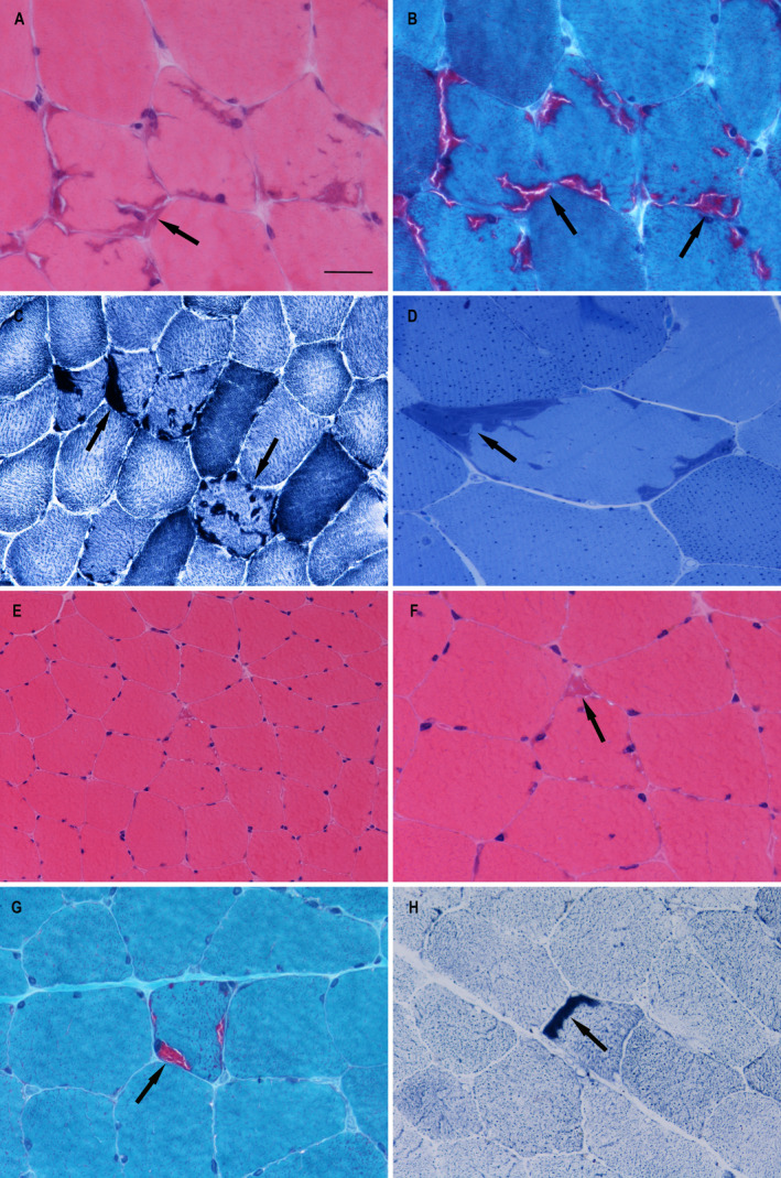
Objective: A group of genes have been reported to be associated with myopathies with tubular aggregates (TAs). Many cases with TAs still lack of genetic clarification. This study aims to explore the genetic background of cases with TAs in order to improve our knowledge of the pathogenesis of these rare pathological structures.
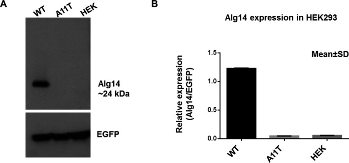
Methods: Thirty-three patients including two family members with biopsy confirmed TAs were collected. Whole-exome sequencing was performed on 31 unrelated index patients and a candidate gene search strategy was conducted. The identified variants were confirmed by Sanger sequencing. The wild-type and the mutant p.Ala11Thr of ALG14 were transfected into human embryonic kidney 293 cells (HEK293), and western blot analysis was performed to quantify protein expression levels.
Results: Eleven index cases (33%) were found to have pathogenic variant or likely pathogenic variants in STIM1, ORAI1, PGAM2, SCN4A, CASQ1 and ALG14. Among them, the c.764A>T (p.Glu255Val) in STIM1 and the c.1333G>C (p.Val445Leu) in SCN4A were novel. Western blot analysis showed that the expression of ALG14 protein was severely reduced in the mutant ALG14 HEK293 cells (p.Ala11Thr) compared with wild type. The ALG14 variants might be associated with TAs in patients with complex multisystem disorders.
Interpretation: This study expands the phenotypic and genotypic spectrums of myopathies with TAs. Our findings further confirm previous hypothesis that genes related with calcium signalling pathway and N-linked glycosylation pathway are the main genetic causes of myopathies with TAs.
© 2021 The Authors. Annals of Clinical and Translational Neurology published by Wiley Periodicals LLC on behalf of American Neurological Association.
Conflict of interest statement
All authors have no competing financial interests.
Figures


References
-
- Engel WK. Mitochondrial aggregates in muscle disease. J Histochem Cytochem. 1964;12(1):46‐48. [published Online First: 1964/01/01] - PubMed
-
- Goebel HH, Sharma MC, Taratuto AL, et al. Disorders of muscle with rare structural abnormalities. In: Goebel HH, Sewry C, Weller RO, eds. Muscle Disease: Pathology and Genetics, 2nd ed. UK: Wiley‐Blackwell; 2013:351‐360.
-
- Chevessier F, Marty I, Paturneau‐Jouas M, et al. Tubular aggregates are from whole sarcoplasmic reticulum origin: alterations in calcium binding protein expression in mouse skeletal muscle during aging. Neuromuscul Disord. 2004;14(3):208‐216. doi:10.1016/j.nmd.2003.11.007 [published Online First: 2004/03/24]. - DOI - PubMed
-
- Pavlovicova M, Novotova M, Zahradnik I. Structure and composition of tubular aggregates of skeletal muscle fibres. Gen Physiol Biophys. 2003;22(4):425‐440. [published Online First: 2004/04/29] - PubMed
Publication types
MeSH terms
Grants and funding
LinkOut - more resources
Full Text Sources
Medical
Miscellaneous
