. 2021 Dec 23;64(24):18025-18053.
doi: 10.1021/acs.jmedchem.1c01382. Epub 2021 Dec 15.

Discovery of Novel Dual-Target Inhibitor of Bromodomain-Containing Protein 4/Casein Kinase 2 Inducing Apoptosis and Autophagy-Associated Cell Death for Triple-Negative Breast Cancer Therapy

Affiliations

Discovery of Novel Dual-Target Inhibitor of Bromodomain-Containing Protein 4/Casein Kinase 2 Inducing Apoptosis and Autophagy-Associated Cell Death for Triple-Negative Breast Cancer Therapy

Jifa Zhang et al. J Med Chem. .

Abstract

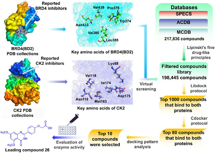
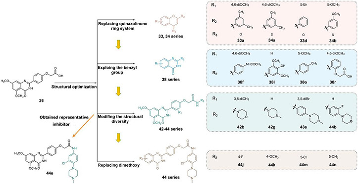
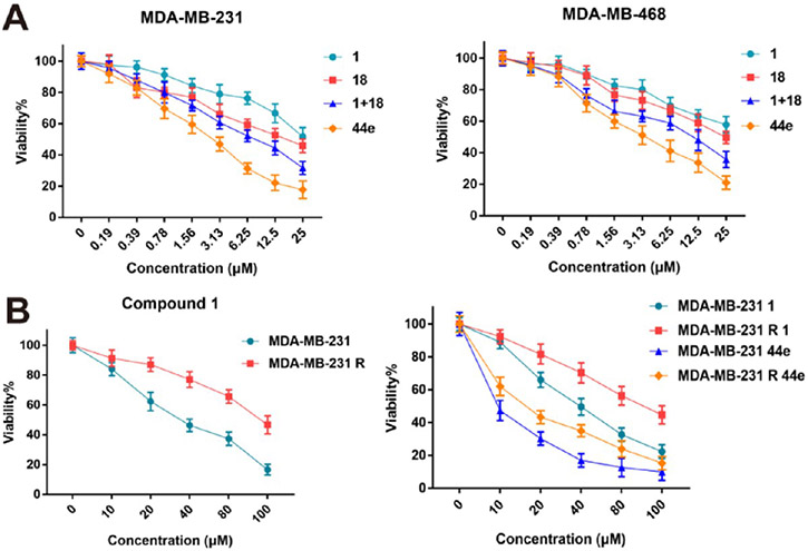
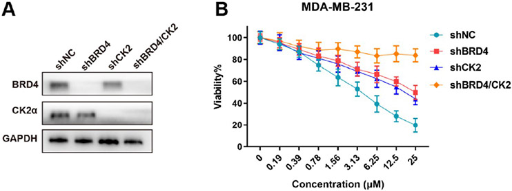
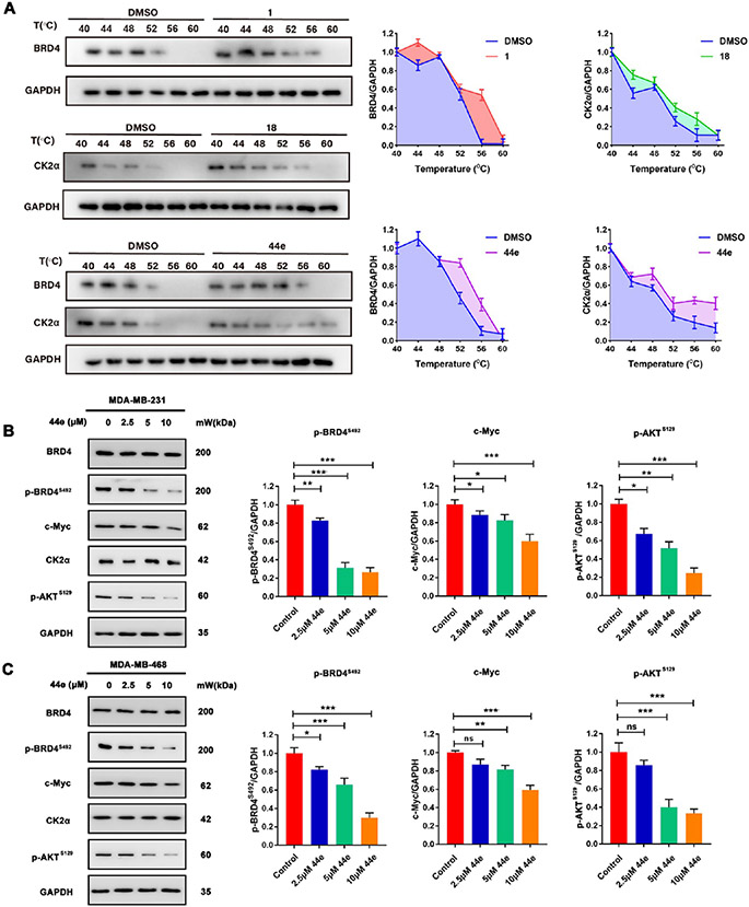
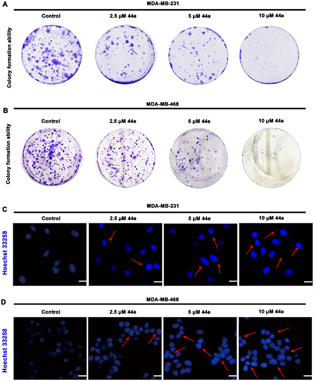
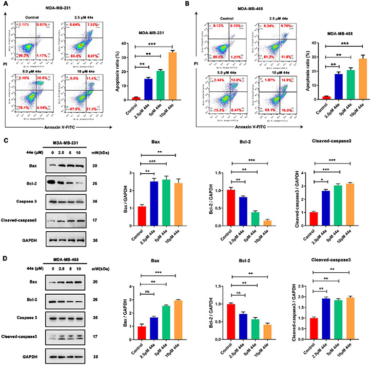
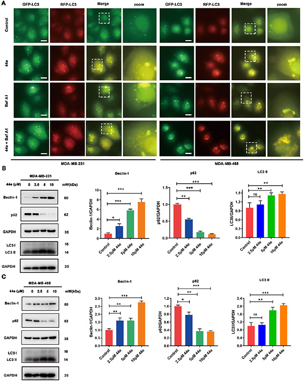
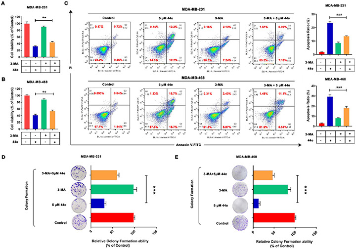
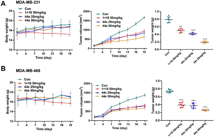
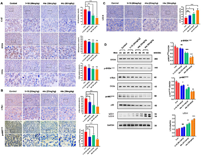
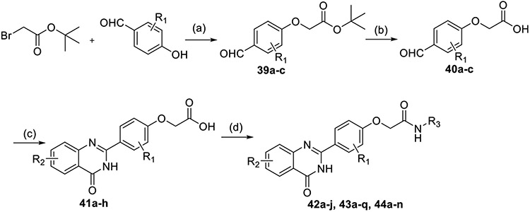
Bromodomain-containing protein 4 (BRD4) is an attractive epigenetic target in human cancers. Inhibiting the phosphorylation of BRD4 by casein kinase 2 (CK2) is a potential strategy to overcome drug resistance in cancer therapy. The present study describes the synthesis of multiple BRD4-CK2 dual inhibitors based on rational drug design, structure-activity relationship, and in vitro and in vivo evaluations, and 44e was identified to possess potent and balanced activities against BRD4 (IC50 = 180 nM) and CK2 (IC50 = 230 nM). In vitro experiments show that 44e could inhibit the proliferation and induce apoptosis and autophagy-associated cell death of MDA-MB-231 and MDA-MB-468 cells. In two in vivo xenograft mouse models, 44e displays potent anticancer activity without obvious toxicities. Taken together, we successfully synthesized the first highly effective BRD4-CK2 dual inhibitor, which is expected to be an attractive therapeutic strategy for triple-negative breast cancer (TNBC).


Figures

Figure 1.
Figure 1.
Structures representing known BRD4 and CK2 inhibitors. (A) Representative published BRD4 inhibitors with diverse scaffolds. (B) Representative published CK2 inhibitors with diverse scaffolds.
Figure 2.
Figure 2.
Bioinformatics analysis. (A) Workflow of bioinformatics analysis of BRD4–CK2 association. (B) Clustering of BRD4 and CK2 interacting protein networks related to cell autophagy and apoptosis. (C) Predicted BRD4 and CK2 related proteins involved in the regulation of cell apoptosis and autophagy-associated cell death. (D) Correlation between BRD4 and CK2 in BC.
Figure 3.
Figure 3.
Workflow of designing BRD4–CK2 inhibitors based on extracted amino acid residues and virtual screening.
Figure 4.
Figure 4.
Binding mode analysis of 26, 42g, and 44e. (A, B) Docking poses show the interaction of 26, 42g, and 44e with BRD4 (BD2) (PDB ID 5UOO) and CK2 (PDB ID 6RFE), respectively. Oxygen atoms are colored in red and nitrogen atoms in blue.
Figure 5.
Figure 5.
Structural optimization and discovery of BRD4–CK2 dual-target inhibitors. Representative compounds are shown.
Figure 6.
Figure 6.
Antiproliferative activity of candidate compound 44e. (A) Antiproliferative activity of 1, 18, 1 + 18, and 44e toward MDA-MB-231 and MDA-MB-468 cells. (B) Antiproliferative activity of 44e toward compound 1-resistant MDA-MB-231R cells.
Figure 7.
Figure 7.
Antiproliferative activity of candidate compound 44e relies on BRD4 and CK2. (A) BRD4, CK2, or BRD4/CK2 knockdown efficiency in MDA-MB-231 cells as determined by Western blots. GAPDH was used as a control. (B) MDA-MB-231 cell viability after 24 h at different concentrations of 44e.
Figure 8.
Figure 8.
Western blotting assay. (A) CETSA assay detected the thermal stability of BRD4 and CK2 in MDA-MB-231 cells treated with 1, 18, and 44e. (B, C) Expression of BRD4, p-BRD4S492, c-Myc, CK2α, and p-AKTS129 in MDA-MB-231 and MDA-MB-468 cells treated with 0, 2.5, 5, and 10 μM of 44e for 24 h. Error bar shows SD; *p < 0.05, **p < 0.01, ***p < 0.001, ****p < 0.0001 compared with the control groups.
Figure 9.
Figure 9.
Compound 44e inhibits colony formation and promotes apoptosis in TNBC cells. (A, B) Colony formation assay. MDA-MB-231 and MDA-MB-468 cells were treated with 0, 2.5, 5, and 10 μM of 44e for 7 days. (C, D) Hoechst 33258 staining assay. MDA-MB-231 and MDA-MB-468 cells treated with 0, 2.5, 5, and 10 μM of 44e for 24 h. Scale bar, 10 μm.
Figure 10.
Figure 10.
Apoptotic effect of 44e on TNBC cells. (A, B) MDA-MB-231 and MDA-MB-468 cells were treated with indicated concentrations of 44e for 24 h. Apoptosis ratios were determined by flow cytometry analysis of Annexin-V/PI double staining. A representative image and quantification of apoptosis are presented. (C, D) Cells were treated with 44e at different concentrations for 24 h. The levels of apoptosis-related proteins, including Bcl-2, Bax, caspase-3, and cleaved caspase-3, were analyzed by Western blotting. GAPDH was used as load control. Error bar shows SD; *p < 0.05, **p < 0.01, ***p < 0.001, ****p < 0.0001 compared with the control groups.
Figure 11.
Figure 11.
Compound 44e induces regulated autophagy in TNBC cells. (A) MDA-MB-231 and MDA-MB-468 cells were transfected with GFP-mRFP-LC3 adenovirus, following co-incubation with 44e (5 μM) in the presence or absence of BafA1 (10 nM). Scale bar, 10 μm. (B, C) Expression levels of Beclin-1, LC3, and p62 in MDA-MB-231 and MDA-MB-468 cells treated with 5 μM 44e, analyzed by Western blotting. GAPDH was used as load control. Error bar shows SD; *p < 0.05, **p < 0.01, ***p < 0.001, ****p < 0.0001 compared with the control groups.
Figure 12.
Figure 12.
Mechanism of autophagy in 44e-induced cell death in TNBC cells. (A, B) MDA-MB-231 and MDA-MB-468 cell viability was measured by MTT assay. Cells were treated with 44e at 5 μM or combined with 3-MA at 1 mM treated for 24 h. 3-MA was added 1 h before treatment with 44e. (C) MDA-MB-231 and MDA-MB-468 cell apoptosis ratios were analyzed by flow cytometry with Annexin-V/PI double staining. Cells were treated with 44e at 5 μM or combined with 3-MA at 1 mM for 24 h. 3-MA was added 1 h before treatment with 44e. (D, E) Colony formation assay in MDA-MB-231 and MDA-MB-468 cells. Cells treated with 44e at 5 μM or combined with 3-MA at 1 mM. Error bar shows SD; *p < 0.05, **p < 0.01, ***p < 0.001 compared with the control groups.
Figure 13.
Figure 13.
Antitumor activity of 44e in the MDA-MB-231 and MDA-MB-468 xenograft models. (A) Body weights, tumor volumes, and tumor weights following treatment by oral administration with 25 or 50 mg/kg 44e or 50 mg/kg 1 + 18 in the MDA-MB-231 tumor xenograft model. (B) Body weights, tumor volumes, and tumor weights following the treatment by oral administration with 25 or 50 mg/kg 44e or 50 mg/kg 1 + 18 in the MDA-MB-468 tumor xenograft model. Error bar shows SD; *p < 0.05, **p < 0.01, ***p < 0.001 compared with the control groups.
Figure 14.
Figure 14.
Potential therapeutic effects of 44e in vivo. (A–C) Representative images of IHC analysis of Ki-67, BRD4, c-Myc, CK2α, p-AKTS129, and LC3II markers in different groups from MDA-MB-231 xenograft tumor model. Scale bar, 40 μm. (D) Two individual tumor tissues (1 and 2) excised from the MDA-MB-231 xenograft tumor model were analyzed. The expression levels of BRD4, c-Myc, CK2α, p-AKTS129, p62, and LC3II were detected by Western blot analysis. Error bar shows SD; *p < 0.05, **p < 0.01, ***p < 0.001, ****p < 0.0001 compared with the control groups.
Scheme 1<sup>a</sup>
Scheme 1a
aReagents and conditions: (a) pyridine, DBU, 80 °C, 1.5 h; (b) pyridine, KOH, 50–80 °C, 2 h; (c) HOAc, 1% H2SO4, 90–110 °C, 2 h; (d) dry toluene, Lawesson’s reagent, 110 °C, 4 h.
Scheme 2<sup>a</sup>
Scheme 2a
aReagents and conditions: (a) HCl/Et2O, rt, overnight; (b) (COCl)2, 160 °C, 2.5 h; (c) NaOH, H2O2, 80 °C, 0.5 h; (d) ammonium hydroxide, HOBt, EDCI, NMM, THF, overnight, rt; (e) benzaldehyde derivatives, DMAC, PTSA, NaHSO3, 120 °C, 4–8 h.
Scheme 3<sup>a</sup>
Scheme 3a
aReagents and conditions: (a) K2CO3, DMF, 80 °C, 3 h; (b) DCM, TFA, rt, 2 h; (c) DMAC, PTSA, NaHSO3, 120 °C, 4–8 h; (d) DMF, Et3N, HOBt, EDCI, rt, 24 h.

References

    1. Siegel RL; Miller KD; Jemal A Cancer statistics, 2020. CA-Cancer. Ca-Cancer J. Clin 2020, 70, 7–30. - PubMed
    1. Xu T; Zhang JF; Yang CC; Pluta R; Wang G; Ye TH; Ouyang L Identifification and optimization of 3-bromo-N’-(4-hydroxybenzylidene)-4-methylbenzohydrazide derivatives as mTOR inhibitors that induce autophagic cell death and apoptosis in triplenegative breast cancer. Eur. J. Med. Chem 2021, 219, 113424. - PubMed
    1. Franzoi MA; Romano E; Piccart M Immunotherapy for early breast cancer: too soon, too superficial, or just right? Ann. Oncol 2021, 32, 323–336. - PubMed
    1. Vagia E; Mahalingam D; Cristofanilli M The landscape of targeted therapies in TNBC. Cancers 2020, 12, 916. - PMC - PubMed
    1. Tang P; Zhang JF; Liu J; Chiang CM; Ouyang L Targeting bromodomain and extraterminal proteins for drug discovery: from current progress to technological development. J. Med. Chem 2021, 64, 2419–2435. - PMC - PubMed

Publication types

MeSH terms

LinkOut - more resources