. 2021 Oct 8;2(4):798-809.
doi: 10.1002/mco2.92. eCollection 2021 Dec.

SARS-CoV-2 spike protein harnesses SNX27-mediated endocytic recycling pathway

Affiliations

SARS-CoV-2 spike protein harnesses SNX27-mediated endocytic recycling pathway

Lin Zhao et al. MedComm (2020). .

Abstract

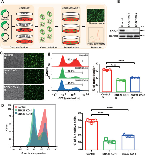
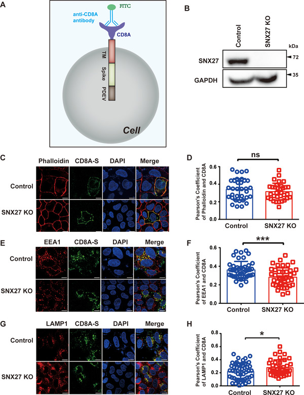
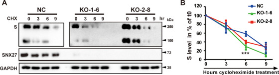
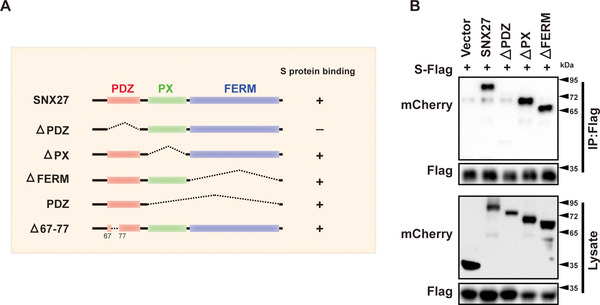
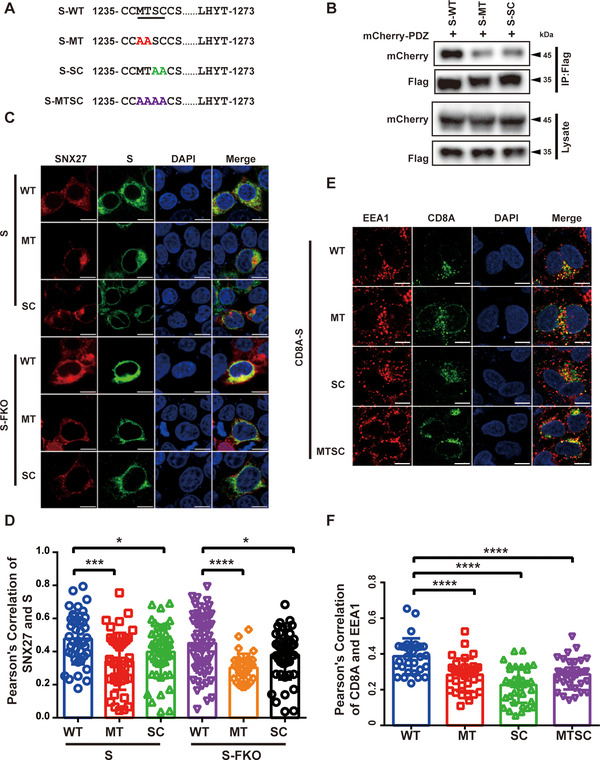
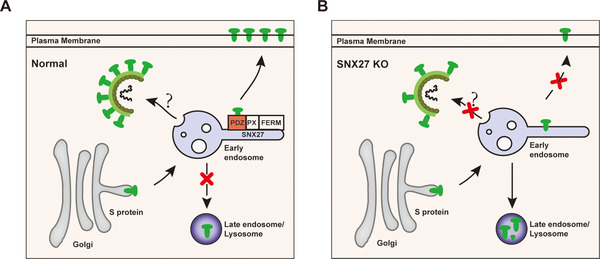
SARS-CoV-2 is an enveloped positive-sense RNA virus that depends on host factors for all stages of its life. Membrane receptor ACE2 is a well-established factor for SARS-CoV-2 docking. In addition to ACE2, whole-genome genetic screens have identified additional proteins, such as endosomal trafficking regulators SNX27 and retromer, as key host factors required for SARS-CoV-2 infection. However, it is poorly understood how SARS-CoV-2 utilize host endocytic transport pathways to produce productive infection. Here, we report that SNX27 interacts with the SARS-CoV-2 spike (S) protein to facilitate S protein surface expression. Interestingly, S protein binds to the PDZ domain of SNX27, although it does not contain a PDZ-binding motif (PDZbm). Either abrogation of the SNX27 PDZ domain or S protein "MTSC" motif, which is critical for SNX27 binding, decreases surface expression of S protein and viral production. Collectively, our study highlights a novel approach utilized by SARS-CoV-2 to facilitate virion trafficking to establish virus infection.

Keywords: S protein; SARS‐CoV‐2; endocytic trafficking; endosome; host–pathogen interaction.


Conflict of interest statement

The authors declare no conflict of interest.

Figures

FIGURE 1
FIGURE 1
SNX27 facilitates transduction efficiency of the S‐bearing pseudovirus and cell surface expression of S protein. (A) Workflow of the S‐bearing pseudovirus transduction efficiency analysis. SARS‐CoV‐2 S‐bearing pseudovirus was produced in WT or SNX27 KO HEK293T cells. Next, the virus‐containing supernatant was harvested and used to transduce ACE2‐expressing HEK293T cells (HEK293T‐ACE2). (B) Immunoblotting analysis of SNX27 protein expression in HEK293T cells transfected with CRISPR‐Cas9 plasmids targeting SNX27 (KO‐1 and KO‐2 employing two different gRNAs), or empty vector (control). (C) Fluorescence and flow cytometry analysis of transduction efficiency of the S‐bearing pseudoviruses produced from cells in b. Left: representative fluorescence images from one experiment. Middle: representative flow cytometry histograms from one experiment. Right: the percent of GFP positive cells, determined by flow cytometry, is used to calculate viral transduction efficiency. Data are mean ±SD of three independent experiments. ****p < 0.0001. Scale bar: 100 µm. (D) Determination of S protein cell surface expression by flow cytometry. HEK293T cells were transiently transfected with plasmids encoding full‐length S. Twenty‐four hours after transduction, cells were collected and stained with anti‐S antibody (which recognizes extracellular domain of S protein), followed by flow cytometer analysis. Left: flow cytometry results from one representative experiment. Right: percent of S‐positive cells. Statistic data represent the results from n = 3 independent experiments and are expressed as mean ± SD. ****p < 0.0001. Scale bar: 100 µm
FIGURE 2
FIGURE 2
SNX27 promotes endocytic recycling of S protein. (A) A cartoon of CD8A‐S chimera used in the experiment. The CD8A‐S chimera construct consists of CD8A (1–205 aa), a linker peptide, S protein (1235–1256 aa), and the PEDV S protein (1374–1387 aa). The PEDV S protein contains an adaptor protein complex 2 (AP‐2) recognition motif, which is used to facilitate endocytosis. (B) Depletion efficiency of SNX27 in HeLa cells, determined by immunoblotting. (C)–(H) Control, SNX27‐KO HeLa cells were transiently transfected with plasmids encoding CD8A‐S for 24 h. Cells were incubated with monoclonal anti‐CD8A antibody on ice for 30 min. Unbound antibodies were removed, and the internalization of antibody‐bound CD8A was chased in DMEM at 37˚C for 0 min (C, D), 30 min (E, F), and 60 min (G, H), respectively. The internalized CD8A–antibody was detected using Alexa‐488 secondary antibodies, with cell membrane stained with phalloidin (C, D), early endosome stained with EEA1 (E, F), lysosomes stained with LAMP1 (G, H). Scale bar: 10 µm. (D) Quantification of CD8A/phalloidin colocalization in cells in C. (F) Quantification of CD8A/EEA1 colocalization in cells in E. (H) Quantification of CD8A/LAMP1 colocalization in cells in G. Each dot represents Pearson's correlation coefficients from one cell. N = 3 independent experiments. p Values were calculated using one‐way ANOVA, Tukey's multiple comparisons test. Ns: no significant difference. *p < 0.05; ***p < 0.001
FIGURE 3
FIGURE 3
Depletion of SNX27 increases degradation of SARS‐CoV‐2 S protein. (A), (B) S protein degradation assays. Two clonal SNX27‐KO cell lines (KO‐1‐6 and KO‐2‐8) were selected from SNX27 KO‐1 and SNX27 KO‐2, respectively. Control and SNX27‐KO cells were transiently transfected with plasmid encoding S protein. Twenty‐four hours after transfection, cells were treated with cycloheximide (CHX, 25 µg/ml) for the indicated time periods. The amount of S protein was expressed relative to the amount of GAPDH (loading control) and then normalized to the sample at 0 h. Graph shows the degradation kinetics (B), with error bars indicating SD. ***< 0.001 comparison of control and SNX27 KO in a one‐way ANOVA test. Experiments were repeated four times
FIGURE 4
FIGURE 4
The PDZ domain of SNX27 is required for interaction with S protein. (A) Schematic representation of full‐length SNX27 and SNX27‐△PDZ, ‐△PX, ‐△FERM, ‐PDZ, and △67‐77 mutants. (B) Vector, SNX27, SNX27‐△PDZ, ‐△PX, and ‐△FERM were cotransfected with S‐Flag (GST‐S1235‐1273‐3×Flag) in HEK293T cells. Cells were lysed, and immunoprecipitation was performed using anti‐Flag magnetic beads. The cell lysate (bottom) and beads‐bound proteins (top) were visualized by immunoblotting
FIGURE 5
FIGURE 5
The PDZ domain of SNX27 is required to promote transduction of the S‐bearing pseudovirus. (A), (B) SNX27 KO HEK293T cells were reexpressed with empty vector, mCherry‐SNX27‐FL and mCherry‐SNX27‐△PDZ, respectively, and were subjected to immunoblotting using antibody against mCherry. (B) Representative fluorescence images of A. (C) Fluorescence and flow cytometry analysis of transduction efficiency of the S‐bearing pseudoviruses produced from cells in A and B. Left: representative fluorescence images from one experiment. Middle: representative flow cytometry results from one experiment. Right: the percent of GFP positive cells, determined by flow cytometry, is used to calculate viral transduction efficiency. Data are mean ± SD of three independent experiments. ****p < 0.001. Scale bar: 100 µm. (D) Fluorescence and flow cytometry analysis of transduction efficiency of the S‐FKO‐bearing pseudoviruses produced from cells in A and B. Left: representative fluorescence images from one experiment. Middle: representative flow cytometry results from one experiment. Right: the percent of GFP positive cells, determined by flow cytometry, is used to calculate viral transduction efficiency. Data are mean ± SD of three independent experiments. ****p < 0.001. Scale bar: 100 µm
FIGURE 6
FIGURE 6
SNX27‐PDZ binds to the “MTSC” motif of S protein to promote the endocytic recycling of S protein. (A) Schematic representation of S protein wildtype and mutants. (B) HEK293T cells were cotransfected with mCherry‐SNX27‐PDZ and S‐Flag. Immunoprecipitation (IP) was performed using anti‐Flag magnetic beads, followed by immunoblotting with indicated antibodies. (C) Confocal immunofluorescence of HEK293T cells cotransfected with S‐Flag‐WT, ‐MT, ‐SC, S‐Flag‐FKO‐WT, ‐MT or ‐SC and mCherry‐ SNX27 for 24 h. S protein was stained with anti‐Flag monoclonal antibody (green), and nuclei stained with DAPI (blue). Scale bar: 10 µm. (D) Colocalization of S protein and mCherry‐SNX27 in cells in a. Pearson's coefficients of S protein and mCherry‐SNX27 were calculated using Image J. Each dot represents the value of one cell. N = 3 independent experiments. p Values were calculated using one‐way ANOVA, Tukey's multiple comparisons test. *p < 0.05; ****p < 0.0001. (E) Confocal immunofluorescence of HeLa cells transfected with CD8A‐S, CD8A‐S‐MT, CD8A‐S‐SC, and CD8A‐S‐MTSC. Cells were incubated with monoclonal anti‐CD8A antibody on ice for 30 min. Unbound antibodies were removed, and the internalization of antibody‐bound CD8A was chased in DMEM at 37˚C for 30 min. The internalized CD8A‐antibody was detected using Alexa‐488 secondary antibodies, early endosome was stained anti‐EEA1antibody (red), and nuclei was stained with DAPI (blue). Scale bar: 10 µm (left). (F) Colocalization of CD8A and EEA1 in cells in C. Pearson's coefficients of CD8A‐S and EEA1 were calculated using Image J. Each dot represents the value of one cell. N = 3 independent experiments. p Values were calculated using one‐way ANOVA, Tukey's multiple comparisons test. *p < 0.05; ****p < 0.001
FIGURE 7
FIGURE 7
Proposed model showing SNX27 promotes intracellular trafficking of S protein and viral production. (A) SNX27, via its PDZ domain, interacts with S protein and promotes endosome‐to‐plasma membrane trafficking of S protein. SNX27 could also promote the production of SARS‐CoV‐2 virions in host cells, although the mechanism remains poorly defined. (B) Depletion of SNX27 impairs endosome‐to‐plasma membrane trafficking of S protein, leading to its lysosomal degradation

References

    1. Jin Y, Yang H, Ji W, et al. Virology, epidemiology, pathogenesis, and control of covid‐19. Viruses. 2020;12(4):372. - PMC - PubMed
    1. Pan X, Chen D, Xia Y, et al. Asymptomatic cases in a family cluster with SARS‐CoV‐2 infection. Lancet Infect Dis. 2020;20(4):410‐411. - PMC - PubMed
    1. Mohamadian M, Chiti H, Shoghli A, Biglari S, Parsamanesh N, Esmaeilzadeh A. COVID‐19: virology, biology and novel laboratory diagnosis. J Gene Med. 2021;23(2):e3303. - PMC - PubMed
    1. Tang T, Bidon M, Jaimes JA, Whittaker GR, Daniel S. Coronavirus membrane fusion mechanism offers a potential target for antiviral development. Antiviral Res. 2020;178:104792. - PMC - PubMed
    1. Zhao P, Praissman JL, Grant OC, et al. Virus‐receptor interactions of glycosylated SARS‐CoV‐2 spike and human Ace2 receptor. Cell Host Microbe. 2020;28(4):586‐601. e6. - PMC - PubMed

LinkOut - more resources