. 2022 Mar 1;7(3):286-297.
doi: 10.1001/jamacardio.2021.5133.

Association of Complement and MAPK Activation With SARS-CoV-2-Associated Myocardial Inflammation

Collaborators, Affiliations

Association of Complement and MAPK Activation With SARS-CoV-2-Associated Myocardial Inflammation

Ludwig T Weckbach et al. JAMA Cardiol. .

Abstract

Importance: Myocardial injury is a common feature of patients with SARS-CoV-2 infection. However, the cardiac inflammatory processes associated with SARS-CoV-2 infection are not completely understood.

Objective: To investigate the inflammatory cardiac phenotype associated with SARS-CoV-2 infection compared with viral myocarditis, immune-mediated myocarditis, and noninflammatory cardiomyopathy by integrating histologic, transcriptomic, and proteomic profiling.

Design, setting, and participants: This case series was a cooperative study between the Ludwig Maximilian University Hospital Munich and the Cardiopathology Referral Center at the University of Tübingen in Germany. A cohort of 19 patients with suspected myocarditis was examined; of those, 5 patients were hospitalized with SARS-CoV-2 infection between March and May 2020. Cardiac tissue specimens from those 5 patients were compared with specimens from 5 patients with immune-mediated myocarditis, 4 patients with non-SARS-CoV-2 viral myocarditis, and 5 patients with noninflammatory cardiomyopathy, collected from January to August 2019.

Exposures: Endomyocardial biopsy.

Main outcomes and measures: The inflammatory cardiac phenotypes were measured by immunohistologic analysis, RNA exome capture sequencing, and mass spectrometry-based proteomic analysis of endomyocardial biopsy specimens.

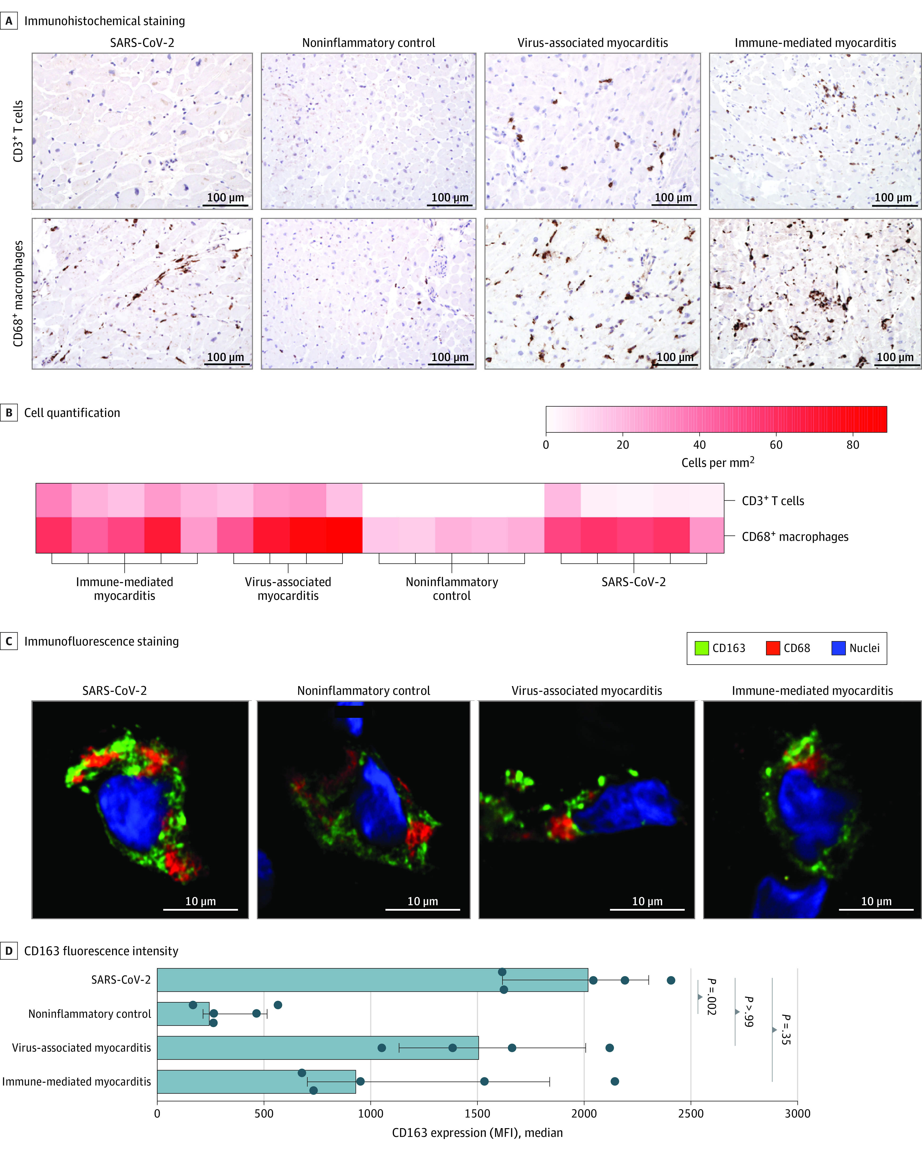
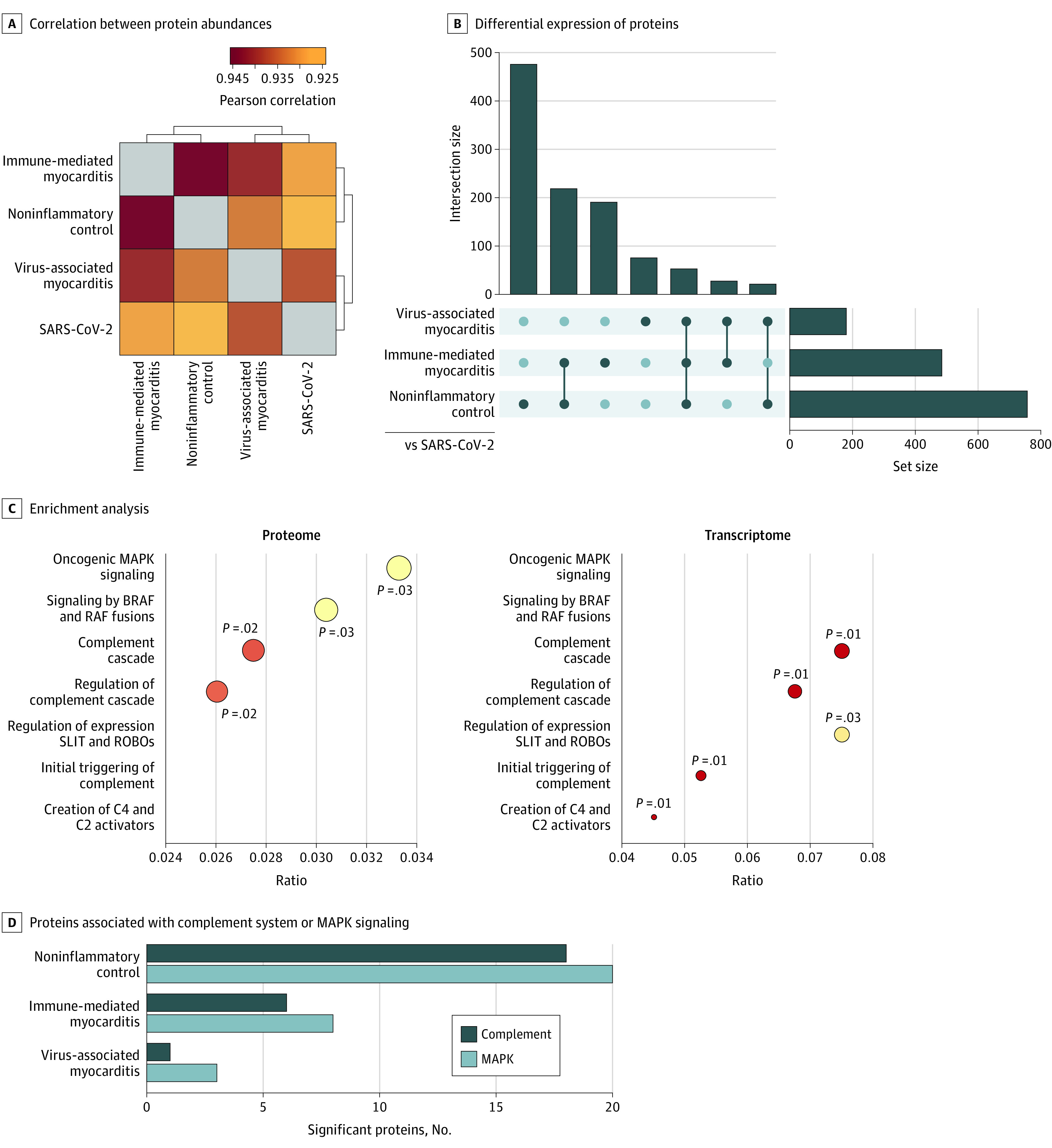
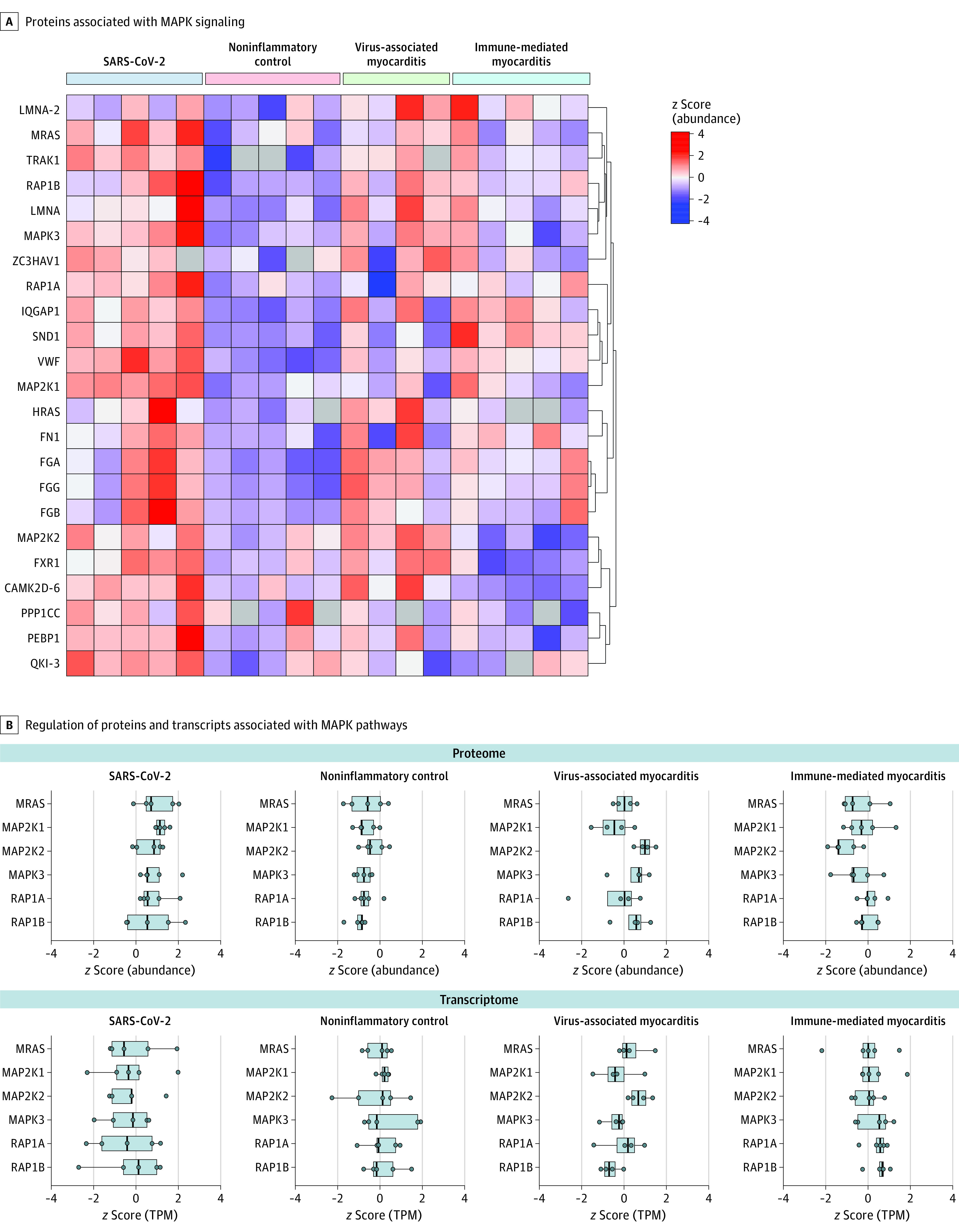
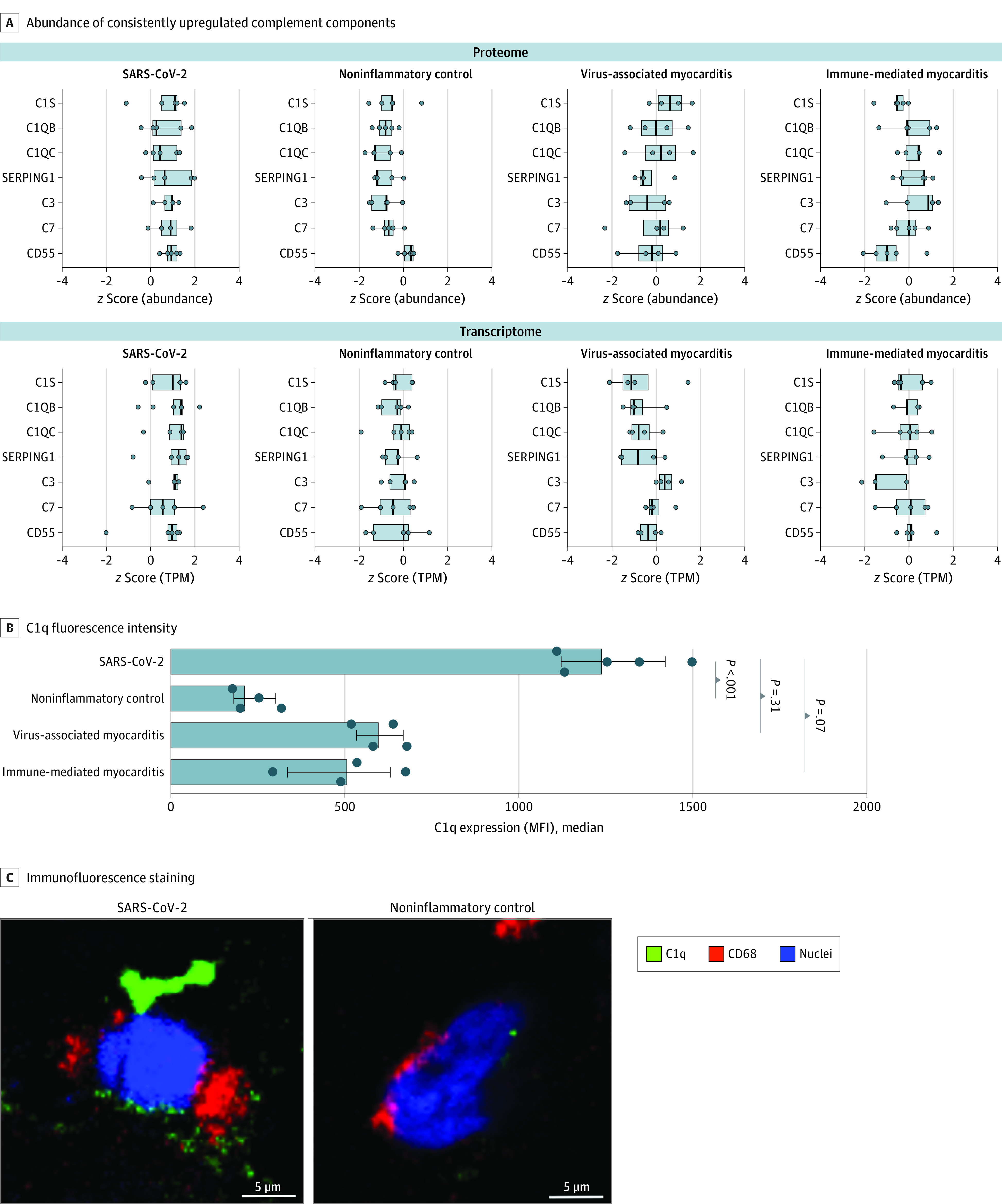
Results: Among 19 participants, the median age was 58 years (range, 37-76 years), and 15 individuals (79%) were male. Data on race and ethnicity were not collected. The abundance of CD163+ macrophages was generally higher in the cardiac tissue of patients with myocarditis, whereas lymphocyte counts were lower in the tissue of patients with SARS-CoV-2 infection vs patients with non-SARS-CoV-2 virus-associated and immune-mediated myocarditis. Among those with SARS-CoV-2 infection, components of the complement cascade, including C1q subunits (transcriptomic analysis: 2.5-fold to 3.6-fold increase; proteomic analysis: 2.0-fold to 3.4-fold increase) and serine/cysteine proteinase inhibitor clade G member 1 (transcriptomic analysis: 1.7-fold increase; proteomic analysis: 2.6-fold increase), belonged to the most commonly upregulated transcripts and differentially abundant proteins. In cardiac macrophages, the abundance of C1q was highest in SARS-CoV-2 infection. Assessment of important signaling cascades identified an upregulation of the serine/threonine mitogen-activated protein kinase pathways.

Conclusions and relevance: This case series found that the cardiac immune signature varied in inflammatory conditions with different etiologic characteristics. Future studies are needed to examine the role of these immune pathways in myocardial inflammation.


Conflict of interest statement

Conflict of Interest Disclosures: Dr Hausleiter reported receiving personal fees from Abbott Vascular and Edwards Lifesciences outside the submitted work. Dr Grabmaier reported receiving grants from the Deutsches Zentrum für Herz-Kreislauf-Forschung and personal fees from AstraZeneca and Lilly Deutschland outside the submitted work. No other disclosures were reported.

Figures

Figure 1.
Figure 1.. Histologic Characterization of Cardiac Specimens
A, Immunohistochemical staining of endomyocardial biopsy (EMB) sections for each group (original magnification, ×200; no hematoxylin-eosin staining was performed). Brown indicates C3+ T cells or CD68+ macrophages. B, CD68+ macrophages and CD3+ T cells per mm2 in each EMB sample. C, Confocal images of immunofluorescence expression in cardiac macrophages. Nuclei were stained with DAPI. D, Quantification of CD163 expression (median fluorescence intensity [MFI]) in macrophages for all 4 groups. Error bars represent IQRs. Analysis of variance and Dunn multiple comparison tests were used to determine P values; bars to the left of each P value indicate the groups being compared. DAPI indicates 4′,6-diamidino-2-phenylindole.
Figure 2.
Figure 2.. Distinct Molecular Signature in SARS-CoV-2–Associated Inflammatory Cardiomyopathy
A, Pearson correlation map of protein abundances indicating similarities and differences of the 4 groups based on proteome. Brackets indicate euclidean hierarchical clustering of samples. B, Shared or unique differential expression of significant proteins (measured using a t test, with significance set at q <0.01 and fold change >2) in the specific comparisons between the SARS-COV-2 group (reference) vs the other study groups. The number of significant proteins within each comparison is depicted by the set size, whereas the intersection size represents the quantity of overlap. C, Gene set enrichment analysis of significantly differential proteins and RNA transcripts in the comparison of SARS-CoV-2 vs noninflammatory control samples in reference to the Reactome pathway database (false discovery rate <0.05). Circle size represents protein or transcript counts for each term, the smallest circle indicating 6, and large circles indicating 23. Circle colors indicate P values (range of .01 [red] to .03 [yellow]). D, Count of significant proteins associated with mitogen-activated protein kinase (MAPK) signaling or the complement system in the respective t test comparisons of specimens from patients from the SARS-Cov-2 group vs the other study groups. BRAF indicates B-Raf protein; RAF, Raf kinase; ROBO, roundabout receptor; and SLIT, Slit protein.
Figure 3.
Figure 3.. Upregulation of MAPK Pathways in SARS-CoV-2 Infection
A, z Score normalized abundances of proteins with significant association to MAPK signaling. Row clustering (indicated by brackets) was determined by euclidean distance. B, Points indicate the distribution of z scored abundances or TPM values within each group of examples shown in A; boxes represent median values (vertical line) and IQRs. TPM indicates transcripts per million. The subchain expansions are given in the eAppendix in Supplement 1.
Figure 4.
Figure 4.. Upregulation of the Complement Cascade in SARS-CoV-2 Infection
A, z Score–normalized abundances or TPM values of complement components that were consistently upregulated in proteome and transcriptome analysis. TPM indicates transcripts per million. B, Quantification of C1q expression (median fluorescence intensity [MFI]) in macrophages for all 4 groups. C1q staining was not possible in 2 samples (1 immune-mediated myocarditis, and 1 noninflammatory control) due to limited sample amounts. Each dot shows the C1q MFI of 1 patient. Whiskers represent IQRs. Analysis of variance and Dunn multiple comparison tests were used to determine P values; bars to the left of each P value indicate the groups being compared. C, Representative confocal images of C1q, CD68, and 4′,6-diamidino-2-phenylindole (DAPI, nuclear staining) immunofluorescence in cardiac macrophages from patients with SARS-CoV-2 infection vs noninflammatory cardiomyopathy. SERPING 1 indicates serine/cysteine proteinase inhibitor clade G member 1.

References

    1. Tschöpe C, Ammirati E, Bozkurt B, et al. . Myocarditis and inflammatory cardiomyopathy: current evidence and future directions. Nature Rev Cardiol. 2021;18(3):169-193. doi:10.1038/s41569-020-00435-x - DOI - PMC - PubMed
    1. Kühl U, Pauschinger M, Noutsias M, et al. . High prevalence of viral genomes and multiple viral infections in the myocardium of adults with “idiopathic” left ventricular dysfunction. Circulation. 2005;111(7):887-893. doi:10.1161/01.CIR.0000155616.07901.35 - DOI - PubMed
    1. Lindner D, Fitzek A, Bräuninger H, et al. . Association of cardiac infection with SARS-CoV-2 in confirmed COVID-19 autopsy cases. JAMA Cardiol. 2020;5(11):1281-1285. doi:10.1001/jamacardio.2020.3551 - DOI - PMC - PubMed
    1. Sandoval Y, Januzzi JL Jr, Jaffe AS. Cardiac troponin for assessment of myocardial injury in COVID-19: JACC review topic of the week. J Am Coll Cardiol. 2020;76(10):1244-1258. doi:10.1016/j.jacc.2020.06.068 - DOI - PMC - PubMed
    1. Puntmann VO, Carerj ML, Wieters I, et al. . Outcomes of cardiovascular magnetic resonance imaging in patients recently recovered from coronavirus disease 2019 (COVID-19). JAMA Cardiol. 2020;5(11):1265-1273. doi:10.1001/jamacardio.2020.3557 - DOI - PMC - PubMed