Review
. 2022 Sep;37(9):2013-2036.
doi: 10.1007/s00467-021-05328-w. Epub 2021 Dec 15.

Rickets guidance: part I-diagnostic workup

Affiliations
Review

Rickets guidance: part I-diagnostic workup

Dieter Haffner et al. Pediatr Nephrol. 2022 Sep.

Abstract

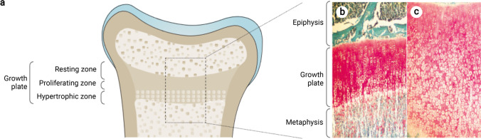
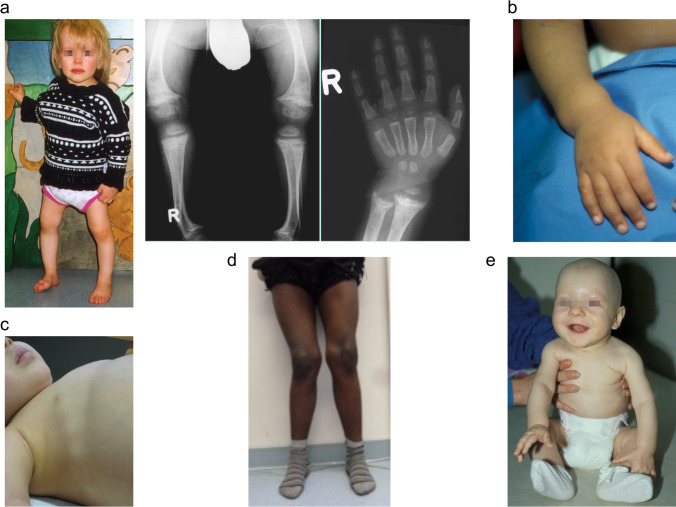
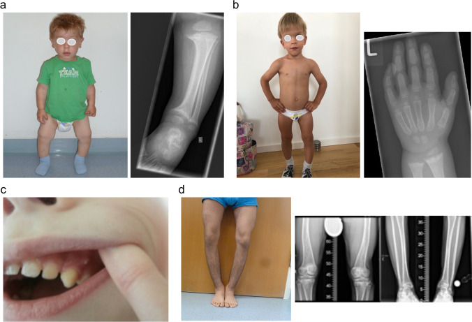
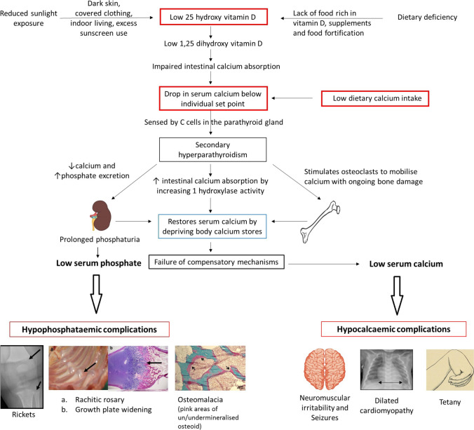
Rickets is a disease of the growing child arising from alterations in calcium and phosphate homeostasis resulting in impaired apoptosis of hypertrophic chondrocytes in the growth plate. Its symptoms depend on the patients' age, duration of disease, and underlying disorder. Common features include thickened wrists and ankles due to widened metaphyses, growth failure, bone pain, muscle weakness, waddling gait, and leg bowing. Affected infants often show delayed closure of the fontanelles, frontal bossing, and craniotabes. The diagnosis of rickets is based on the presence of these typical clinical symptoms and radiological findings on X-rays of the wrist or knee, showing metaphyseal fraying and widening of growth plates, in conjunction with elevated serum levels of alkaline phosphatase. Nutritional rickets due to vitamin D deficiency and/or dietary calcium deficiency is the most common cause of rickets. Currently, more than 20 acquired or hereditary causes of rickets are known. The latter are due to mutations in genes involved in vitamin D metabolism or action, renal phosphate reabsorption, or synthesis, or degradation of the phosphaturic hormone fibroblast growth factor 23 (FGF23). There is a substantial overlap in the clinical features between the various entities, requiring a thorough workup using biochemical analyses and, if necessary, genetic tests. Part I of this review focuses on the etiology, pathophysiology and clinical findings of rickets followed by the presentation of a diagnostic approach for correct diagnosis. Part II focuses on the management of rickets, including new therapeutic approaches based on recent clinical practice guidelines.

Keywords: Fibroblast growth factor 23; Nutritional rickets; Osteomalacia; Rickets; Vitamin D; Vitamin D-dependent rickets; X-linked hypophosphatemia.


Conflict of interest statement

D.H. and D.S. received speaker fees, consultation fees, and research grants from Kyowa Kirin. All other authors declare no conflict of interest.

Figures

Fig. 1
Fig. 1
Morphology of the growth plate in rickets. (a,b) Morphology of a healthy, human growth plate (physis). The growth plate is characterized by maturation of chondrocytes (cartilage cells) occurring progressively from the epiphysis to the metaphysis. The border between the metaphysis and the growth plate is marked by a provisional zone of calcification of the cartilage matrix (red–pink staining) which undergoes resorption and replacement with mineralized bone (turquoise staining). (c) A rachitic growth plate showing a marked increase in longitudinal width, marked by the persistence of the zone of hypertrophic chondrocytes with lost columnar arrangement. The growth plate abnormalities are the consequence of impaired chondrocyte apoptosis and impaired mineralization of the cartilage matrix surrounding the apoptotic chondrocytes. Apoptosis of hypertrophic chondrocytes is induced by extracellular phosphate via phosphorylation of mitogen-activated protein kinase (MAPK) pathway intermediates and downstream inhibition of the caspase-9-dependent mitochondrial apoptotic pathway. Thus, reduction in ambient phosphate availability to the chondrocyte, which is common to all forms of rickets, seems to be central to the impaired apoptosis. The ligand 1,25-dihydroxyvitamin D and its receptor may also be involved. Finally, expansion of the hypertrophic chondrocyte zone can be induced by impaired vascularization, influenced by vascular endothelial growth factor, which is regulated by MAPK pathway intermediates. Figure reproduced from Carpenter et al. with permission [1]
Fig. 2
Fig. 2
Clinical features of calcipenic rickets. (a) 18-month-old girl presenting with genu vara, and widening of the growth plates and metaphyseal fraying on X-rays, caused by nutritional rickets. (b, c) Two infants with widening of the wrist and rachitic rosary, respectively, due to nutritional rickets. (d) 14-year-old boy with genu valga due to nutritional rickets. (e) Infant with alopecia due to vitamin D-dependent rickets type 2A. Figure 2a, b, and e are reproduced from Schnabel and Haffner with permission [109]
Fig. 3
Fig. 3
Clinical features of phosphopenic rickets. (a) 2-year-old boy diagnosed with X-linked hypophosphatemia (XLH) at the age of 2 years, presenting with disproportionate short stature (–2.3 SD score), genu vara, and widening of growth plates and metaphyseal fraying on X-rays. (b) 3-year-old patient with XLH started on treatment with active vitamin D and phosphate at the age of 2 years showing disproportionate short stature (height, –2.4 SD score), frontal bossing, dolichocephalus and mild signs of rickets on X-ray. (c) Dental abscess on an apparently healthy tooth in a child with XLH. (d) 16-year-old boy with autosomal-recessive hypophosphatemic rickets type 2 (ARHR2) showing genu vara and mild ricketic signs on X-ray. Figure 3c reproduced with permission from Haffner and Linglart [110]
Fig. 4
Fig. 4
Regulation of calcium (A) phosphate (B) homeostasis. (A) The parathyroid gland senses extracellular calcium (Ca++) levels and secretes parathyroid hormone (PTH). PTH secretion is stimulated by low Ca++ and suppressed by high Ca++ plasma concentrations, respectively. PTH stimulates resorption of Ca++ from the bone, as well as renal Ca++ reabsorption. PTH also stimulates renal 1,25(OH)2D synthesis, and thereby enhances osteoclastic resorption of Ca++ from bone, as well as renal calcium reabsorption via TRPV5 and suppresses PTH synthesis. Circulating fibroblast growth factor 23 (FGF23) originates mainly from osteocytes. FGF23 suppresses both renal 1,25(OH)2D production and PTH. Both Ca++ and 1,25(OH)2D stimulate FGF23 production. The sites of defects of the different causes of hypocalcemic disorders (also called calcipenic rickets) are given in the purple boxes. This includes nutritional rickets due to vitamin D deficiency and/or impaired dietary calcium availability and genetic defects in vitamin D metabolism or action (vitamin D-dependent rickets (VDDR types 1–3)). Note: hypophosphatemia due to secondary hyperparathyroidism-associated renal phosphate wasting, rather than hypocalcemia ultimately causes rickets in calcipenic rickets. (B) FGF23 and PTH reduce renal tubular phosphate (Pi) reabsorption by reducing the apical expression of the sodium–phosphate cotransporters NaPi IIa and NaPi IIc. PTH stimulates, while FGF23 inhibits 1,25(OH)2D production. 1,25(OH)2D increases intestinal absorption of dietary Pi by enhancing NaPi IIb expression and stimulates FGF23 synthesis. PTH and FGF23 affect each other’s production through a negative feedback loop by as yet unknown mechanisms. The sites of defects of the different causes of hypophosphatemic disorders (“phosphopenic rickets”) are given in the purple boxes. This includes impaired dietary phosphate availability and genetic defects: XLH, X-linked hypophosphatemia; ARHR1/2/3, autosomal recessive hypophosphatemic rickets 1/2/3; FD/MAS, Fibrous dysplasia/McCune-Albright syndrome; HHRH, hereditary hypophosphatemic rickets with hypercalciuria; IIH, idiopathic hypercalcemia; ADHR, autosomal dominant hypophosphatemic rickets. *FGF23-protein resistant to degradation. Created with BioRender.com
Fig. 5
Fig. 5
Vitamin D homeostasis and hereditary causes of impaired VDDR function. The overall metabolic control of vitamin D homeostasis is shown. Synthesis of calciferols via sunlight exposure or dietary intake of vitamin D2 and D3 is given on the left-hand side. The genes involved in the different forms of VDRR are indicated in italics in the rounded boxes. Created with BioRender.com
Fig. 6
Fig. 6
Algorithm for the evaluation of a child presenting with rickets. The differential diagnoses are based on the mechanisms leading to hypophosphatemia, i.e., high parathyroid hormone (PTH) activity (calcipenic rickets), inadequate intestinal phosphate absorption, or renal phosphate wasting (phosphopenic rickets). The latter may be due to either primary tubular defects or high serum FGF23 concentratons. Further details are given in Table 1; FGF23, fibroblast growth factor 23. Created with BioRender.com
Fig. 7
Fig. 7
Pathophysiology of nutritional rickets, due to vitamin D deficiency and/or dietary calcium deficiency. Both etiologies result in calcium deprivation and hyperparathyroidism occurs in an attempt to maintain normal serum calcium levels. In the long run, calcium deprivation and phosphate loss result in hypocalcaemic and hypophosphataemic complications. Figure reproduced from Uday and Högler with permission [29]

References

    1. Carpenter TO, Shaw NJ, Portale AA, Ward LM, Abrams SA, Pettifor JM. Rickets Nat Rev Dis Primers. 2017;3:17101. doi: 10.1038/nrdp.2017.101. - DOI - PubMed
    1. Sabbagh Y, Carpenter TO, Demay MB. Hypophosphatemia leads to rickets by impairing caspase-mediated apoptosis of hypertrophic chondrocytes. Proc Natl Acad Sci U S A. 2005;102:9637–9642. doi: 10.1073/pnas.0502249102. - DOI - PMC - PubMed
    1. Penido MG, Alon US. Hypophosphatemic rickets due to perturbations in renal tubular function. Pediatr Nephrol. 2013;29:361–373. doi: 10.1007/s00467-013-2466-z. - DOI - PubMed
    1. Tiosano D, Hochberg Z. Hypophosphatemia: The common denominator of all rickets. J Bone Miner Metab. 2009;27:392–401. doi: 10.1007/s00774-009-0079-1. - DOI - PubMed
    1. Shore RM, Chesney RW. Rickets: Part I. Pediatr Radiol. 2013;43:140–151. doi: 10.1007/s00247-012-2532-x. - DOI - PubMed