

Three-dimensional printing of patient-specific lung phantoms for CT imaging: Emulating lung tissue with accurate attenuation profiles and textures
- PMID: 34910309
- PMCID: PMC8828694
- DOI: 10.1002/mp.15407
Three-dimensional printing of patient-specific lung phantoms for CT imaging: Emulating lung tissue with accurate attenuation profiles and textures
Abstract
Purpose: Phantoms are a basic tool for assessing and verifying performance in CT research and clinical practice. Patient-based realistic lung phantoms accurately representing textures and densities are essential in developing and evaluating novel CT hardware and software. This study introduces PixelPrint, a 3D printing solution to create patient-based lung phantoms with accurate attenuation profiles and textures.
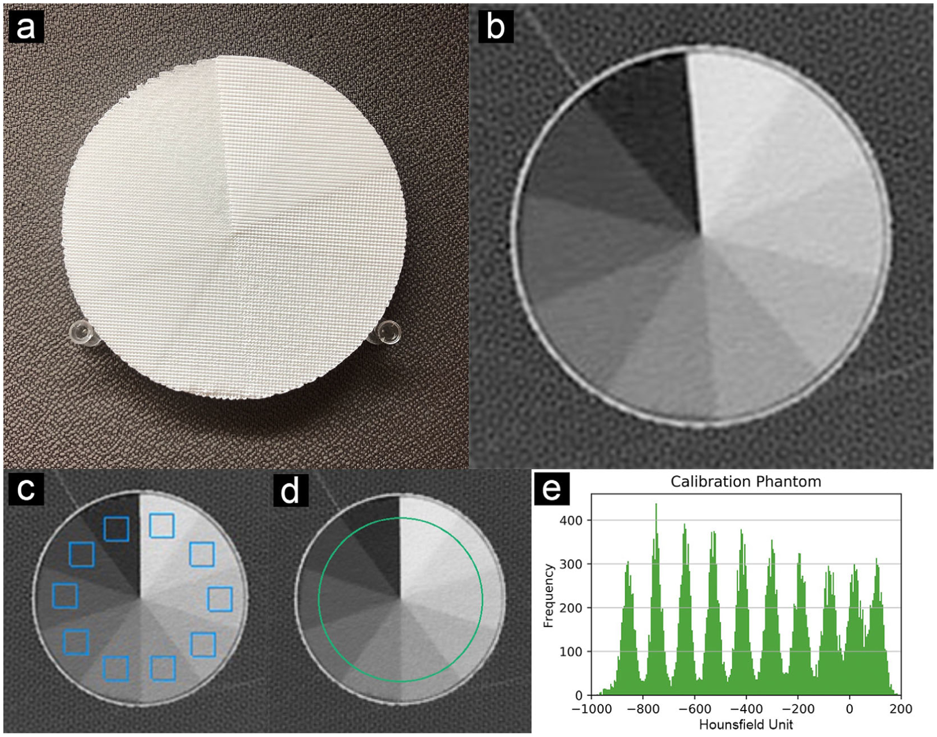
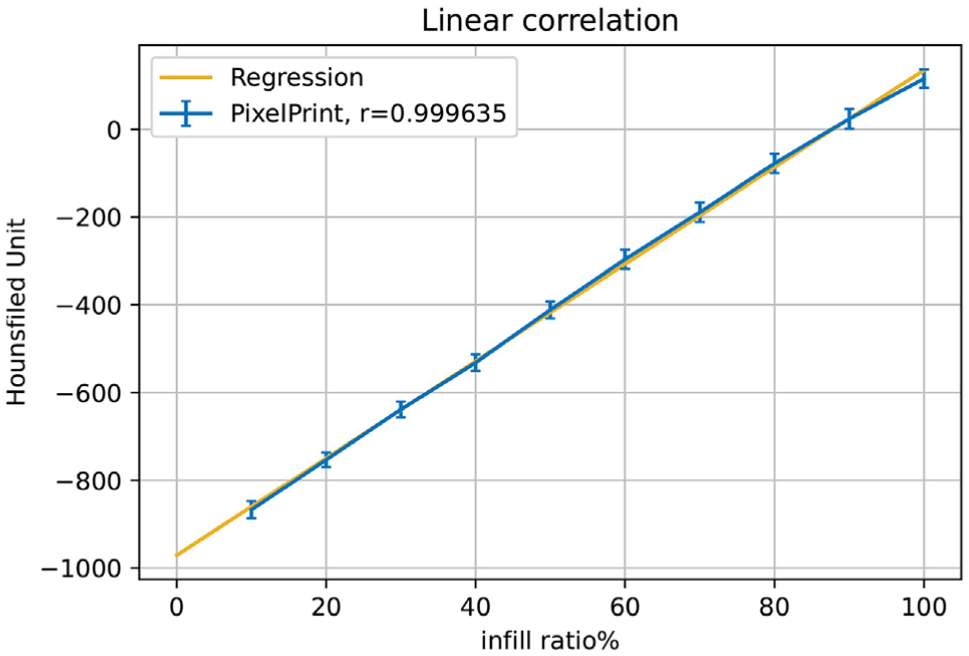
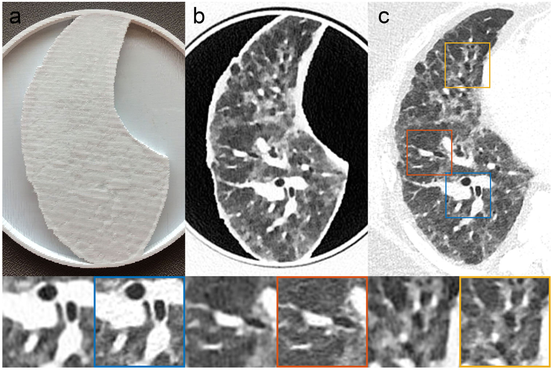
Methods: PixelPrint, a software tool, was developed to convert patient digital imaging and communications in medicine (DICOM) images directly into FDM printer instructions (G-code). Density was modeled as the ratio of filament to voxel volume to emulate attenuation profiles for each voxel, with the filament ratio controlled through continuous modification of the printing speed. A calibration phantom was designed to determine the mapping between filament line width and Hounsfield units (HU) within the range of human lungs. For evaluation of PixelPrint, a phantom based on a single human lung slice was manufactured and scanned with the same CT scanner and protocol used for the patient scan. Density and geometrical accuracy between phantom and patient CT data were evaluated for various anatomical features in the lung.
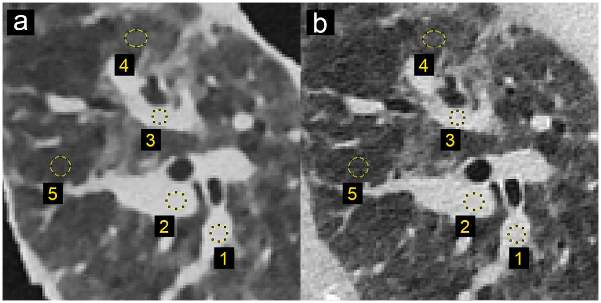
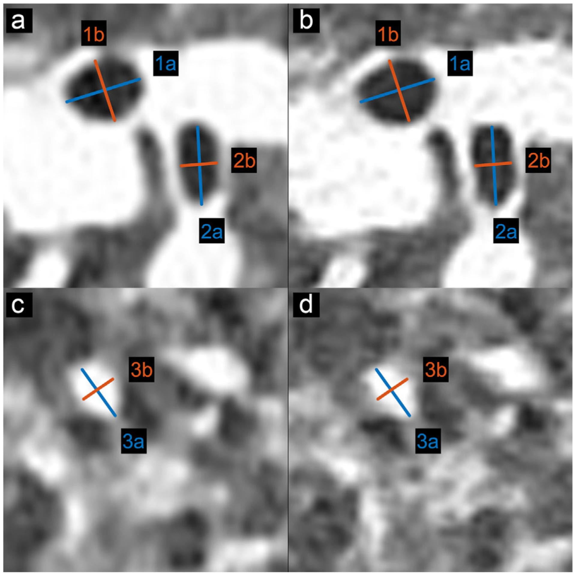
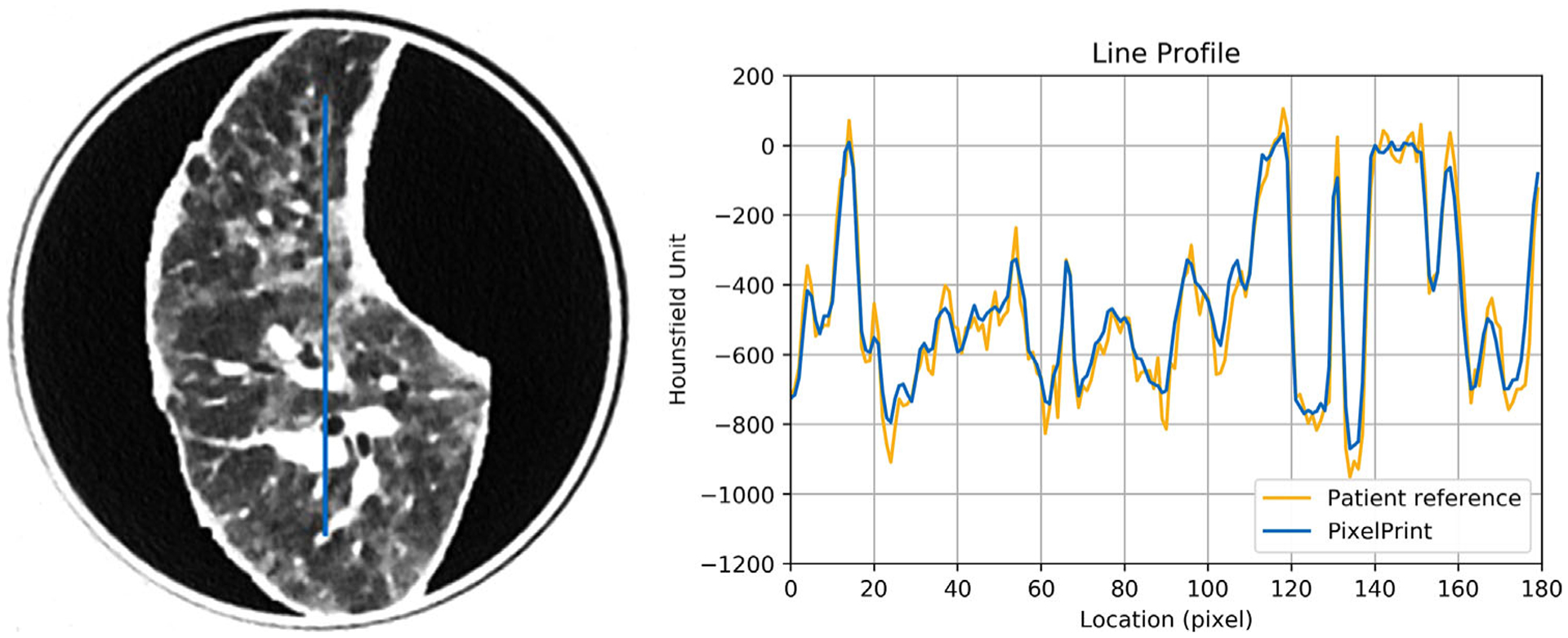
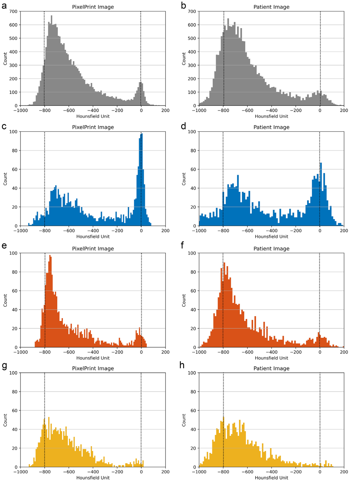
Results: For the calibration phantom, measured mean HU show a very high level of linear correlation with respect to the utilized filament line widths, (r > 0.999). Qualitatively, the CT image of the patient-based phantom closely resembles the original CT image both in texture and contrast levels (from -800 to 0 HU), with clearly visible vascular and parenchymal structures. Regions of interest comparing attenuation illustrated differences below 15 HU. Manual size measurements performed by an experienced thoracic radiologist reveal a high degree of geometrical correlation of details between identical patient and phantom features, with differences smaller than the intrinsic spatial resolution of the scans.
Conclusion: The present study demonstrates the feasibility of 3D-printed patient-based lung phantoms with accurate organ geometry, image texture, and attenuation profiles. PixelPrint will enable applications in the research and development of CT technology, including further development in radiomics.
Keywords: 3D printing; computed tomography; image quality; lung; quality assurance; radiomics.
© 2021 American Association of Physicists in Medicine.
Conflict of interest statement
CONFLICT OF INTEREST
The authors declare no conflict of interest.
Figures
References
-
- Shahrubudin N, Lee TC, Ramlan R. An overview on 3D printing technology: technological, materials, and applications. Procedia Manuf. 2019;35:1286–1296. 10.1016/j.promfg.2019.06.089 - DOI
