Review
. 2021 Dec 15;9(2):eESP00222021.
doi: 10.1128/ecosalplus.ESP-0022-2021. Epub 2021 Dec 13.

Localization, Assembly, and Activation of the Escherichia coli Cell Division Machinery

Affiliations
Review

Localization, Assembly, and Activation of the Escherichia coli Cell Division Machinery

Petra Anne Levin et al. EcoSal Plus. .

Abstract

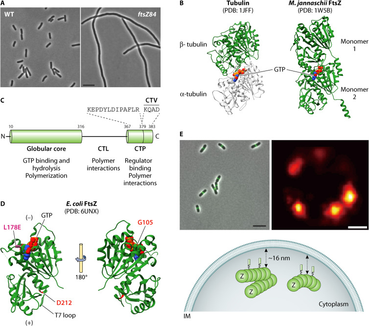
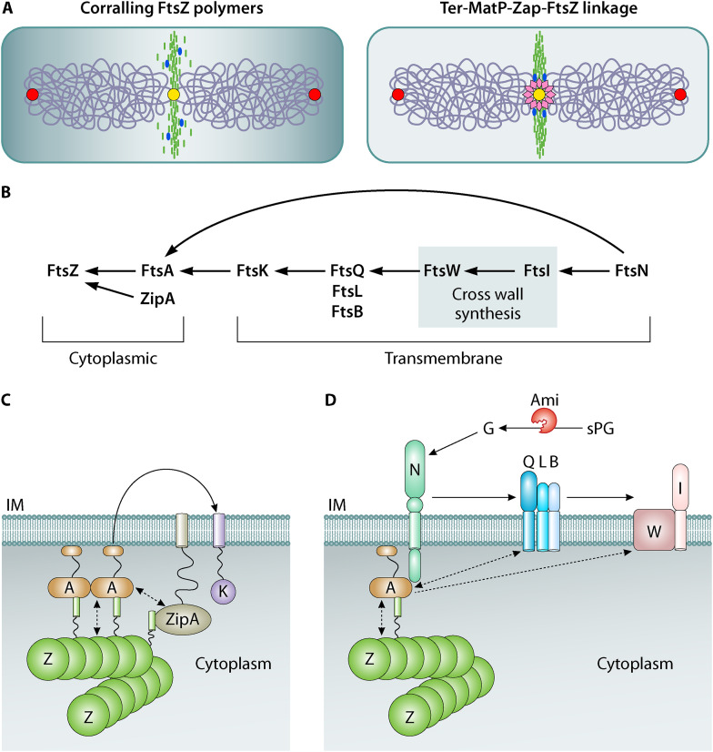
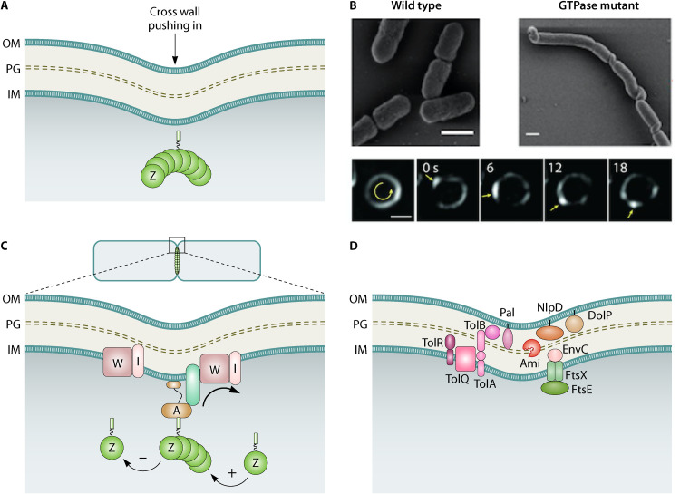
Decades of research, much of it in Escherichia coli, have yielded a wealth of insight into bacterial cell division. Here, we provide an overview of the E. coli division machinery with an emphasis on recent findings. We begin with a short historical perspective into the discovery of FtsZ, the tubulin homolog that is essential for division in bacteria and archaea. We then discuss assembly of the divisome, an FtsZ-dependent multiprotein platform, at the midcell septal site. Not simply a scaffold, the dynamic properties of polymeric FtsZ ensure the efficient and uniform synthesis of septal peptidoglycan. Next, we describe the remodeling of the cell wall, invagination of the cell envelope, and disassembly of the division apparatus culminating in scission of the mother cell into two daughter cells. We conclude this review by highlighting some of the open questions in the cell division field, emphasizing that much remains to be discovered, even in an organism as extensively studied as E. coli.

Keywords: Escherichia coli; FtsZ; cell division; divisome; peptidoglycan.


Figures

FIG 1
FIG 1
The tubulin-like FtsZ plays a foundational role in E. coli division. (A) Phase images of wild-type E. coli under permissive (30°C) and ftsZ84 (Ts) mutant cells under restrictive (42°C) growth temperatures (M. Buczek, unpublished data). FtsZ84 fails to localize to the nascent division site under nonpermissive growth conditions. Bar, 5 μm. (B) Three-dimensional structures of Tubulin and FtsZ: an α/β heterodimer of tubulin (PDB 1JFF) (left); Methanococcus jannaschii FtsZ (PDB 1W5B) dimer made up of two identical monomers (right). The C-terminal linker (CTL) and C-terminal peptide (CTP) domains are not present in the structure. Sandwiched in the dimeric units of each protein is a space-filled model of GTP. The two structures were modeled using UCSF Chimera (https://www.cgl.ucsf.edu/chimera/). (C) The domain architecture of FtsZ consists of a short disordered N-terminal end followed by the globular core, which contains the tubulin-signature motif responsible for binding GTP and GTP hydrolysis residues. The core is linked to the C-terminal peptide (CTP) by an intrinsically disordered linker region (CTL). The 14 aa CTP serves as a common binding site for various FtsZ binding proteins and can be further split into conserved and variable (CTV) regions. (D) The structure of E. coli FtsZ (ftsZL178E; residue L178 is shown in pink; PDB 6UNX) monomer bound to GTP is shown and was generated using UCSF Chimera. FtsZ L178E is incompetent for assembly in the presence of GTP (32). Other critical residues D212 and G105 are labeled and are mutated in strains ftsZ2 (D212G) and ftsZ84 (G105S) alleles referred to in the main text. In the polymer, the T7 synergy loop at the bottom surface is inserted into the nucleotide binding pocket of the second subunit resulting in GTP hydrolysis. Monomers are added toward the T7 loop-end of the growing polymer referred to as the “+” end. Monomers disperse from the end to which GTP is bound, which is referred to as the “−” end of a treadmilling FtsZ polymer. (E) Conventional fluorescence images of FtsZ polymers at midcell in wild-type E. coli cells. ZapA-GFP is used as a proxy for Z ring localization in these cells (A. Cardenas Arevalo, unpublished data). Bar, 5 μm; super resolution PALM microscopy image of FtsZ polymer assemblies in a cross section of the “ring” in a wild-type cell reveals a heterogeneous wreath-like arrangement (image courtesy of Z. Jason Lyu and Jie Xiao). Bar, 200 nm. A diagrammatic interpretation of cytoplasmic FtsZ polymers arranged in a random, heterogenous structure 16 nm away from the inner membrane (IM). The FtsZ polymers are tethered to the IM by FtsA or ZipA (not shown).
FIG 2
FIG 2
Assembly and activation of the divisome. (A) Left, FtsZ polymers (green) are corralled at midcell by a number of factors. Negative positional factors Min and nucleoid occlusion (NO), which together generate a high to low gradient of inhibitor concentration from poles to the cell center (gray), prevent FtsZ assembly elsewhere in the cell. The replicating ori regions (red ovals) and the ter region (yellow) on a duplicating chromosome are marked. FtsZ cross-linking proteins, including ZapA, ZapC, and ZapD (blue), are postulated to condense FtsZ polymers through lateral interactions to ensure efficient recruitment of cell wall synthesis enzymes to midcell. The FtsZ CTL domain is also considered to contribute to FtsZ lateral interactions. Right, the Ter-MatP linkage provides a positive positional signal that possibly links chromosome replication and segregation with FtsZ placement. The DNA-binding protein MatP (pink diamonds) binds both the terminus (ter) region on the chromosome and the ZapB-ZapA complex. (B) The genetically defined assembly of the essential E. coli divisome proteins is shown. The direction of the arrows represents the presence of a protein that helps in the recruitment of the next protein in the sequence. In this scheme, the proteins that constitute the cytokinetic ring at the cytoplasmic face of the IM can be visualized initially followed by the integral membrane proteins which predominantly play structural, regulatory, and synthesis roles in cell wall assembly. FtsN, the last protein to be recruited, can in certain genetically defined backgrounds arrive early to the divisome and through its interactions with FtsA (arrow) back recruit the other proteins. (C) The cytoplasmic core of the cytokinetic ring is composed of three proteins: FtsZ, FtsA, and ZipA. Together, FtsA and ZipA anchor FtsZ filaments to the membrane. Of the two, FtsA plays the primary role in division progress. ZipA influences FtsA polymerization (dotted double arrow) and enhances the recruitment of other division proteins to the midcell site (curved arrow; only FtsK is included in this figure for clarity). FtsZ and FtsA polymer architecture are considered to be influenced by each other (dotted double arrow). (D) In dividing cells, high levels of FtsN accumulate at the septum where it interacts with FtsA on the cytoplasmic side. The periplasmic essential (E) subdomain of FtsN is involved in activating (arrow) the FtsQLB complex. The resulting conformational changes in FtsQLB allow it to transition from a recruitment state to an activated form. At this point, FtsL interacts with FtsI to stimulate the transglycosylase and transpeptidase activities of FtsW and FtsI, respectively, leading to septal PG (sPG) synthesis. The periplasmic SPOR domain of FtsN also interacts with denuded glycan strands (G) at the septum, formed due to the action of amidases (Ami) cleaving the sPG. This invokes a septal PG feedback loop and the recruitment of additional FtsN molecules to midcell. Signals from both cytoplasmic (FtsA-FtsN) and periplasmic (FtsN-PG and FtsN-FtsQLB) compartments lead to the activation of cross wall synthesis. FtsA and FtsQ interactions may help stabilize FtsQLB directly or indirectly (dotted double arrow). FtsA oligomerization state may also influence FtsWI activity (dotted arrow). IM, inner membrane; PG, peptidoglycan; OM, outer membrane.
FIG 3
FIG 3
FtsZ treadmilling drives uniform distribution of cross wall synthases leading to septal PG (sPG) synthesis, constriction, and scission. (A) Accumulating evidence shows that the “pushing” force of sPG synthesis is the major driver of constriction and has long been known to be the rate-limiting step. FtsZ membrane anchors FtsA and ZipA are not shown. (B) Super resolution microscopy images of E. coli FtsZ treadmilling across the septum at different time points is shown (reprinted from reference with permission of the publisher). Treadmilling is GTP dependent and influences the distribution of septal cross wall synthesis enzymes (reprinted from reference with permission of the publisher). (C) Cartoon of treadmilling FtsZ polymers guiding sPG synthesis. The polarity of the FtsZ polymer is shown with growing (+) and shrinking (−) ends. The treadmilling FtsZ polymer is linked (via essential divisomal factors represented as a green rod) to sPG synthases and can move bidirectionally around the septal plane. The essential septal transglycosylase FtsW and transpeptidase FtsI are shown in two tracks. The FtsZ track (shown associated via other divisiomal proteins in green) promotes uniform distribution of the two synthases around the septum (arrow). Independent of FtsZ (shown alone), the enzymatically active synthases generate new cross wall exhibiting slower directional movements. (D) PG and OM remodeling is coordinated during division. The activity of the periplasmic amidases (Ami) involved in PG hydrolysis is controlled from the IM by FtsEX-linked activation of the periplasmic EnvC and from the OM by lipoproteins NlpD-DolP. The Tol-Pal complex spanning the cell envelope layers is energized by the proton motive force across the IM and plays a major role in constriction. This protein complex coordinates the constriction of the OM with the restructuring of the PG as well. IM, inner membrane; PG, peptidoglycan; OM, outer membrane.

References

    1. Nanninga N. 1991. Cell division and peptidoglycan assembly in Escherichia coli. Mol Microbiol 5:791–795. doi:10.1111/j.1365-2958.1991.tb00751.x. - DOI - PubMed
    1. de Boer PAJ. 2010. Advances in understanding E. coli cell fission. Curr Opin Microbiol 13:730–737. doi:10.1016/j.mib.2010.09.015. - DOI - PMC - PubMed
    1. Haeusser DP, Margolin W. 2016. Splitsville: structural and functional insights into the dynamic bacterial Z ring. Nat Rev Microbiol 14:305–319. doi:10.1038/nrmicro.2016.26. - DOI - PMC - PubMed
    1. Du S, Lutkenhaus J. 2019. At the heart of bacterial cytokinesis: the Z ring. Trends Microbiol 27:781–791. doi:10.1016/j.tim.2019.04.011. - DOI - PMC - PubMed
    1. Söderström B, Daley DO. 2017. The bacterial divisome: more than a ring? Curr Genet 63:161–164. doi:10.1007/s00294-016-0630-2. - DOI - PubMed

Publication types

MeSH terms

LinkOut - more resources