. 2021 Dec:74:103745.
doi: 10.1016/j.ebiom.2021.103745. Epub 2021 Dec 12.

Elevated Wnt2 and Wnt4 activate NF-κB signaling to promote cardiac fibrosis by cooperation of Fzd4/2 and LRP6 following myocardial infarction

Affiliations

Elevated Wnt2 and Wnt4 activate NF-κB signaling to promote cardiac fibrosis by cooperation of Fzd4/2 and LRP6 following myocardial infarction

Chao Yin et al. EBioMedicine. 2021 Dec.

Abstract

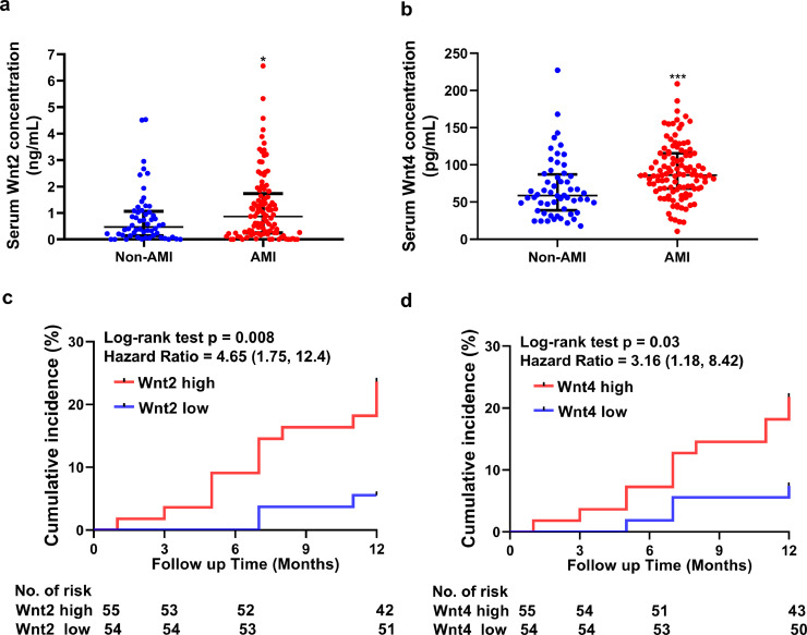
Background: Acute myocardial infarction (AMI)-induced excessive myocardial fibrosis exaggerates cardiac dysfunction. However, serum Wnt2 or Wnt4 level in AMI patients, and the roles in cardiac fibrosis are largely unkown.

Methods: AMI and non-AMI patients were enrolled to examine serum Wnt2 and Wnt4 levels by ELISA analysis. The AMI patients were followed-up for one year. MI mouse model was built by ligation of left anterior descending branch (LAD).

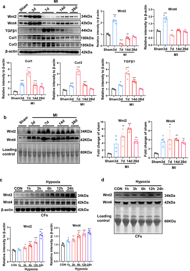
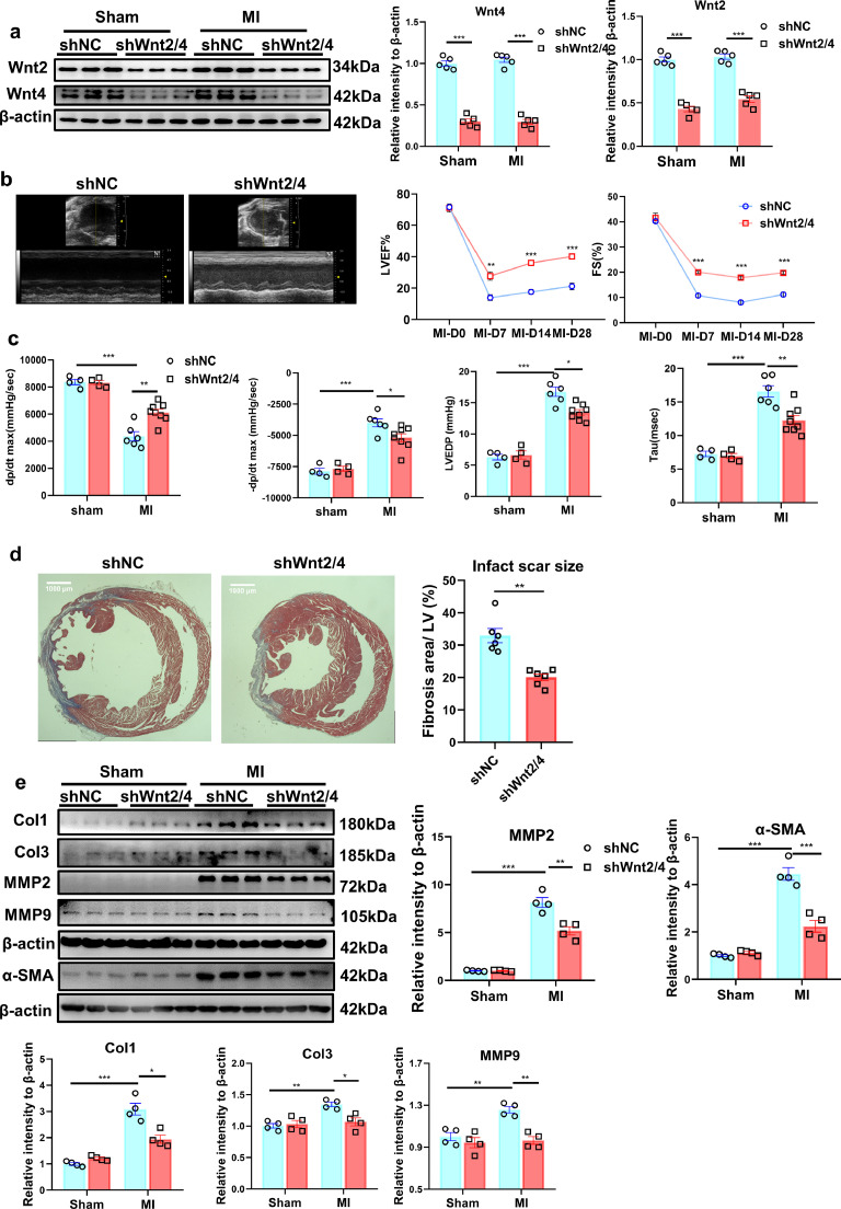
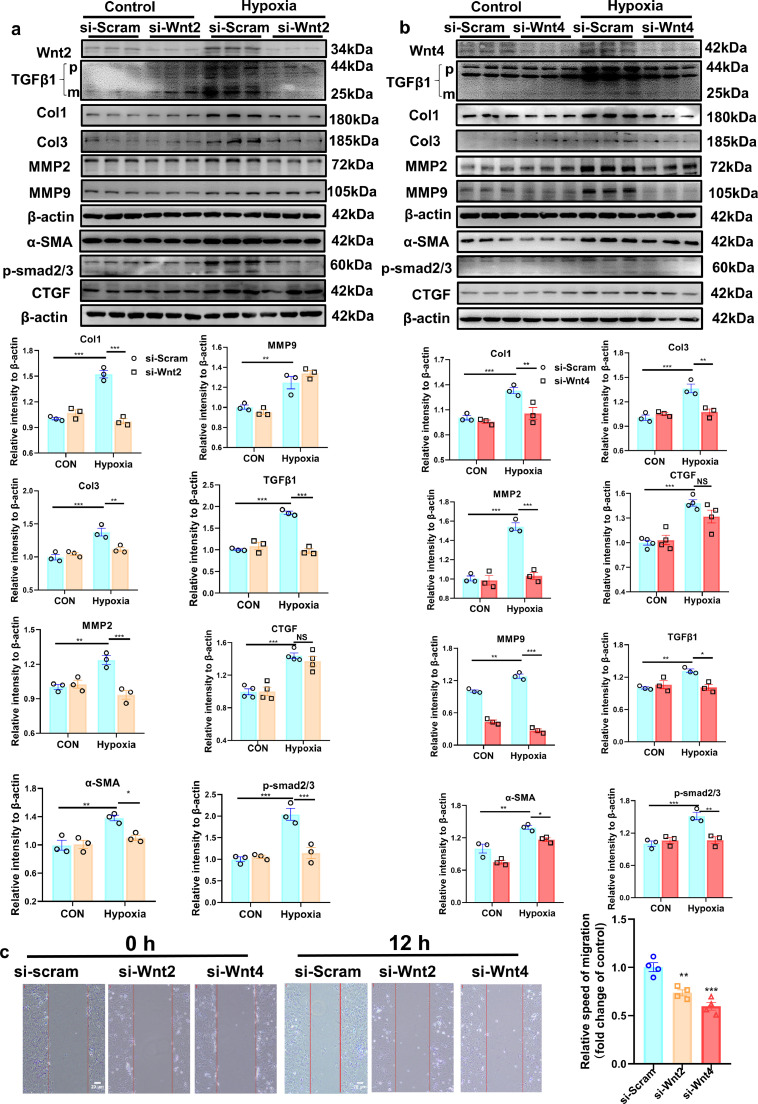
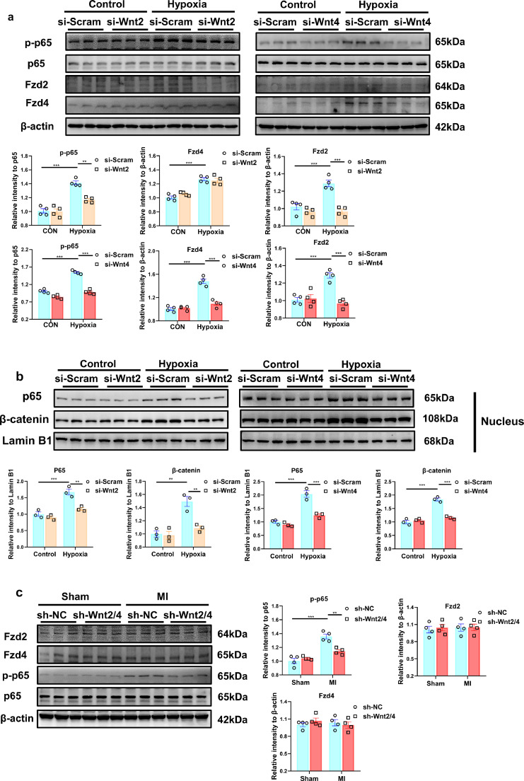
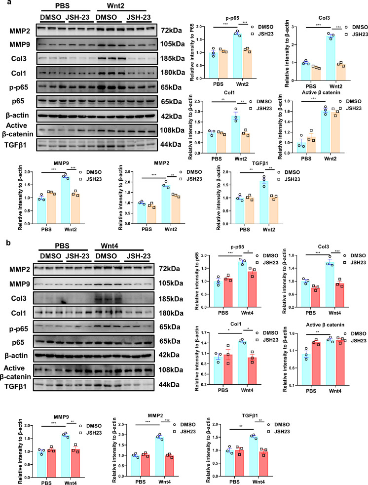
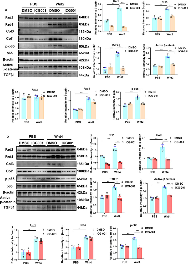
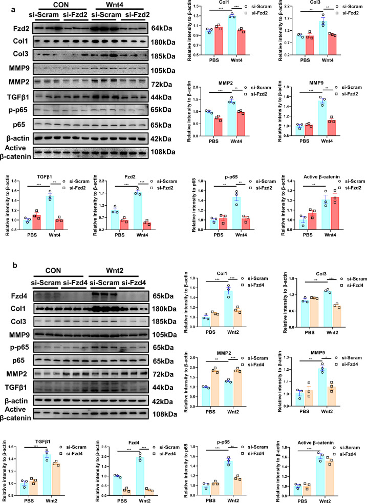
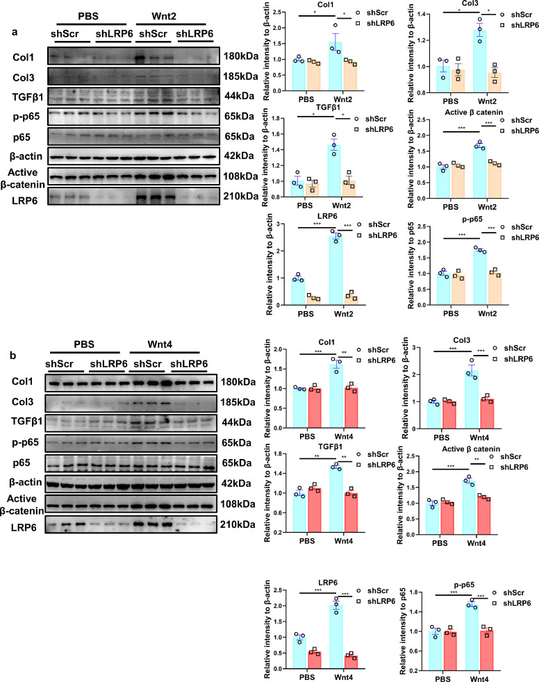
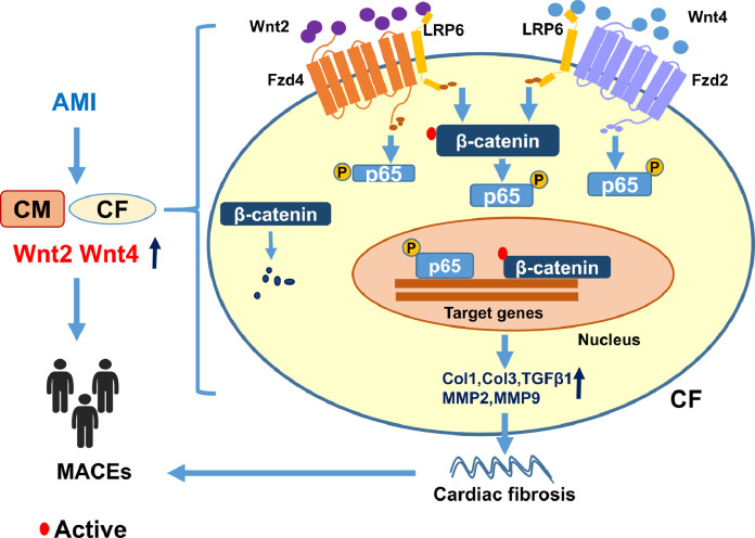
Findings: Serum Wnt2 or Wnt4 level was increased in patients with AMI, and the elevated Wnt2 and Wnt4 were correlated to adverse outcome of these patients. Knockdown of Wnt2 and Wnt4 significantly attenuated myocardial remodeling and cardiac dysfunction following experimental MI. In vitro, hypoxia enhanced the secretion and expression of Wnt2 and Wnt4 in neonatal rat cardiac myocytes (NRCMs) or fibroblasts (NRCFs). Mechanistically, the elevated Wnt2 or Wnt4 activated β-catenin /NF-κB signaling to promote pro-fibrotic effects in cultured NRCFs. In addition, Wnt2 or Wnt4 upregulated the expression of these Wnt co-receptors, frizzled (Fzd) 2, Fzd4 and (low-density lipoprotein receptor-related protein 6 (LRP6). Further analysis revealed that Wnt2 or Wnt4 activated β-catenin /NF-κB by the co-operation of Fzd4 or Fzd2 and LRP6 signaling, respectively.

Interpretation: Elevated Wnt2 and Wnt4 activate β-catenin/NF-κB signaling to promote cardiac fibrosis by cooperation of Fzd4/2 and LRP6 in fibroblasts, which contributes to adverse outcome of patients with AMI, suggesting that systemic inhibition of Wnt2 and Wnt4 may improve cardiac dysfunction after MI.

Keywords: Cardiac fibrosis; Frizzled; Myocardial infarction; NF-κB; Wnt.


Conflict of interest statement

Declaration of Competing Interest The authors declare no conflict of interest.

Figures

Fig 1
Fig. 1
Serum Wnt2 and Wnt4 are increased in patients with AMI and correlated to the increased risk of adverse outcomes of patients (a, b) ELISA analysis of serum Wnt2 and Wnt4 level in a total of 109 patients with AMI and 56 non-AMI patients. Data are expressed as means±SD, *p < 0.05, ** p < 0.01, *** p < 0.001 by Student's t test. (c, d) Kaplan–Meier incidence of MACEs in one year according to high or low level of serum Wnt2 or Wnt4. Wnt2 and Wnt4 were dichotomized into 2 categories with categorical analysis including higher than median and lower than median. Wnt2 high: ≥0.86 (ng/mL); Wnt2 low: < 0.86(ng/mL); Wnt4 high:≥86.2(pg/mL); Wnt4 low: < 86.2(pg/mL). MACEs: major adverse cardiovascular events. Estimated HR, 95% CIs, and p values were calculated. Statistics: The cumulative incidence of the MACE was determined by the Kaplan–Meier method, and the difference between groups was compared using the log-rank test. MACEs: major adverse cardiovascular events. Estimated HR, 95% CIs, and p values were calculated.
Fig 2
Fig. 2
Serum and cardiac Wnt2 and Wnt4 levels are increased in mice following MI. (a) Western blot analysis of the expression of Wnt2, Wnt4, Col1, Col3 and TGF- β1 in the border zone of infarcted area at different time-points (3d, 7d, 14d, 28d) following MI. Sham operation was used as control. Values were expressed as means±S.E.M; * p < 0.05, ** p < 0.01, *** p < 0.001 vs sham group, n = 5/group. (b) Western blot analysis of serum Wnt2 and Wnt4 from sham group and different time-points (3d,7d,14d,28d) after MI. Values were expressed as means±S.E.M; * p < 0.05, ** p < 0.01, *** p < 0.001 vs sham group, n = 4/group. (c) Wnt2 and Wnt4 expression were analyzed by Western blot analysis in cultured neonatal rat cardiac fibroblasts (NRCFs) at different time-points (1 h,3 h,6 h,12 h,24  h) in response to hypoxia. Normaxia group was used as control. Values were expressed as means±S.E.M; * p < 0.05, ** p < 0.01, *** p < 0.001 vs control group. n = 4/group. (d) The representative images of Wnt2 and Wnt4 expression with Western blot analysis in conditional medium from neonatal rat cardiac fibroblasts (NRCFs) of control group and at different time-points (1 h,3 h,6 h,12 h,24 h) after hypoxia. In (b) and (d), a conventional Coomassie staining of polyvinylidene fluoride (PVDF) membranes showed the total protein load which was as the internal control (Loading control). The experiment was repeated for three times. MI: Myocardial infarction; CON: control. Statistics: One-way ANOVA with post-hoc Tukey test.
Fig 3
Fig. 3
Knockdown of Wnt2/Wnt4 suppresses cardiac dysfunction and fibrosis post-MI. 8-10 weeks old male mice were injected with shWnt2/4-AAV9 (shWnt2/4) or shScramble-AAV9 (shNC, as control) by tail vein at 3 weeks before sham or MI operation. (a) Wnt2 and Wnt4 levels were determined in the non-infarcted area by western blot method on day 28 post-MI. Values were expressed as means±S.E.M; *** p < 0.001 vs shNC group; n = 5/each group. Statistics: Two-way ANOVA with a Bonferroni post hoc test. (b) Echocardiographic analysis of mice at day 0, day7, day14 and day28 after MI. Left lane: Representative images of M-mode echocardiogram captured on the 28th day post-MI. Right lane: quantitative analysis of LVEF, FS; LVEF: Left ventricular ejection fraction; FS, fraction shortening. Values were presented as means±S.E.M. * p < 0.05, ** p < 0.01, *** p < 0.001 vs shNC group; n = 6/group. Statistics: Student's t test. (c) Hemodynamics analysis of dp/dt, -dp/dt, LVEDP and Tau at 21 days after MI in mice. Values were expressed as means±S.E.M. * p < 0.05;** p < 0.01;*** p < 0.001, n = 4-8 in each group. Statistics: Two-way ANOVA with a Bonferroni post hoc test. (d) Cardiac fibrosis was examined by Masson staining at 28 days after MI in mice. Bar =1000 μm. Values were expressed as means±S.E.M. ** p < 0.01 vs shNC group, n = 6 in each group. Statistics: Student's t test. (e) Col1, Col3, MMP2, MMP9 and α-SMA protein levels were analyzed by Western blot in border zone of infarcted area on day 28 post-MI. Values were presented as means±S.E.M .* p < 0.05, ** p < 0.01,*** p < 0.001; n = 4 in each group. Statistics: Two-way ANOVA with a Bonferroni post hoc test.
Fig 4
Fig. 4
Wnt2 or Wnt4 is involved in fibroblasts activation in response to hypoxia. (a,b) Western blot analysis of the expressions of Wnt2, Wnt4, Col1, Col3, TGFβ1, MMP9 MMP2, p-smad2/3, CTGF and α-SMA protein in neonatal rat cardiac fibroblasts (NRCFs). These NRCFs were pretreated with siRNA targeted Wnt2 or Wnt4 (si-Wnt2 or si-Wnt4) or si-Scramble (si-Scram) for 24 h, and then exposed to hypoxia or normoxia (Control) for 3 h. p: pro-TGF-β1; m: mature or active TGF-β1; TGF-β1 mature form was analyzed. Values were described as means±S.E.M; * p < 0.05, ** p < 0.01, *** p < 0.001; n = 3/group. Statistics: Two-way ANOVA with a Bonferroni post hoc test. c: Scratch assay for migratory ability of cardiac fibroblasts pre-transfected with si-scram (CON), si-Wnt2 or si-Wnt4 followed by hypoxia. The representative images were showed at 0 h and 12 h after hypoxia. The bar=20 um; Values were quantitified and expressed as means±S.E.M. ** p < 0.01, *** p < 0.001 vs si-scram group; n = 4/group. Statistics: One-way ANOVA with post-hoc Tukey test.
Fig 5
Fig. 5
Wnt2 or Wnt4 activates NF-κB pathway signaling and enhances the expression of Fzd4/Fzd2 in neonatal rat cardiac fibroblasts (NRCFs) in response to hypoxia. (a) The expressions of p-p65, P65, Fzd2 and Fzd4 in NRCFs were detected by Western blot analysis. n = 4/group. (b) The expressions of p65 and β-catenin were detected in nucleus of NRCFs by western blot. n = 3/group. (a,b) These NRCFs were pretreated with siRNA targeted Wnt2 or Wnt4 (si-Wnt2 or si-Wnt4) or si-Scramble (si-Scram) for 24 h, and then exposed to hypoxia or normoxia (Control) for 3 h. Values were expressed as means±S.E.M. * p < 0.05, ** p < 0.01, *** p < 0.001. (c) The expressions of p-p65, p65, Fzd 2 and Fzd 4 were detected in non-infarcted area by Western blot analysis. 8-10 weeks old male mice were intravenously injected with shWnt2/4-AAV9 (shWnt2/4) or shScramble-AAV9 (shNC) at 3 weeks before sham or MI operation. These proteins were detected at day 28 post MI or sham operation. n = 4 in each group. Values were expressed as means±S.E.M; ** p < 0.01, *** p < 0.001. Statistics: Two-way ANOVA with a Bonferroni post hoc test.
Fig 6
Fig. 6
NF-κB inhibitor ameliorates Wnt2/Wnt4-induced pro-fibrotic effects in neonatal rat cardiac fibroblasts (NRCFs). (a,b) The expressions of Col1, Col3, active β-catenin, MMP2, MMP9, p65, p-p65 and TGFβ1 were measured in NRCFs by Western blot analysis. These NRCFs were pretreated with JSH-23(NF-κB inhibitor, 10μM) or the same volume of DMSO for 1 h, followed by PBS, human recombinant Wnt2 (20 ng/ml) or Wnt4 (50 ng/ml) treatment for 24 h. Values were expressed as means±S.E.M. * p < 0.05, ** p < 0.01, *** p < 0.001; n = 3 in each group. Statistics: Two-way ANOVA with a Bonferroni post hoc test.
Fig 7
Fig. 7
Wnt/β-catenin inhibitor relieves Wnt2/Wnt4-induced pro-fibrotic effects in neonatal rat cardiac fibroblasts (NRCFs). (a,b) The expressions of Col1, Col3, active β-catenin, p65, TGFβ1, Fzd2, Fzd4 and p-p65 were measured in NRCFs by Western blot analysis. These NRCFs were pretreated with ICG-001(β-catenin inhibitor, 10 μM) or the same volume of DMSO for 1 h, followed by PBS, human recombinant Wnt2 (20 ng/ml) or Wnt4 (50 ng/ml) treatment for 24 h. Values were expressed as means±S.E.M. * p < 0.05, ** p < 0.01, *** p < 0.001, n = 3 in each group. Statistics: Two-way ANOVA with a Bonferroni post hoc test.
Fig 8
Fig. 8
Knockdown of Fzd2 or Fzd4 attenuates Wnt4- or Wnt2-induced pro-fibrotic effects in neonatal rat cardiac fibroblasts (NRCFs). (a,b) Western blot analysis of the expressions of Fzd2, Col1, Col3, active β-catenin, MMP2, MMP9, TGFβ1, and p-65 in NRCFs. Values were expressed as means±S.E.M. * p < 0.05, ** p < 0.01, *** p < 0.001; n = 3 in each group. Statistics: Two-way ANOVA with a Bonferroni post hoc test. (a) NRCFs were pretreated with siRNA targeted Fzd2 (si- Fzd2) or si-Scramble (si-Scram) for 24 h, followed by PBS, human recombinant Wnt4 (50 ng/ml) treatment for another 24 h. (b) NRCFs were pretreated with siRNA targeted Fzd4 (si- Fzd4) or si-Scramble (si-Scram) for 24 h, followed by PBS, human recombinant Wnt2 (20 ng/ml) treatment for another 24 h.
Fig 9
Fig. 9
Knockdown of LRP6 attenuates Wnt2- or Wnt4-induced pro-fibrotic effects in neonatal rat cardiac fibroblasts (NRCFs). (a,b) Western blot analysis of the expressions of Col1, Col3, active β-catenin, TGFβ1, LRP6 and p-65 in NRCFs. Cultured NRCFs were transfected with lenti-shScr (shScr) or lenti-LRP6(shLRP6) for 48 h and then followed by PBS, human recombinant Wnt2 (20 ng/ml) or Wnt4(50 ng/ml) treatment for another 24 h. Values were expressed as means±S.E.M. * p < 0.05, ** p < 0.01, *** p < 0.001. n = 3 /group. Statistics: Two-way ANOVA with a Bonferroni post hoc test.
Fig 10
Fig. 10
A proposed model of showing how Wnt2 and Wnt4 promote cardiac fibrosis by activation of β-catenin/NF-κB trough LRP6 and Fzds signaling following MI. MI induced the increased expression or secretion of Wnt2 and Wnt4 in cardiaomyocytes (CMs) or cardiac fibroblasts (CFs), elevated Wnt2 and Wnt4 promote fibrotic effects by activation of β-catenin/NF-κB signaling dependently on the cooperation of Fzd4 or Fzd2 and LRP6 signaling in cardiac fibroblasts, which contributes to cardiac fibrosis and dysfunction post-MI. Thus high Wnt2 and Wnt4 are independently associated with adverse outcome in AMI patients.

Similar articles

Cited by

References

    1. Townsend N., Wilson L., Bhatnagar P., Wickramasinghe K., Rayner M., Nichols M. Cardiovascular disease in Europe: epidemiological update 2016. Eur Heart J. 2016;37(42):3232–3245. - PubMed
    1. Seropian I.M., Toldo S., Van Tassell B.W., Abbate A. Anti-inflammatory strategies for ventricular remodeling following ST-segment elevation acute myocardial infarction. J Am Coll Cardiol. 2014;63(16):1593–1603. - PubMed
    1. van den Borne S.W., Diez J., Blankesteijn W.M., Verjans J., Hofstra L., Narula J. Myocardial remodeling after infarction: the role of myofibroblasts. Nat Rev Cardiol. 2010;7(1):30–37. - PubMed
    1. Chistiakov D.A., Orekhov A.N., Bobryshev Y.V. The role of cardiac fibroblasts in post-myocardial heart tissue repair. Exp Mol Pathol. 2016;101(2):231–240. - PubMed
    1. Clevers H. Wnt/beta-catenin signaling in development and disease. Cell. 2006;127(3):469–480. - PubMed

MeSH terms