Review
. 2022 Jan 12;19(2):10.1088/1478-3975/ac4390.
doi: 10.1088/1478-3975/ac4390.

Cell-extracellular matrix dynamics

Affiliations
Review

Cell-extracellular matrix dynamics

Andrew D Doyle et al. Phys Biol. .

Abstract

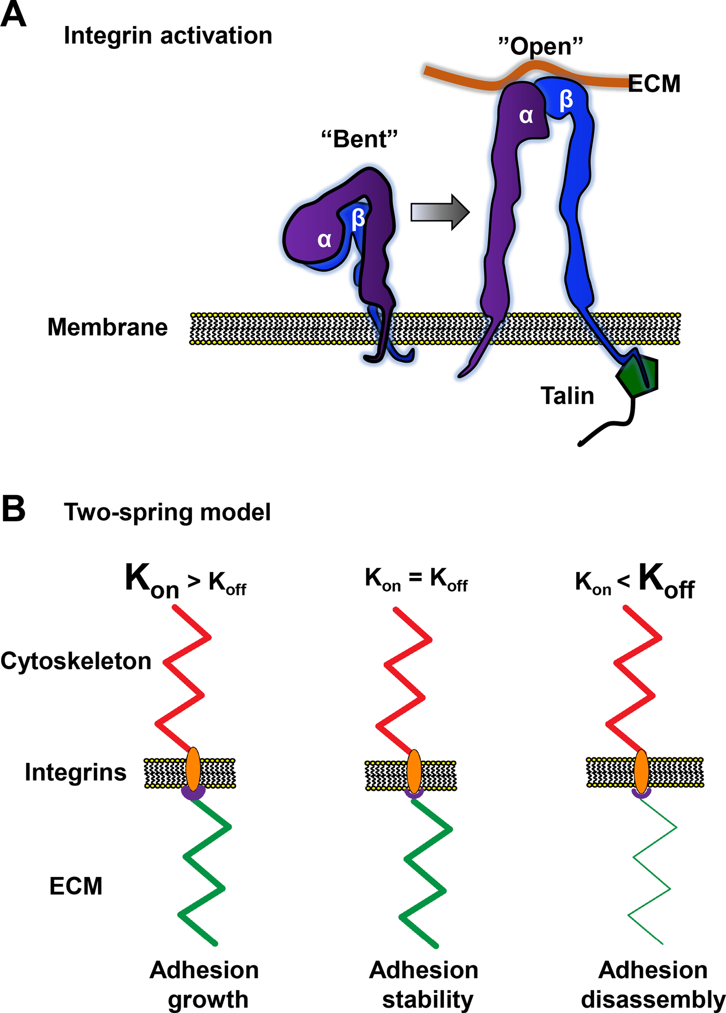
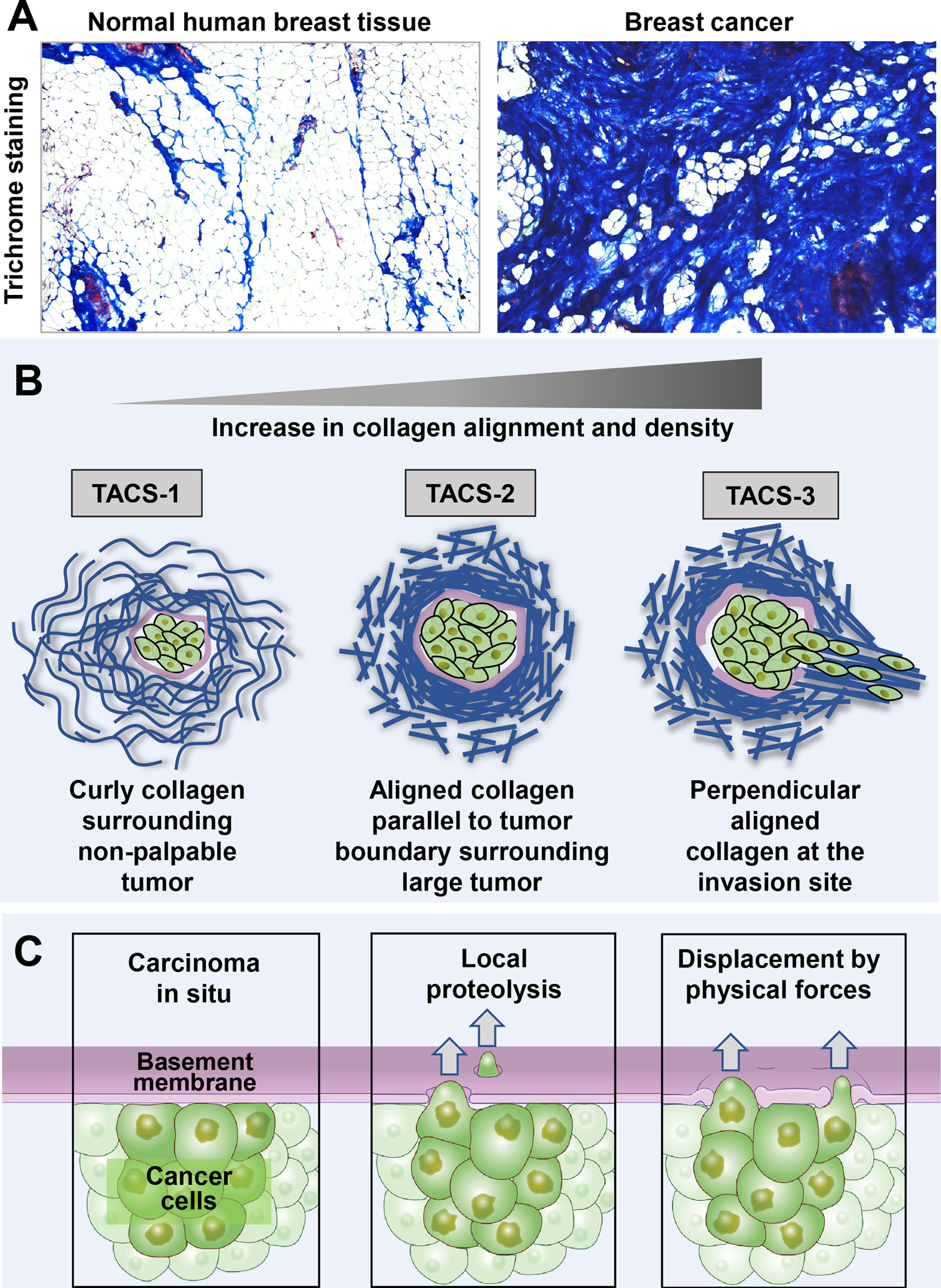
The sites of interaction between a cell and its surrounding microenvironment serve as dynamic signaling hubs that regulate cellular adaptations during developmental processes, immune functions, wound healing, cell migration, cancer invasion and metastasis, as well as in many other disease states. For most cell types, these interactions are established by integrin receptors binding directly to extracellular matrix proteins, such as the numerous collagens or fibronectin. For the cell, these points of contact provide vital cues by sampling environmental conditions, both chemical and physical. The overall regulation of this dynamic interaction involves both extracellular and intracellular components and can be highly variable. In this review, we highlight recent advances and hypotheses about the mechanisms and regulation of cell-ECM interactions, from the molecular to the tissue level, with a particular focus on cell migration. We then explore how cancer cell invasion and metastasis are deeply rooted in altered regulation of this vital interaction.

Keywords: cell migration; dynamics; extracellular matrix; focal adhesions.


Figures

Figure 1:
Figure 1:. Focal adhesions are mechanical signal hubs.
A) Schematic side view of integrins (orange) in the plasma membrane binding ECM and clustering together with intracellular focal adhesion proteins. The mechanosensitive proteins vinculin (blue) and talin (green) are shown interacting with actin monomers (light blue) that have assembled into stress fibers and are crosslinked with myosin II (red). Inset: focal adhesion proteins are organized in layers. B) TIRF images of EGFP-paxillin in a fibroblast showing numerous nascent (NA: yellow arrowheads) and focal (FA: blue arrowheads) adhesions that have formed on a 2D ECM. C) Paxillin (green), α5 integrin (red) and fibronectin (blue)-containing fibrillar adhesions (FX: white arrowheads). Fibroblasts were plated on Matrigel and medium was supplemented with labeled soluble fibronectin to observe the process of fibronectin fibrillogenesis. D) Fibroblast expressing EGFP-VASP (green) and mCherry-actin (magenta) on a fibronectin-coated 2D surface. Blue arrowheads indicate numerous focal adhesions associated with lamellipodia (LA). The kymograph at the right from the white-dashed region shows LA expansion followed by FA formation FAs, establishing the new leading edge. After FA elongation and maturation, a new LA can form. Time is in seconds. Scale bars: 10 μm.
Figure 2:
Figure 2:. Integrin activation and the two-spring model.
A) Schematic representation of an α and β integrin heterodimer in the “Bent” and “Open” configuration. In the “Bent” configuration, the integrin pair has weak ECM-binding affinity. In the “Open” or activated configuration, both subunits extend away from the membrane and the transmembrane domains and cytoplasmic tails separate. Talin is shown binding to the β cytoplasmic tail, which may assist in the activation process. B) Schematic representation of three scenarios in the two spring-model of cell adhesion, where the “springs” represent the ECM (green) and the cytoskeleton (red) with integrin/focal adhesion in between. In the 1st condition, Kon (associated with high ligand binding affinity: purple region) is larger than Koff (associated with contractility). ECM stiffness matches the cytoskeletal “spring,” leading to adhesion growth. In the 2nd scenario, both “springs” (Kon and Koff rates) are equal, resulting in adhesion stability. The 3rd situation shows high Koff (contractility higher than the binding of integrins can withstand) together with a softer ECM “spring,” which leads to focal adhesion disassembly.
Figure 3:
Figure 3:. The molecular clutch.
A side view of a cell on a 2D ECM (purple) showing the leading edge and crucial components of the molecular clutch. Three scenarios are shown. 1) In the absence of integrin-linked clutch proteins (green) engaging actin (yellow), the retrograde flow of F-actin associated with myosin-II contractility is high, greatly exceeding the rate of G-actin polymerization and no protrusion occurs (actin cannot push against the membrane). 2) With partial clutch engagement, the binding of clutch proteins to F-actin helps to slow retrograde flow at adhesion sites and allow the local actin polymerization rate to equal and then surpass rearward flow to weakly promote protrusion. 3) A fully engaged molecular clutch causes a significant reduction in local actin retrograde flow, promoting high leading-edge protrusion, advancing the lamella and forming a nascent adhesion in the lamellipodia.
Figure 4:
Figure 4:. Fibrillar adhesion dynamics and 3D cell migration.
A) A fibroblast expressing EGFP-paxillin (green) pulls soluble fibronectin (magenta) into fibrils shortly after plating on Matrigel. Inset kymograph of the white-dashed box shows the dynamic movement of paxillin upward toward the cell center, polymerizing fibronectin fibrils behind it. Time is in minutes. B) Schematic representation of how fibrillar adhesions can slide inwards using a contractile “winch”. C) A fibroblast stained for activated β1 integrin (left image; magenta in right) crawling through a 3D collagen hydrogel (green). D) A fibroblast expressing TagGFP2-LifeAct crawling through a collagen gel. Right image shows the ECM strain map, where warmer colors depict higher strain at the leading edge. Arrow indicates migration direction. Scale bars: A and C, 10 μm; D, 20 μm
Figure 5:
Figure 5:. Matrix remodeling during tumor progression and mechanisms of cancer cell invasion.
A) Breast cancer progression correlates with higher collagen density demonstrated by trichrome blue staining to measure collagen density in normal compared to tumor tissue from the same patient. B) Matrix remodeling closely correlates with tumor progression. Normal curly collagen fibrils surround a non-palpable tumor with localized collagen density around the periphery of the tumor mass (TACS-1). As the tumor enlarges, collagen is remodeled and appears more linear, dense, and aligned parallel to the tumor boundary (TACS-2). During cancer cell invasion, collagen becomes perpendicular to the tumor boundary at the invasion site (TACS-3). C) During cancer invasion, cancer cells can breach the basement membrane barrier by chemically degrading the matrix using proteolysis or physically displacing the matrix by pushing through the basement membrane using invasive protrusions.

References

    1. Case LB, Waterman CM. Integration of actin dynamics and cell adhesion by a three-dimensional, mechanosensitive molecular clutch. Nat Cell Biol. 2015;17(8):955–63. - PMC - PubMed
    1. Adebowale K, Gong Z, Hou JC, Wisdom KM, Garbett D, Lee HP, et al. Enhanced substrate stress relaxation promotes filopodia-mediated cell migration. Nat Mater. 2021. - PMC - PubMed
    1. Ridley AJ, Schwartz MA, Burridge K, Firtel RA, Ginsberg MH, Borisy G, et al. Cell migration: integrating signals from front to back. Science. 2003;302(5651):1704–9. - PubMed
    1. Chen WT. Mechanism of retraction of the trailing edge during fibroblast movement. J Cell Biol. 1981;90(1):187–200. - PMC - PubMed
    1. Kanchanawong P, Shtengel G, Pasapera AM, Ramko EB, Davidson MW, Hess HF, et al. Nanoscale architecture of integrin-based cell adhesions. Nature. 2010;468(7323):580–4. - PMC - PubMed

Publication types

LinkOut - more resources