. 2022 Feb 16;60(2):e0173721.
doi: 10.1128/JCM.01737-21. Epub 2021 Dec 15.

Comparison of Whole-Genome Sequence-Based Methods and PCR Ribotyping for Subtyping of Clostridioides difficile

Affiliations

Comparison of Whole-Genome Sequence-Based Methods and PCR Ribotyping for Subtyping of Clostridioides difficile

A Baktash et al. J Clin Microbiol. .

Abstract

Clostridioides difficile is the most common cause of antibiotic-associated gastrointestinal infections. Capillary electrophoresis (CE)-PCR ribotyping is currently the gold standard for C. difficile typing but lacks the discriminatory power to study transmission and outbreaks in detail. New molecular methods have the capacity to differentiate better and provide standardized and interlaboratory exchangeable data. Using a well-characterized collection of diverse strains (N = 630; 100 unique ribotypes [RTs]), we compared the discriminatory power of core genome multilocus sequence typing (cgMLST) (SeqSphere and EnteroBase), whole-genome MLST (wgMLST) (EnteroBase), and single-nucleotide polymorphism (SNP) analysis. A unique cgMLST profile (more than six allele differences) was observed in 82 of 100 RTs, indicating that cgMLST could distinguish most, but not all, RTs. Application of cgMLST in two outbreak settings with RT078 and RT181 (known to have low intra-RT allele differences) showed no distinction between outbreak and nonoutbreak strains in contrast to wgMLST and SNP analysis. We conclude that cgMLST has the potential to be an alternative to CE-PCR ribotyping. The method is reproducible, easy to standardize, and offers higher discrimination. However, adjusted cutoff thresholds and epidemiological data are necessary to recognize outbreaks of some specific RTs. We propose to use an allelic threshold of three alleles to identify outbreaks.

Keywords: Clostridioides difficile; core-genome MLST; typing methods; whole-genome MLST; whole-genome sequencing.


Conflict of interest statement

The authors declare a conflict of interest. D.W.E.: lecture fees from Gilead, outside the submitted work; E.J.K.: unrestricted research grant from Vedanta Bioscience, Boston.

D.W.E. received lecture fees from Gilead, outside the submitted work. E.J.K. received an unrestricted research grant from Vedanta Bioscience (Boston, MA). The other authors declare no conflict of interest.

Figures
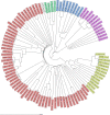
FIG 1
FIG 1
Neighbor joining tree from 100 unique ribotypes (RTs) based on SeqSphere cgMLST allele difference. Each RT is depicted with “RTn” followed by “reference” (belonging to the Leeds-Leiden collection) or “clinical” (non-Leeds-Leiden strain). RTs from multilocus sequence typing (MLST) clades 1, 2, 3, 4, and 5 are colored red, yellow, green, blue, and purple, respectively. RT131 has no designated MLST clade and is shown in white. The distance is given as the absolute allelic difference.
FIG 2
FIG 2
Clustering of different PCR RTs at different thresholds using SeqSphere+ core genome multilocus sequence typing (cgMLST; zero to six allelic difference). The number of clustering RTs is shown in blue, and the number of clusters at every threshold is shown in pink; e.g., at three allele differences, nine different RTs belong to seven clusters.
FIG 3
FIG 3
(A) Mean intra-RT allele and single-nucleotide polymorphism (SNP) difference shown for individual RTs from MLST clade 1 (RT001 to RT056), clade 2 (RT027 to RT244), clade 3 (RT023), clade 4 (RT017), and clade 5 (RT033 to RT127). Mean intra-RT allele difference per RT is shown in light green, turquois, and orange for SeqSphere+ core genome multilocus sequence typing (cgMLST), EnteroBase (EB) cgMLST1, and EB whole-genome multilocus sequence typing (wgMLST), respectively. Mean intra-RT SNP difference per RT is shown in red. (B) Mean intra-RT allele and SNP difference shown for MLST clade 1, clade 2, and clade 5. Mean intra-RT allele difference per clade is shown in light green, turquois, and orange for SeqSphere+ cgMLST, EB cgMLST, and EB wgMLST, respectively. Mean intra-RT SNP difference per clade is shown in red.
FIG 4
FIG 4
SeqSphere+ cgMLST analysis with minimum-spanning trees of two suspected CDI outbreaks of RT078 and RT181. (A) Minimum-spanning tree of RT078 (clade 5) CDI suspected outbreak with six cases (RT078, shown in red and in green) of which three were confirmed (shown in largest septated red circle) and added control strains of RTs belonging to clade 5 (RT033, RT045, RT066, RT078, RT126, and RT127 shown in blue). (B) Minimum-spanning tree of RT181 (clade 2) CDI suspected outbreak with 15 suspected and 15 confirmed cases (RT181, shown in septated red circles) and added control strains of RTs belonging to clade 2 (RT016, RT027, RT036, RT176, RT181 and RT198 shown in blue). The size and septation of the circle in the minimum-spanning trees corresponds to the number of included strains. The numbers between each circle correspond to the number of different alleles between the strains. The colored shadowing of circles represents a cluster with at most six allele differences that are genetically related. One or more strains inside a circle means that these strains have zero allele differences.
FIG 5
FIG 5
Visualized distance matrices of strain pairs based on cgMLST, wgMLST, and SNP analysis of isolates of cluster 1 as described in Fig. 4. (A) Visualized distance matrix of strain pairs belonging to cluster 1 of RT078. (B) Visualized distance matrix of strain pairs belonging to cluster 1 of RT181. Allele difference per pair of strains is shown in light green, turquois, and orange for SeqSphere+ cgMLST, EB cgMLST, and wgMLST, respectively. SNP difference per pair of strains is shown in red.

References

    1. Keller JJ, Kuijper EJ. 2015. Treatment of recurrent and severe Clostridium difficile infection. Annu Rev Med 66:373–386. 10.1146/annurev-med-070813-114317. - DOI - PubMed
    1. Smits WK, Lyras D, Lacy DB, Wilcox MH, Kuijper EJ. 2016. Clostridium difficile infection. Nat Rev Dis Primers 2:16020. 10.1038/nrdp.2016.20. - DOI - PMC - PubMed
    1. Indra A, Huhulescu S, Schneeweis M, Hasenberger P, Kernbichler S, Fiedler A, Wewalka G, Allerberger F, Kuijper EJ. 2008. Characterization of Clostridium difficile isolates using capillary gel electrophoresis-based PCR ribotyping. J Med Microbiol 57:1377–1382. 10.1099/jmm.0.47714-0. - DOI - PMC - PubMed
    1. Fawley WN, Knetsch CW, MacCannell DR, Harmanus C, Du T, Mulvey MR, Paulick A, Anderson L, Kuijper EJ, Wilcox MH. 2015. Development and validation of an internationally-standardized, high-resolution capillary gel-based electrophoresis PCR-ribotyping protocol for Clostridium difficile. PLoS One 10:e0118150. 10.1371/journal.pone.0118150. - DOI - PMC - PubMed
    1. Griffiths D, Fawley W, Kachrimanidou M, Bowden R, Crook DW, Fung R, Golubchik T, Harding RM, Jeffery KJ, Jolley KA, Kirton R, Peto TE, Rees G, Stoesser N, Vaughan A, Walker AS, Young BC, Wilcox M, Dingle KE. 2010. Multilocus sequence typing of Clostridium difficile. J Clin Microbiol 48:770–778. 10.1128/JCM.01796-09. - DOI - PMC - PubMed

LinkOut - more resources